INTRODUCTION
Microsporidia are obligate intracellular unicellular organisms, which are kinds of spore-forming fungal pathogens. Microsporidia can infect a wide variety of animals ranging from vertebrate to invertebrate species, including human beings (Didier et al. Reference Didier, Snowden and Shadduck1998). To date, more than 1300 species are identified. Of these, at least 13 species have been described as pathogens in humans, especially the AIDS patients (Didier et al. Reference Didier, Snowden and Shadduck1998; Corradi et al. Reference Corradi, Gangaeva and Keeling2008). Also, more than 150 species are reported using insect as host (Becnel and Andreadis, Reference Becnel, Andreadis, Wittner and Weiss1999), such as Nosema bombycis, which is the first identified microsporidia infecting Bombyx mori (Nageli, Reference Nageli1857; Becnel and Andreadis, Reference Becnel, Andreadis, Wittner and Weiss1999). In 2013, the genome of N. bombycis was released (Pan et al. Reference Pan, Xu, Li, Xia, Liu, Zhang, Li, Li, Liu, Yang, Liu, Zhang, Wu, Fan, Dang, Xiang, Tao, Li, Hu, Li, Lin, Luo, Geng, Wang, Long, Wan, He, Zhang, Lu and Patrick2013). There is no doubt that it will facilitate strongly to unveil the mystery underlying this parasite, such as the process of infection and invasion.
Now, it is reported that the spore wall proteins play key roles in the host–pathogens interaction process, including the pathogen infection, invasion and other pathogenic process (Weidner et al. Reference Weidner, Avila and Harris1992; Weidner and Halonen, Reference Weidner and Halonen1993). Some researchers discovered that spore wall proteins adhere to host cell using a mechanism involving host cell surface glycosaminoglycan (GAGs) (Hayman et al. Reference Hayman, Southern and Nash2005; Southern et al. Reference Southern, Jolly and Russell Hayman2006, Reference Southern, Jolly, Lester and Hayman2007). In Encephalitozoon cuniculi, 2 exospores SWP1, SWP2, 1 endospore SWP3, and a protein named EcEnP1 had been identified. It was indicated that EcEnP1 could function as a potential spore adherence ligand and be involved in host cell adherence and infection (Bohne et al. Reference Bohne, Ferguson, Kohler and Gross2000; Peuvel-Fanget et al. Reference Peuvel-Fanget, Polonais, Brosson, Texier, Kuhn, Peyret, Vivarè and Delbac2006; Xu et al. Reference Xu, Peter, Ann, Wang, Zhang, George and Louis2006; Southern et al. Reference Southern, Jolly, Lester and Hayman2007). In 2001, 2 exospore proteins, SWP1 and SWP2, were described in Encephalitozoon intestinalis (Hayman et al. Reference Hayman, Hayes, Amon and Nash2001). Recently, 14 hypothetical spore wall proteins were identified using proteomic-based approach in N. bombycis (Wu et al. Reference Wu, Li, Pan, Tan, Hu, Zhou and Xiang2008). To date, SWP1, SWP2 and SWP26 were expressed mainly on the endospore, other 2 proteins SWP3 and SWP5 were expressed on exospores in N. bombycis (Wu et al. Reference Wu, Li, Pan, Tan, Hu, Zhou and Xiang2008; Li et al. Reference Li, Wu, Pan, He, Zhang, Hu and Zhou2009, Reference Li, Pan, Li, Huang, Chen, Geng, Yang, Wang and Zhou2012). In function analysis, SWP26 was proved to be associated with the host cell adherence via the heparin binding motif (HBMs) (Li et al. Reference Li, Wu, Pan, He, Zhang, Hu and Zhou2009). SWP5 can interact with the polar tube proteins PTP2 and PTP3 and mediate the infection of N. bombycis (Li et al. Reference Li, Pan, Li, Huang, Chen, Geng, Yang, Wang and Zhou2012) or host cell invasion (Cai et al. Reference Cai, Lu, Qiu, Li and Feng2011). In addition, SWP26, SWP1, SWP3 and SWP12 were proved to be able of binding to deproteinated chitin spore coat of N. bombycis (Yang et al. Reference Yang, Dang, Tian, Long, Li, Li, Chen, Li, Pan and Zhou2014; Chen et al. Reference Chen, Long, Li, Geng, Li, Yang, Ma, Wu, Ma, Li, Pan and Zhou2013). However, among these reported proteins, only 2 spore wall proteins having tandem repeats were reported, including SWP1 of E. cuniculi and SWP2 of E. intestinalis. In these 2 proteins, SWP1 of E. cuniculi has 5 glycine- and serine-rich repeats at C-terminal and SWP2 of E. intestinalis has 50 repeats with 12-or 15-amino acids length at C-terminal , both of them having no proline in the tandem repeats (Bohne et al. Reference Bohne, Ferguson, Kohler and Gross2000; Hayman et al. Reference Hayman, Hayes, Amon and Nash2001). In this paper, a new spore wall protein NbSWP16 with 3 proline-rich tandem repeats was identified in N. bombycis.
MATERIALS AND METHODS
Microsporidian spore production and purification
Nosema bombycis CQ1, isolated from infected silkworms in Chongqing, China, was conserved in the China Veterinary Culture Collection Center (CVCC No.102059). The production and purification procedure of the spores was as described previously (Wu et al. Reference Wu, Li, Pan, Tan, Hu, Zhou and Xiang2008).
Sequence analysis
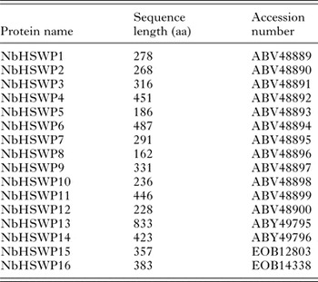
To date, 15 spore wall proteins have been described in N. bombycis (Appendix, Table A1). Here, by scanning the genome of N. bombycis, a protein (GenBank NO: EOB14338) annotated as spore wall protein and anchoring dick complex was found, we named it as NbHSWP16. The signal peptide of protein sequence was predicted using online tools (http://www.cbs.dtu.dk/services/SignalP) (Julenius et al. Reference Julenius, Mølgaard, Gupta and Brunak2005). Protein motif prediction was carried out using SMART proteomic tools (http://smart.embl-heidelberg.de). The glycosylation sites (http://www.cbs.dtu.dk/services/NetNGlyc) and phosphorylation sites (http://www.cbs.dtu.dk/services/NetPhos) were predicted. Glycosylphosphatidylinositol (GPI) anchor motif was predicted using the online program big-PI predictor (http://mendel.imp.ac.at/sat/gpi/gpi_server.html). Subcellular localization predication was performed using the PSORT server (http://psort.hgc.jp/). To find the homology proteins of NbHSWP16, BLASTP analysis was performed on the local database. Amino acid sequence alignments were generated using the ClustalX program. Multiple sequence alignment of NbHSWP16 and its homologous proteins were analysed phylogenetically using MEGA5 software to prepare a phylogenetic tree. The tree was constructed using the neighbour-joining method.
Analysis of gene transcription pattern
RT-PCR was performed to analyse the transcription of gene Nbhswp16. The midguts of infected silkworms were collected at 1–8 days and stored at −80°C. The methods used to extract the total RNA were as described previously (Li et al. Reference Li, Pan, Li, Huang, Chen, Geng, Yang, Wang and Zhou2012). The primers used to amplify the Nbhswp16 gene are as followed: forward primer: 5′-TTTAGCACCAATCCTCAACC-3′ and reverse primer: 5-TTGCCATAATAATGAAGGAG-3′. The tubulin-β gene used as an internal control was amplified using the primers: forward primer: 5′-TGGACGCCATTAGACAAG-3′ and reverse primer: 5′-GGCTCGAGTTCATAACATCTAACC-3′. PCR conditions of tubulin-β and Nbhswp16 were as follows: 94°C for 4 min, 25 cycles of 94°C for 45 s, 55°C for 45 s and 72°C for 1 min.
Expression, purification and polyclonal antibody production
The NbHSWP16 protein was expressed as a truncated form without the signal peptide in the N-terminal. Based on the genomic DNA sequence, polymerase chain reaction primers were designed as follows: forward primer: 5′-cgcggatccTCTGTGGAAATAAGAAAAGGC-3′ and reverse primer: 5′-gcgtcgacGTCAAAGGAAACACCATAAA-3′. The forward primer and the reverse primer contained a BamHI and SalI restriction site, respectively. When the PCR finished, a genomic DNA fragment that coded the amino acid region of 26–383 of NbHSWP16 protein was obtained. Then, the amplified fragment was cloned into the pMD19T vector (Invitrogen). After the DNA fragment was verified by sequence, it was cloned into prokaryotic expression vectors pET30a. The resulting recombinant plasmids were introduced into the Escherichia coli BL21 strain. The recombinant NbHSWP16 (rNbHSWP16) was induced at 37°C for 6 h in the presence of 0·4 mm IPTG. The expressed fusion protein was purified with a Ni-NTA Superflow Cartridge (Qiagen). Four mice were immunized with rNbHSWP16 mixed with Freund's adjuvant (1:1; sigma) and one more mice was injected PBS using as negative control. One week after the 4th injection, the mice were sacrificed and their sera were collected and stored at −20°C.
Western-blotting analysis
The spore protein of mature spores was extracted. Briefly, spores (1×10⁹ spores mL−1) were disrupted with 0·4 g acid-washed glass beads in buffer PBS pH 7·4 containing PMSF for 5 min. After centrifugation at 12 000 r m−1 for 10 min at 4°C, the supernatant was removed and the pellet was treated with 2·5% SDS, 0·1 m DTT and 2-Mercaptoethanol for 4 h at 4°C, then centrifuged and captured supernatant as spore protein sample. For immunoblotting analysis, about 10 μg protein sample was subjected to SDS-PAGE and transferred to a PVDF membrane (Millipore). After blocked with 5% (w/v) skim milk overnight at 4°C, the membrane was incubated with anti-NbHSWP16 or negative sera (1:1000) for 1 h at 37°C, then membranes were reacted with horseradish peroxidase-labelled goat anti-mouse IgG (Sigma). Following several washes, the membranes were developed with ECL Western blot detection kit (Thermo).
Immunofluorescence analysis
The purified mature spores were treated with 1% tritonX-100 for 30 min, then washed 3 times with PBS pH 7·4 and fixed with 4% paraformaldehyde at room temperature for 30 min. After rinsed 3 times with PBS, the fixed spores were blocked and incubated with polyclonal antibody or a negative serum for 60 min at 37°C. After 3 PBS washing, the spores were treated with FITC-conjugated goat anti-mouse IgG (1:100, Sigma). DNA was stained with 5 μg mL−1 DAPI for 10 min at room temperature. After several rinsed, spores were examined using OLYMPUS BX50 fluorescence microscope. The germinated spore coats were obtained by inducing the purified mature spores with 0·1 m K2CO3 for 2 h at 28°C in vitro and then the following treatment method used was similar as above.
Immunoelectron microscopy (IEM) analysis
Purified spores were fixed with 4% paraformaldehyde and 0·1% glutaraldehyde in 0·1 m sodium cacodylate buffer, pH 7·2, rinsed with PBS, dehydrated with graded ethanol and sequentially permeabilized in K4M at −20°C. The samples were then embedded and photopolymerized in K4M resin. Ultrathin sections were placed on nickel grids and immune-staining was performed. After blocked with standard blocking solution, the grids were incubated with anti-NbHSWP16 serum (1:20) or negative serum as primary antibody for 1 h at room temperature. Followed by incubation with gold conjugated anti-mouse IgG (1:100, Sigma), grids were rinsed with sterile water, dried, examined and photographed with an HITACHI H-7500 transmission electron microscope.
Spore adherence analysis
To analyse the role of NbSWP16 in spore adherence, antibody blocking was performed. Briefly, BmE-SWU1 cells that were used as host cells were seeded on a 24-well plate and allowed to grow for 16 h at 28°C. After the culture was removed, spores incubated with anti-NbSWP16 sera (dilution 1:50) as well as the negative sera were added onto the monolayer and incubated for 2 h at 28°C. After removing the unbounded spores and several washing, the average numbers of bounded spores per host cell were counted in at least 20 fields for each sample. Meanwhile, the anti-NbSWP5 was added together with anti-NbSWP16 to determine whether the antibodies have synergistic effect. The significance of the difference between the control and experimental assays was measured using t-test. Experiments were repeated at least 3 times.
Ethics statement
The approval for animal experiments was obtained from The Laboratory Animal Welfare and Ethics Committee of The Third Medical University, Chongqing, China. The agreement number is SYXK-PLA-20120031.
RESULTS
Characterization of the gene coding for the hypothetical spore wall protein NbHSWP16
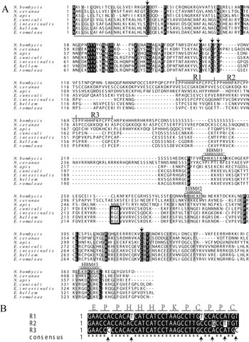
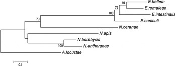
The gene Nbhswp16 is 1152 bp and encodes a 383 amino acid protein which has a calculated molecular mass of 44 kDa and a theoretical pI of 9·17. One copy gene (GenBank accession number: EOB12060) having 99% identity with Nbhswp16 was also found in the genome of N. bombycis. As shown in Fig. 1A, a 25-amino acid signal peptide was predicted at the N-terminal NbHSWP16, but no GPI anchor sequence signals or transmembrane domain was predicted. According to NetNglyc and NetOglyc analysis, NbHSWP16 contained 2 potential N-glycosylation sites. More than 30 phosphorylation sites, most of which occurred on the residue serine, were predicted. It is worth to notice that there are 3 predicted HBMs at the C-terminal NbHSWP16. More interestingly, a region consisting of 3 proline-rich tandem repeats with no homology to any known protein was also predicted in NbHSWP16. In DNA sequence, not only it was noticed that these tandem sequences are conserved, but also single nucleotide polymorphism (SNP) sites were found in partial synonymous codes (Fig. 1B). By BLASTP analysis, several homologous proteins of NbHSWP16 were found in several microsporidia, including Nosema apis, Nosema antheraeae, Nosema ceranae, E. cuniculi, E. intestinalis, Encephalitozoon hellem, Encephalitozoon romaleae and Antonospora locustae, but no homologous was found in Encephalitozoon bieneusi. These proteins share 26–34% identity with NbHSWP16. The phylogenetic analysis indicated that NbHSWP16 were conserved comparatively in the evolution process (Fig. 2).
The sequence character of NbHSWP16. (A) Amino acid sequence alignment of HSWP16 from several microsporidia. The identical amino acids are shaded. The signal peptide is indicated by a thin underline. The conserved cysteines are indicated by arrows. The predicted HBMs and the ‘RGD/E’ motif are boxed in thin and bold rectangle, respectively. The 3 proline-rich tandem repeats are bracketed and numbered as R1, R2 and R3; (B) the DNA sequence alignment of 3 tandem repeats in gene Nbhswp16. The SNP sites are indicated by arrow heads. The amino acids are shown as single letters.

The phylogenetic analysis of NbHSWP16 in microsporidia. Neighbour-joining tree was constructed with the MEGA5 software. The GenBank accession numbers of the protein sequences used are as following: N. bombycis: EOB14338, N. apis: EQB62243, N. ceranae: EEQ82233, E. cuniculi: CAD24952, E. intestinalis: ABU24317, E. romaleae: AFN82405. The protein sequence of A. locustae is from database (http://gmod.mbl.edu/cgi-bin/site/antonospora01) and N. antheraeae is from database (http://silkpathdb.swu.edu.cn/silkpathdb/).

Transcriptional analysis of Nbhswp16 gene
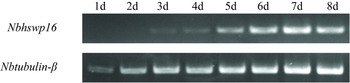
To investigate the transcriptional pattern of Nbhswp16 gene, RT-PCR was performed using RNA from the midguts of infected silkworms at 1–8 days post-infection. As shown in Fig. 3, Nbhswp16 started to transcribe at the third day post-infection and the transcriptional level is with a slow increment at 3–8 days post-infection. Nbhswp16 was not detected during the first 2 days post-infection, the reason may be that the template using for RT-PCR is not enough.
Analysis of transcriptional pattern of Nbhswp16 gene. Transcriptional analysis was performed by RT-PCR using the RNA from the midguts of infected silkworms at 1–8 days post-infection. The tubulin-β gene was used as an internal control.

Expression of rNbHSWP16 and western-blotting
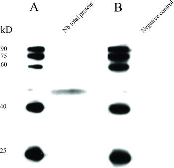
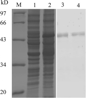
To study NbHSWP16 in N. bombycis, a recombinant NbHSWP16 without the signal peptide was expressed in E. coli BL21 strain and purified by Ni-NTA Superflow Cartridge (Qiagen) (Appendix, Fig. A1). A polyclonal antibody against rNbHSWP16 was generated by immunizing mice with the purified fusion protein and used in immunoblotting analysis. A special band about 44 kDa consistent with the calculated molecular weight of NbHSWP16 was detected using anti-NbHSWP16 as primary antibody and no signals was detected in the negative control (Fig. 4).
Western blot analysis of N. bombycis NbHSWP16. (A) NbHSWP16 was detected from N. bombycis total protein preparation using mouse anti-NbHSWP16; (B) negative control.

Localization of NbHSWP16 in N. bombycis
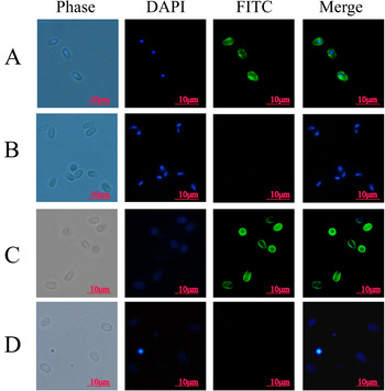
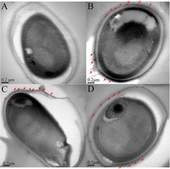
In order to survey NbHSWP16 subcellular localization, IFA was performed. From the result of IFA, we noticed that fluorescence signals were detected on the surface of the mature spores as well as on the germinated spore coats (Fig. 5). To further determine the cellular location of NbHSWP16, IEM was employed. The results of IEM demonstrated that labelled gold particles were mainly distributed on the exospore regions of the mature spores. These results suggested that the NbHSWP16 is localized mainly on the exospore wall of the mature spore (Fig. 6). So, we renamed NbHSWP16 as NbSWP16.
Indirect immunofluorescence of NbHSWP16 in mature spore and germinated spore coats of N. bombycis. (A) NbHSWP16 was detected on the mature spores with anti-NbHswp16; (B) negative control; (C) NbHSWP16 was detected on the germinated spore coats with antibody against NbHSWP16. (D): negative control. Anti-NbHSWP16 was diluted at 1:100. FITC-conjugated goat anti-mouse IgG was diluted at 1:64. The scale bar represents 10 μ m.

IEM of NbHSWP16 in mature N. bombycis. (A) Negative control; (B), (C), (D) spore labelled on the exospore region. Anti-NbSWP16 serum was used at 1:20 dilution. Secondary antibody was goat anti-mouse IgG conjugated with 10 nm colloidal gold particles (sigma). The labelled gold particles were marked with arrowheads. The scale bar represents 0·2 μ m. Ex, exospore; En, endospore.

Anti-NbSWP16 decrease the N. bombycis adherence to host cell
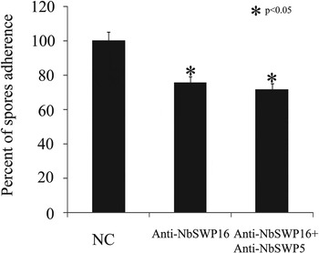
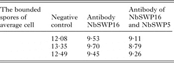
Based on the sequence analysis and subcellular localization of NbSWP16 above, we discovered that NbSWP16 had 3 protein–heparin protein interaction motifs (HBMs), but also can be localized on the exospore of mature spore. To dissect whether NbSWP16 protein is involved in spore adherence or not, spore adherence analysis was performed. The statistical result (Appendix, Table A2) showed that the rate of spore adherence to host cell was decreased more than 20% by anti-NbSWP16 blocking compared with the negative control in vitro. But in contrast with anti-NbSWP16 used alone, no remarkable decrement inhibition was detected when antibodies of NbSWP16 and NbSWP5 were used simultaneously (Fig. 7).
Antibody of NbSWP16 inhibiting spores adherence to host cell in vitro. The spore adherence analysis of NbSWP16 was performed used BmE-SWU1 as host cell. Antibodies of NbSWP16 and NbSWP5 as well as negative mouse sera were used at 1:50 dilution. Statistically significant difference is indicated with asterisk (P<0·05). NC, negative control.

DISCUSSION
As an obligate unicellular eukaryotic parasite, one of the prominent features of microsporidia is that it could deposit a rigid spore wall on the surface. The spore wall is capable of protecting microsporidia against the environment stress (Shadduck and Polley, Reference Shadduck and Polley1978). Now it is documented that the spore wall proteins of microsporidia may function in spore adherence and infection process (Weidner et al. Reference Weidner, Avila and Harris1992; Weidner and Halonen, Reference Weidner and Halonen1993). However, although 10 years has passed since the publication of the first microsporidian genome of E. cuniculi (Katinka, Reference Katinka2001), the available information about the spore wall protein of microsporidia is still limited, especially the ones having tandem repeat.
At the present study, a new spore wall protein NbSWP16 was identified in N. bombycis. A 25-amino acid signal peptide was predicted at the N-terminal NbSWP16, but no transmembrane domain was predicted. It is proposed that this protein may be secreted. In the western-blotting analysis, NbSWP16 could only be detected in the protein sample obtained in the presence of high concentration of thiolreducing agents dithiothreitol and 2-mercaptoethanol, but not be detected in the protein sample prepared with PBS or SDS. This result indicated that disulfide bridges may exist in NbSWP16 and may be required to retain the stability of the spore wall.
To date, 13 proteins have been described having HBMs in microsporidia, including EcEnP1 in E. cuniculi (Southern et al. Reference Southern, Jolly, Lester and Hayman2007), EiEnP1 in E. intestinalis (Southern et al. Reference Southern, Jolly, Lester and Hayman2007), SWP2, SWP26, SWP3 and 8 other hypothetical spore wall proteins in N. bombycis (Wu et al. Reference Wu, Li, Pan, Tan, Hu, Zhou and Xiang2008; Li et al. Reference Li, Wu, Pan, He, Zhang, Hu and Zhou2009). Among these proteins, EcEnP1 and SWP26 were proved to be involved in spore adherence process (Southern et al. Reference Southern, Jolly, Lester and Hayman2007; Li et al. Reference Li, Wu, Pan, He, Zhang, Hu and Zhou2009). In this paper, 3 HBMs were predicted in NbSWP16, it suggested the HBMs motifs may contribute to the spore adherence of NbSWP16. From the result of spore adherence analysis, we discovered that anti-NbSWP16 could decrease the spore adherence rate more than 20%. In addition, in contrast with anti-NbSWP16 used alone, no remarkable decrement inhibition was detected when antibodies of NbSWP16 and NbSWP5 were used simultaneously, indicating the anti-NbSWP16 combining anti-NbSWP5 may not have synergistic effect in inhibition. It is still a long way to clarify the molecular mechanism of spore adherence. Unfortunately, it is not success to obtain the soluble protein of NbSWP16. To confirm whether the HBMs is responsible for the spore adherence function of NbSWP16, further work is need.
A previous data survey using tandem repeats in protein sequence (TRIPS) revealed that proteins with tandem repeats are conspicuous in structural and cell surface proteins (Katti et al. Reference Katti, Sami-Subbu, Ranjekar and Gupta2000). Moreover, tandem repeats functioning in some species were described previously. In Listeria monocytogenes, an ActA protein containing 4 repeats, which are 11 amino acids in length and consist of 3 or 4 consecutive prolines, can contribute to the movement rate of this species (Smith et al. Reference Smith, Theriot and Portnoy1996). The Saccharomyces cerevisiae FlO1 gene consisting of tandem repeated units encodes a cell-surface adhesin that mediates cell–cell and cell-surface adhesion and phenotypic tests illustrated that the intensity of the adhesion directly correlated with the length of the repeat (Verstrepen et al. Reference Verstrepen, Jansen, Lewitter and Fink2005). Here, 3 proline-rich tandem repeats were found in NbSWP16 and the results of IFA and IEM proved that NbSWP16 is an exospore wall protein. It suggests that the tandem repeats existing in NbSWP16 might be responsible for the function to this protein. Besides, in DNA sequence, though the tandem repeats are conserved in gene nbswp16, 6 SNP sites were noticed in some synonymous codes (Fig. 1B). This may be associated with the evolution and adaptation of N. bombycis. Because some evidence previously mentioned that the variations in tandem repeats can influence organism's phenotypes and facilitate evolution and rapid adaptation (Gemayel et al. Reference Gemayel, Cho, Boeynaems and Verstrepen2012).
In conclusion, a novel exospore protein NbSWP16 with 3 proline-rich tandem repeats was characterized in N. bombycis. Spore adherence analysis demonstrated NbSWP16 might contribute to the spore adherence in vitro. The works focusing on the function of NbSWP16 will be carried out in the future.
ACKNOWLEDGEMENTS
We are of pleasure to say thanks to those people who provided the means for us to access software free of charge used and cited in this article.
FINANCIAL SUPPORT
This work was supported by Key Project of Natural Science Foundation of China under grant no. 30930067; National Basic Research Program of China (973 Program) under grant no. 2012CB114604; Project of Natural Science Foundation of China under grant no. 31270138; Chongqing Natural Science Foundation of China under grant no. cstc2013jcyja0363; Natural Science Foundation of China (grant no. 31101770) and the Natural Science Foundation Project of CQ CSTC (grant no. cstc2011jjA1111).
SUPPLEMENTARY MATERIAL
To view supplementary material for this article, please visit http://dx.doi.org/10.1017/S0031182014001565.
APPENDIX
The spore wall proteins studied in our group

To analyse the role of NbSWP16 in spore adherence, antibody blocking analysis was performed. In each group data (C1/S1, C2/S2 and C3/S3), the spores bounded to 5 selected randomly host cells were counted in 21 independent field and it was repeated 3 times. S1, S2 and S3 represent the 3 times of anti-NbSWP16 blocking analysis, respectively. C1, C2 and C3 represent the control. The raw data of spore counting for each experimental analysis was shown in Tables 1, 2 and 3, respectively.
The raw data of spore counting with 3 times. Please see supplementary material online (Excel spreadsheet: go to http://journals.cambridge.org/PAR.).
SDS-PAGE (12%) analysis of expression and purification of rNbHSWP16 M, protein molecular weight marker; lane 1, expression product of pET30a (+)-NbHSWP16 without induction; lane 2, expression product of pET30a (+)-NbHSWP16 induced with IPTG; lanes 3 and 4, purified rNbHSWP16 inclusion body.

The statistical results of spore adherence analysis with antibody of NbSWP16 and NbSWP5