1. Overview of the functions and properties of the APC/C
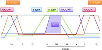
The anaphase promoting complex or cyclosome (APC/C) is a multi-subunit cullin-RING E3 ubiquitin ligase that functions to regulate progression through the mitotic phase of the cell cycle and controls entry into S phase (Irniger et al. Reference Irniger, Piatti, Michaelis and Nasmyth1995; King et al. Reference King, Peters, Tugendreich, Rolfe, Hieter and Kirschner1995; Sudakin et al. Reference Sudakin, Ganoth, Dahan, Heller, Hershko, Luca, Ruderman and Hershko1995; Tugendreich et al. Reference Tugendreich, Tomkiel, Earnshaw and Hieter1995), reviewed by (Harper et al. Reference Harper, Burton and Solomon2002; Peters, Reference Peters2006; Pines, Reference Pines2006; Sullivan & Morgan, Reference Sullivan and Morgan2007b; Thornton & Toczyski, Reference Thornton and Toczyski2006). The APC/C also plays a role to regulate meiosis, and has been implicated in post-mitotic functions including dendrite formation in neurons, metabolic, and learning and memory processes (Herrero-Mendez et al. Reference Herrero-Mendez, Almeida, Fernandez, Maestre, Moncada and Bolanos2009; Kim et al. Reference Kim, Puram, Bilimoria, Ikeuchi, Keough, Wong, Rowitch and Bonni2009; Li et al. Reference Li, Shin, Hou, Huang, Wei, Klann and Zhang2008; Van Roessel et al. Reference Van Roessel, Elliott, Robinson, Prokop and Brand2004). APC/C-mediated coordination of cell cycle progression is achieved through the temporal regulation of APC/C activity and substrate specificity (Fig. 1). The APC/C becomes activated at the onset of mitosis, and at prometaphase, ubiquitylates Nek2A and cyclin A (S-phase cyclin). At metaphase, the APC/C ubiquitylates two inhibitors of the transition to anaphase, namely, securin and cyclin B (M-phase cyclin) (Clute & Pines, Reference Clute and Pines1999; Cohen-Fix et al. Reference Cohen-Fix, Peters, Kirschner and Koshland1996). Securin is a protein inhibitor of separase, a protease that cleaves the cohesin subunit SCC1 (Uhlmann et al. Reference Uhlmann, Wernic, Poupart, Koonin and Nasmyth2000). Cleavage of SCC1 causes disassembly of cohesin, necessary to allow sister chromatid segregation (Ciosk et al. Reference Ciosk, Zachariae, Michaelis, Shevchenko, Mann and Nasmyth1998; Funabiki et al. Reference Funabiki, Yamano, Kumada, Nagao, Hunt and Yanagida1996; Michaelis et al. Reference Michaelis, Ciosk and Nasmyth1997), reviewed by Nasmyth (Reference Nasmyth2002) . Reduced cyclin levels are also required for entry into anaphase, since cyclin-dependent kinase (Cdk) activity inhibits separase (Gorr et al. Reference Gorr, Boos and Stemmann2005; Holland & Taylor, Reference Holland and Taylor2006; Stemmann et al. Reference Stemmann, Zou, Gerber, Gygi and Kirschner2001). After anaphase, cyclin destruction continues to maintain negligible Cdk activity, necessary for the cell to disassemble the mitotic spindle and exit mitosis (Murray et al. Reference Murray, Solomon and Kirschner1989; Shirayama et al. Reference Shirayama, Toth, Galova and Nasmyth1999; Wasch & Cross, Reference Wasch and Cross2002). During G1, the main role of the APC/C is to sustain low levels of mitotic Cdk activity to allow for resetting of replication origins as a prelude to a new round of DNA replication in S phase (Irniger & Nasmyth, Reference Irniger and Nasmyth1997; Noton & Diffley, Reference Noton and Diffley2000) (Fig. 1).
Schematic of the cell cycle, depicting oscillations of APC/CCdc20 and APC/CCdh1 activities, together with G1/S-, S- and M-phase cyclins. During prophase and prometaphase the SAC inactivates APC/CCdc20 towards most APC/C substrates, allowing only specific substrates, for example cyclin A and Nek2A, to be ubiquitylated. P: prophase, PM: prometaphase, M: metaphase, A: anaphase, T: telophase.

The temporal regulation of APC/C activity is achieved through a combination of two structurally related co-activator subunits, Cdc20 and Cdh1 (Cdc20 homology 1/fizzy related/Hct1 (APC/C co-activator); Dawson et al. Reference Dawson, Roth and Artavanis-Tsakonas1995; Fang et al. Reference Fang, Yu and Kirschner1998a; Kramer et al. Reference Kramer, Gieffers, Holzl, Hengstschlager and Peters1998a; Lim et al. Reference Lim, Goh and Surana1998; Lorca et al. Reference Lorca, Castro, Martinez, Vigneron, Morin, Sigrist, Lehner, Doree and Labbe1998; Schwab et al. Reference Schwab, Lutum and Seufert1997; Shirayama et al. Reference Shirayama, Zachariae, Ciosk and Nasmyth1998; Sigrist et al. Reference Sigrist, Jacobs, Stratmann and Lehner1995; Sigrist & Lehner, Reference Sigrist and Lehner1997; Visintin et al. Reference Visintin, Prinz and Amon1997; Zachariae et al. Reference Zachariae, Schwab, Nasmyth and Seufert1998a), coupled to protein phosphorylation, APC/C inhibitors, differential affinity for APC/C substrates and auto-ubiquitylation of its cognate E2. The two APC/C co-activators have opposing activity profiles (Fig. 1). Cdc20 activates the APC/C during early mitosis, when APC/C is phosphorylated and Cdh1 activity is low due to its Cdk-dependent phosphorylation, whereas APC/CCdc20-mediated reduction of Cdk activity stimulates Cdh1. In turn, APC/CCdh1 ubiquitylates Cdc20, leading to APC/CCdc20 inactivation. Thus, Cdc20 activates Cdh1 that in turn antagonises Cdc20 activity. The switching between APC/CCdc20 and APC/CCdh1 fulfils two main functions. First, APC/CCdc20 and APC/CCdh1 have overlapping but nevertheless distinct substrate specificities. Therefore, specific cell cycle regulators are degraded during the separate phases of APC/CCdc20 and APC/CCdh1 activities, allowing for ordered progression through the cell cycle. Second, Cdc20 and Cdh1 are subject to control by different regulatory mechanisms. Cdc20 activates APC/C phosphorylated by Cdk and polo-like kinase 1 (Plk1) protein kinases during early mitosis, whereas Cdh1 is inhibited by its Cdk-mediated phosphorylation. Importantly, APC/CCdc20 activity towards securin and cyclin B is inhibited by the mitotic checkpoint complex (MCC), a multi-protein complex activated by the spindle assembly checkpoint (SAC), reviewed by Musacchio & Salmon (Reference Musacchio and Salmon2007) . The SAC ensures that anaphase is delayed until every chromosome is aligned on the mitotic spindle.
2. Subunit organisation and APC/C structure
2.1 Subunit organisation
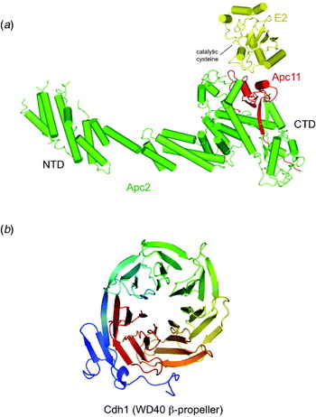
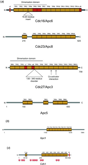
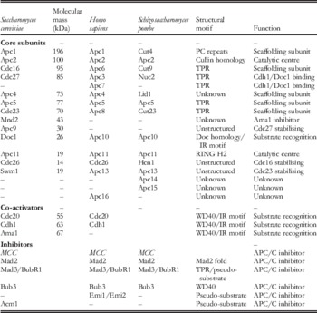
Structural information on the APC/C is currently limited, and surprisingly even the subunit stoichiometry and accurate molecular mass of the APC/C core are currently poorly defined. In budding yeast, 13 genes encode core APC/C subunits, most of which are highly conserved in humans and other eukaryotes, and are essential for activity (Table 1) (Hutchins et al. Reference Hutchins, Toyoda, Hegemann, Poser, Heriche, Sykora, Augsburg, Hudecz, Buschhorn, Bulkescher, Conrad, Comartin, Schleiffer, Sarov, Pozniakovsky, Slabicki, Schloissnig, Steinmacher, Leuschner, Ssykor, Lawo, Pelletier, Stark, Nasmyth, Ellenberg, Durbin, Buchholz, Mechtler, Hyman and Peters2010; Kops et al. Reference Kops, Voet, Manak, Van Osch, Naini, Brear, Mcleod, Hentschel, Yates, van Den Heuvel and Shah2010; Kramer et al. Reference Kramer, Fesquet, Johnson and Johnston1998b; Passmore et al. Reference Passmore, Mccormack, Au, Paul, Willison, Harper and Barford2003; Peters et al. Reference Peters, King, Hoog and Kirschner1996; Schwickart et al. Reference Schwickart, Havlis, Habermann, Bogdanova, Camasses, Oelschlaegel, Shevchenko and Zachariae2004; Yoon et al. Reference Yoon, Feoktistova, Wolfe, Jennings, Link and Gould2002; Yu et al. Reference Yu, Peters, King, Page, Hieter and Kirschner1998; Zachariae et al. Reference Zachariae, Shin, Galova, Obermaier and Nasmyth1996, Reference Zachariae, Shevchenko, Andrews, Ciosk, Galova, Stark, Mann and Nasmyth1998b). The catalytic core of the APC/C is composed of the cullin subunit Apc2 and RING H2 domain subunit Apc11, analogous to the cullin and Rbx1 subunits, respectively, of cullin-RING ligases (CRLs) of the SCF superfamily (Zheng et al. Reference Zheng, Schulman, Song, Miller, Jeffrey, Wang, Chu, Koepp, Elledge, Pagano, Conaway, Conaway, Harper and Pavletich2002) (Fig. 2a). Apc2 and Apc11 form a tight complex and together are competent to catalyse ubiquitylation, although they are devoid of substrate selectivity (Gmachl et al. Reference Gmachl, Gieffers, Podtelejnikov, Mann and Peters2000; Leverson et al. Reference Leverson, Joazeiro, Page, Huang, Hieter and Hunter2000; Tang et al. Reference Tang, Li, Bharadwaj, Zhu, Ozkan, Hakala, Deisenhofer and Yu2001b). The largest APC/C subunit, Apc1 is ~200 kDa in size. Interestingly, the C-terminal region of Apc1 is composed of between seven to eight tandem repeats of a 35–40 amino acid motif shared with the Rpn1 and Rpn2 subunits of the proteasome regulatory particle (Lupas et al. Reference Lupas, Baumeister and Hofmann1997) (Fig. 3b). Such motifs, termed proteasome/cyclosome (PC) repeats, for which no definitive structural data are available, are intriguing given the close functional relationship between the APC/C and proteasome. Suggestions that PC repeats adopt α/β architectures similar to leucine-rich repeats (Lupas et al. Reference Lupas, Baumeister and Hofmann1997), or α/α motifs typical of helical solenoids (Kajava, Reference Kajava2002), thus far lack experimental evidence. The most abundant structural motif observed in APC/C subunits is the tetratricopeptide repeat (TPR) with four yeast and five vertebrate APC/C subunits composed almost exclusively of 12–15 copies of this 34-amino-acid motif arranged in tandem (Table 1) (Fig. 3a). The TPR motif, originally discovered within proteins subsequently identified as components of the APC/C (Hirano et al. Reference Hirano, Kinoshita, Morikawa and Yanagida1990; Lamb et al. Reference Lamb, Michaud, Sikorski and Hieter1994; Sikorski et al. Reference Sikorski, Boguski, Goebl and Hieter1990; Zachariae & Nasmyth, Reference Zachariae and Nasmyth1996), is one of the most abundant protein motifs present in eukaryotic genomes, and functions in scaffolding proteins to mediate protein–protein interactions, and the assembly of multi-protein complexes. As discussed later, the super-helical architecture generated by consecutive TPR motifs facilitates protein–protein interactions. Finally, the functionally and structurally related co-activators Cdc20 and Cdh1 contain WD40 β-propeller domains that contribute to substrate recognition (Table 1) (Figs 2b and 3c).
(a) Model of Apc2/11 bound to an E2 based on the SFC structure of (Zheng et al. Reference Zheng, Schulman, Song, Miller, Jeffrey, Wang, Chu, Koepp, Elledge, Pagano, Conaway, Conaway, Harper and Pavletich2002). (b) Model of the WD40 β-propeller of Cdh1.

Primary structure of selective APC/C subunits. (a) Conserved TPR subunits of the APC/C: Apc6/Cdc16, Apc8/Cdc23, Apc3/Cdc27 and Apc5. Schematic of Apc3/Cdc27 and Apc6/Cdc16 based on Zhang et al. (Reference Zhang, Roe, Diogon, Kong, El Alaoui and Barford2010a, Reference Zhang, Kulkarni, Hanrahan, Thompson and Barfordb), respectively. (b) Apc1 showing the PC (proteasome-cyclosome repeats). (c) Cdh1. ‘P’ indicates consensus Cdk-phosphorylation sites that are responsible for blocking APC/C interaction with Cdh1.

Core and regulatory subunits of the APC/C

Molecular mass corresponds to S. cerevisiae subunits.
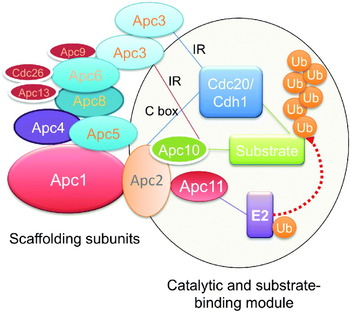
Subunit deletion experiments using modified yeast strains provided important insights into the architectural topology of APC/C subunits (Thornton et al. Reference Thornton, Ng, Matyskiela, Carroll, Morgan and Toczyski2006). Thornton & Toczyski (Reference Thornton and Toczyski2003) developed a budding yeast strain (deletion of securin/Pds1 and the S-phase cyclin Clb5, and overexpression of the Cdk inhibitor Sic1, based on earlier findings of Nasmyth and co-workers (Shirayama et al. Reference Shirayama, Toth, Galova and Nasmyth1999)) that is viable in the absence of functional APC/C. Such a strain allows deletion of essential APC/C subunits and subsequent analysis of the consequences on APC/C structure and activity. Importantly, this study indicated that Apc1 functions as a scaffolding-like assembly subunit linking the catalytic sub-complex formed from Apc2, Apc11 and Apc10/destruction of cyclin 1 (Doc1) with the TPR subunits Apc6/Cdc16, Apc8/Cdc23 and Apc3/Cdc27 via Apc4 and Apc5. The subunits Apc4, Apc5, Apc8/Cdc23 and Apc1 associate interdependently (Fig. 4). TPR subunits (Apc3, Apc6 and Apc8) display a sequential binding dependency, since the interaction of Apc3/Cdc27 with APC/C (tagged Apc1) was dependent on Apc6/Cdc16, whereas the interaction of both Apc3/Cdc27 and Apc6/Cdc16 was dependent on Apc8/Cdc23 (Thornton et al. Reference Thornton, Ng, Matyskiela, Carroll, Morgan and Toczyski2006). Thus, Apc3/Cdc27 is the most peripheral TPR subunit, Apc8/Cdc23 the most internal and Apc6/Cdc16 connects both Apc3/Cdc27 and Apc8/Cdc23 (Fig. 4). However, there is also evidence that Apc3/Cdc27 and Apc8/Cdc23 interact directly. A Cdc27-663 mutation (a temperature sensitive Gly to Asp mutation in TPR10 of Apc3/Cdc27 (Hirano et al. Reference Hirano, Kinoshita, Morikawa and Yanagida1990; Sikorski et al. Reference Sikorski, Michaud, Wootton, Boguski, Connelly and Hieter1991)) compromised the interaction of Apc3/Cdc27 with Apc8/Cdc23, but had no effect on either Apc3/Cdc27 self-association or on Apc3/Cdc27–Apc6/Cdc16 interactions (Lamb et al. Reference Lamb, Michaud, Sikorski and Hieter1994). Significantly this study, indicating that all three TPR subunits were found to self-associate, provided the first evidence that Apc3/Cdc27, Apc6/Cdc16 and Apc8/Cdc23 are present at supra-stoichiometric amounts within the APC/C. Intragenic complementation between heteroalleles of cdc23 had earlier suggested that Cdc23 polypeptides may interact directly with each other as part of a supramolecular complex (Sikorski et al. Reference Sikorski, Michaud and Hieter1993).
Schematic of APC/C subunit topology based on biochemical and genetic data cited in the text. The catalytic centre is formed from Apc2/Apc11, which engages activated E2–Ub, together with the substrate recognition subunit Apc10/Doc1, and the substrate recruiting and catalytic co-activator subunits Cdc20 and Cdh1 (S. cerevisiae Ama1 in meiosis).

In yeast, it has been demonstrated that the small and mainly unstructured subunits Apc9, Apc13 and Cdc26 appear to function to stabilise the overall subunit organisation of the APC/C, specifically the TPR sub-complex (Passmore et al. Reference Passmore, Mccormack, Au, Paul, Willison, Harper and Barford2003; Thornton et al. Reference Thornton, Ng, Matyskiela, Carroll, Morgan and Toczyski2006; Zachariae et al. Reference Zachariae, Shevchenko, Andrews, Ciosk, Galova, Stark, Mann and Nasmyth1998b). Apc9, for example, is required for stable Apc3/Cdc27 association (Passmore et al. Reference Passmore, Mccormack, Au, Paul, Willison, Harper and Barford2003; Zachariae et al. Reference Zachariae, Shevchenko, Andrews, Ciosk, Galova, Stark, Mann and Nasmyth1998b), whereas at the restrictive temperature, deletion of Cdc26 results in reduced levels of Apc6/Cdc16, Apc3/Cdc27 and Apc9 within the complex (Zachariae et al. Reference Zachariae, Shevchenko, Andrews, Ciosk, Galova, Stark, Mann and Nasmyth1998b), in agreement with the finding that overexpression of the fission yeast homolog of Cdc26 (Hcn1) suppresses the assembly defect caused by a cut9 (cdc16) mutation (Yamada et al. Reference Yamada, Kumada and Yanagida1997). Notably, repression of hcn1+ expression in Schizosaccharomyces pombe (Cdc26) resulted in the formation of catalytic sub-complex of Apc1, Apc2, Apc11, Apc4, Apc5 and Apc15 and a distinct and undefined Cdc23-containing sub-complex (Yoon et al. Reference Yoon, Feoktistova, Chen, Jennings, Link and Gould2006) in agreement with the subunit architecture defined for Saccharomyces cerevisiae APC/C (Thornton et al. Reference Thornton, Ng, Matyskiela, Carroll, Morgan and Toczyski2006). Pairwise interactions between S. pombe subunits indicated that Apc2 interacts with Apc11, Apc4 interacts with Apc5, Apc3/Cdc27 interacts with Apc10/Doc1 and Apc8/Cdc23 interacts with Apc13 (Yoon et al. Reference Yoon, Feoktistova, Chen, Jennings, Link and Gould2006). Finally, Apc13/Swm1 was shown to stabilise the association of Apc3/Cdc27, Apc6/Cdc16, Apc9 and Cdc26 with other subunits of yeast APC/C (Schwickart et al. Reference Schwickart, Havlis, Habermann, Bogdanova, Camasses, Oelschlaegel, Shevchenko and Zachariae2004). This study, using tagged Apc13/Swm1, also demonstrated a TPR sub-complex comprising Apc8/Cdc16, Apc3/Cdc27, Cdc26, Apc9 and Apc13/Swm1 isolated from cdc23-ts mutant cells.
The architectural map of the budding yeast APC/C obtained via systematic subunit deletions is in broad agreement with the composition of vertebrate APC/C sub-complexes identified during biochemical isolation of human APC/C (Vodermaier et al. Reference Vodermaier, Gieffers, Maurer-Stroh, Eisenhaber and Peters2003). These authors isolated a catalytic sub-complex comprising Apc1, Apc2, Apc11, Apc4 and Apc5 that was distinct from TPR-containing subunits, and lacked Apc10/Doc1. In contrast to budding yeast APC/C, Apc10/Doc1 was found to associate through its C-terminal Ile-Arg (IR) motif with the TPR subunit Apc3/Cdc27 (Wendt et al. Reference Wendt, Vodermaier, Jacob, Gieffers, Gmachl, Peters, Huber and Sondermann2001). However, consistent with findings using S. cerevisiae APC/C, Vodermaier et al. (Reference Vodermaier, Gieffers, Maurer-Stroh, Eisenhaber and Peters2003) noted that in their APC/C devoid of the Apc2/Apc11 heterodimer (Δ2/11-APC/C), levels of Apc10/Doc1 were reduced, implicating a contribution of Apc2 to bind Apc10/Doc1 within the complex. Altogether, through a combination of genetic and biochemical data, a detailed and informative topographic map of APC/C subunits has now emerged (Fig. 4). It is interesting to note, however, that unlike the large multi-subunit proteasome complex, composed of regulatory and core (catalytic) particles, no evidence for the existence of specific APC/C chaperones or assembly factors has been defined (Funakoshi et al. Reference Funakoshi, Tomko, Kobayashi and Hochstrasser2009; Kaneko et al. Reference Kaneko, Hamazaki, Iemura, Sasaki, Furuyama, Natsume, Tanaka and Murata2009; Murata et al. Reference Murata, Yashiroda and Tanaka2009; Roelofs et al. Reference Roelofs, Park, Haas, Tian, Mcallister, Huo, Lee, Zhang, Shi, Gygi and Finley2009; Saeki et al. Reference Saeki, Toh, Kudo, Kawamura and Tanaka2009b), and little is known concerning the assembly pathway of the APC/C.
Finally, Mnd2, a subunit unique to S. cerevisiae APC/C, functions to inhibit the meiotic co-activator Ama1, and prevents premature activation of APC/CAma1 in meiosis and mitosis (Oelschlaegel et al. Reference Oelschlaegel, Schwickart, Matos, Bogdanova, Camasses, Havlis, Shevchenko and Zachariae2005; Penkner et al. Reference Penkner, Prinz, Ferscha and Klein2005).
2.2 Co-activators bind APC/C through an N-terminal C box and C-terminal IR motif
Co-activators utilise two conserved sequence motifs for APC/C binding (Figs 3 and 4) a C box, located toward the N-terminus (Schwab et al. Reference Schwab, Neutzner, Mocker and Seufert2001; Vodermaier et al. Reference Vodermaier, Gieffers, Maurer-Stroh, Eisenhaber and Peters2003) and an extreme C-terminal IR motif, also shared with Apc10/Doc1 (Kraft et al. Reference Kraft, Vodermaier, Maurer-Stroh, Eisenhaber and Peters2005; Matyskiela & Morgan, Reference Matyskiela and Morgan2009; Passmore et al. Reference Passmore, Mccormack, Au, Paul, Willison, Harper and Barford2003; Thornton et al. Reference Thornton, Ng, Matyskiela, Carroll, Morgan and Toczyski2006; Vodermaier et al. Reference Vodermaier, Gieffers, Maurer-Stroh, Eisenhaber and Peters2003; Wendt et al. Reference Wendt, Vodermaier, Jacob, Gieffers, Gmachl, Peters, Huber and Sondermann2001). Co-activator binding to APC/C mediated via its IR motif requires Apc3/Cdc27 and the C-terminal region of Apc7 (the Apc3/Cdc27 paralog found only in vertebrates) (Kraft et al. Reference Kraft, Vodermaier, Maurer-Stroh, Eisenhaber and Peters2005; Matyskiela & Morgan, Reference Matyskiela and Morgan2009; Passmore et al. Reference Passmore, Mccormack, Au, Paul, Willison, Harper and Barford2003; Thornton et al. Reference Thornton, Ng, Matyskiela, Carroll, Morgan and Toczyski2006; Vodermaier et al. Reference Vodermaier, Gieffers, Maurer-Stroh, Eisenhaber and Peters2003). Whereas Apc2/Apc11 was dispensable for recruiting Cdh1 to human APC/C (Vodermaier et al. Reference Vodermaier, Gieffers, Maurer-Stroh, Eisenhaber and Peters2003), Thornton et al. (Reference Thornton, Ng, Matyskiela, Carroll, Morgan and Toczyski2006) showed that deletion of Apc2 caused a 10-fold reduction in affinity for Cdh1, strongly implicating Apc2 as a Cdh1-interacting protein. Moreover, they found that a mutation in the C-box of Cdh1 that only has a mild affect on Cdh1 interactions with wild-type APC/C, was unable to stimulate APC/C lacking the Cdc27 subunit, suggesting the possibility that the C box mediates interactions with Apc2 (Thornton et al. Reference Thornton, Ng, Matyskiela, Carroll, Morgan and Toczyski2006).
2.3 Structure of the APC/C and its subunits
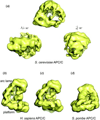
Our knowledge of the structure of the intact APC/C is based on electron microscopy single-particle three-dimensional reconstruction studies of budding and fission yeast, human and Xenopus APC/C (Fig. 5). Yeast APC/C structures were determined using cryo-electron microscopy, whereas the human and Xenopus structures were determined using the approach of both cryo- and cryo-negative stain electron microscopy (Dube et al. Reference Dube, Herzog, Gieffers, Sander, Riedel, Muller, Engel, Peters and Stark2005; Herzog et al. Reference Herzog, Primorac, Dube, Lenart, Sander, Mechtler, Stark and Peters2009; Ohi et al. Reference Ohi, Feoktistova, Ren, Yip, Cheng, Chen, Yoon, Wall, Huang, Penczek, Gould and Walz2007; Passmore et al. Reference Passmore, Booth, Venien-Bryan, Ludtke, Fioretto, Johnson, Chiu and Barford2005). The effective resolution is of the order of 20–27 Å. A combination of antibody, Ni-NTA nano-gold labelling and Apc2/11 subunit deletion has been used to locate the positions of the majority of APC/C subunits within the context of the intact particle. Whereas details of the structures from different species differ, all studies reveal that the basic overall shape of the APC/C resembles a triangular structure of ~200–230 Å in dimensions, featuring a central cavity measuring 50 Å×75 Å×100 Å (in budding yeast APC/C) surrounded by a protein shell. Subunit mapping studies are consistent with the proposal for the APC/C subunit topology based on genetic and biochemical data (Thornton et al. Reference Thornton, Ng, Matyskiela, Carroll, Morgan and Toczyski2006; Vodermaier et al. Reference Vodermaier, Gieffers, Maurer-Stroh, Eisenhaber and Peters2003). The catalytic subunits Apc2 and Apc11 are in close proximity to Apc1, whereas the TPR-containing subunits (Apc6/Cdc16, Apc8/Cdc23 and Apc3/Cdc27) co-localise to a more distal region of the molecule. These two regions of the APC/C have been defined as the platform and arc lamp, respectively, for human and Xenopus APC/C (Dube et al. Reference Dube, Herzog, Gieffers, Sander, Riedel, Muller, Engel, Peters and Stark2005). In the human apo-APC/C electron microscopy study, conformational flexibility involving rotation of the arc lamp domain relative to the platform was observed, with three main states being populated. While similar flexibility was less evident in the apo-APC/C from Xenopus (and not observed for yeast APC/C), engagement of the co-activator Cdh1 to Xenopus APC/C promoted a 10° rotation of the arc lamp domain. The co-activator Cdh1 was located on the outer protein wall of the APC/C in close proximity to Apc2 (Dube et al. Reference Dube, Herzog, Gieffers, Sander, Riedel, Muller, Engel, Peters and Stark2005).
Structures of the APC/C determined using electron microscopy. (a) Orthogonal views of a 25-cryo-EM structure of S. cerevisiae APC/C (Passmore et al. Reference Passmore, Booth, Venien-Bryan, Ludtke, Fioretto, Johnson, Chiu and Barford2005). (b) Cryo-negative stain structure of human APC/C and (c) human APC/C-MCC complex (Dube et al. Reference Dube, Herzog, Gieffers, Sander, Riedel, Muller, Engel, Peters and Stark2005; Herzog et al. Reference Herzog, Primorac, Dube, Lenart, Sander, Mechtler, Stark and Peters2009). (d) Cryo-EM structure of S. pombe APC/C (Ohi et al. Reference Ohi, Feoktistova, Ren, Yip, Cheng, Chen, Yoon, Wall, Huang, Penczek, Gould and Walz2007).

MCC binds and inhibits APC/CCdc20. Electron microscopy has been used to visualise the position of MCC bound to human APC/C isolated from cells arrested by the SAC at prometaphase using the microtubule stabilising agent Taxol (Fig. 5c) (Herzog et al. Reference Herzog, Primorac, Dube, Lenart, Sander, Mechtler, Stark and Peters2009). MCC was localised to the front-end of the platform domain. Cdc20 is located in a site that partially overlaps with MCC, suggesting that the precise location of Cdc20 may change on MCC binding. However, in general, the co-localisation of MCC to the site of co-activator binding is consistent with the notion that co-activators function to recruit substrate to the APC/C and that MCC inhibits APC/C activity by blocking selective Cdc20–substrate interactions. As discussed below, Mad3, a component subunit of MCC, contains KEN boxes (Lys-Glu-Asn APC/C destruction motifs), necessary for Cdc20 binding, suggesting that Mad3 functions as a pseudo-substrate inhibitor directly antagonising substrate binding (Burton & Solomon, Reference Burton and Solomon2007). Moreover, the MCC promotes a more closed rigid conformation of the APC/C, concomitant with rearrangement of Apc4, which might prevent the conformational changes potentially required for the catalytic cycle.
Mass estimates of the APC/C derived from scanning tunnelling electron microscopy (STEM), calibrated size exclusion and analytical ultracentrifugation indicate a mass of between ~1.4 and 1.5 MDa for vertebrate and budding yeast APC/C and ~0.9 MDa for fission yeast APC/C (Dube et al. Reference Dube, Herzog, Gieffers, Sander, Riedel, Muller, Engel, Peters and Stark2005; Ohi et al. Reference Ohi, Feoktistova, Ren, Yip, Cheng, Chen, Yoon, Wall, Huang, Penczek, Gould and Walz2007; Passmore et al. Reference Passmore, Booth, Venien-Bryan, Ludtke, Fioretto, Johnson, Chiu and Barford2005). For vertebrate and budding yeast, this exceeds the mass of all APC/C subunits at unit stoichiometry (S. cerevisiae mass of 850 kDa, for example), consistent with findings that three TPR subunits (Apc6/Cdc16, Apc8/Cdc23 and Apc3/Cdc27) are present in 2–3 copies per complex (Dube et al. Reference Dube, Herzog, Gieffers, Sander, Riedel, Muller, Engel, Peters and Stark2005; Ohi et al. Reference Ohi, Feoktistova, Ren, Yip, Cheng, Chen, Yoon, Wall, Huang, Penczek, Gould and Walz2007; Passmore et al. Reference Passmore, Booth, Venien-Bryan, Ludtke, Fioretto, Johnson, Chiu and Barford2005). Since Apc5 is also a TPR-containing subunit, approximately 50% of the APC/C by mass would be composed of TPR motifs (Passmore et al. Reference Passmore, Booth, Venien-Bryan, Ludtke, Fioretto, Johnson, Chiu and Barford2005). Thus, consistent with the role of TPR proteins as assembly subunits, the TPR-containing subunits (Apc6/Cdc16, Apc8/Cdc23, Apc3/Cdc27, Apc5 and in vertebrates, Apc7) may form a scaffold to bind other APC/C subunits, co-activators or substrates. Supporting this hypothesis, co-activators and the APC/C subunit Apc10/Doc1 bind to TPR subunits via their C-terminal Ile-Arg motifs (Kraft et al. Reference Kraft, Vodermaier, Maurer-Stroh, Eisenhaber and Peters2005; Passmore et al. Reference Passmore, Mccormack, Au, Paul, Willison, Harper and Barford2003; Vodermaier et al. Reference Vodermaier, Gieffers, Maurer-Stroh, Eisenhaber and Peters2003; Wendt et al. Reference Wendt, Vodermaier, Jacob, Gieffers, Gmachl, Peters, Huber and Sondermann2001).
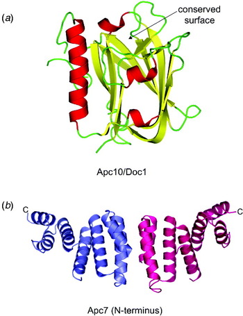
Until recently, the only conserved subunit for which X-ray crystallographic data were available was Apc10/Doc1 (Au et al. Reference Au, Leng, Harper and Barford2002; Wendt et al. Reference Wendt, Vodermaier, Jacob, Gieffers, Gmachl, Peters, Huber and Sondermann2001); however, recently the structures of numerous TPR subunits have been determined. Apc10/Doc1 is composed of a Doc homology domain that has also been identified in putative E3 ubiquitin ligases characterised by cullin and HECT domains (Grossberger et al. Reference Grossberger, Gieffers, Zachariae, Podtelejnikov, Schleiffer, Nasmyth, Mann and Peters1999), including a variant SCF composed of CUL7 (Dias et al. Reference Dias, Dolios, Wang and Pan2002). Apc10/Doc1 was originally discovered as an APC/C-associated protein in a genetic screen in S. cerevisiae as a mutant defective in cyclin destruction (destruction of cyclin) (Hwang & Murray, Reference Hwang and Murray1997), with other groups identifying Apc10/Doc1 as an essential component of S. pombe and human APC/C (Grossberger et al. Reference Grossberger, Gieffers, Zachariae, Podtelejnikov, Schleiffer, Nasmyth, Mann and Peters1999; Kominami et al. Reference Kominami, Seth-Smith and Toda1998; Kurasawa & Todokoro, Reference Kurasawa and Todokoro1999). The Doc homology domain forms a β-sandwich structure that is related in architecture to the galactose-binding domain of galactose oxidase, the coagulation factor C2 domain and an XRCC1 domain (Fig. 6a). Residues that are invariant among Apc10/Doc1 sequences, including a temperature-sensitive mitotic arrest mutant, map to a β-sheet region of the molecule, whose counterpart in galactose oxidase, the coagulation factor C2 domains and XRCC1, mediate bio-molecular interactions. Interestingly, mutations of this region abrogate APC/C E3 ubiquitin ligase activity in a D box (APC/C destruction box [RxxLxx(I/V)xN])-dependent manner, indicating a role for this region in mediating D box recognition, although whether this is direct or indirect is unknown (Carroll et al. Reference Carroll, Enquist-Newman and Morgan2005).
Crystal structures of APC/C subunits. (a) Apc10/Doc1 (S. cerevisiae) (Au et al. Reference Au, Leng, Harper and Barford2002). (b) N-terminal dimerisation domain of human Apc7 shown as a homo-dimer (Han et al. Reference Han, Kim, Kim, Kang, Lee and Kim2009).

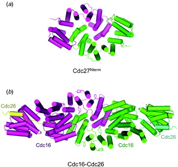
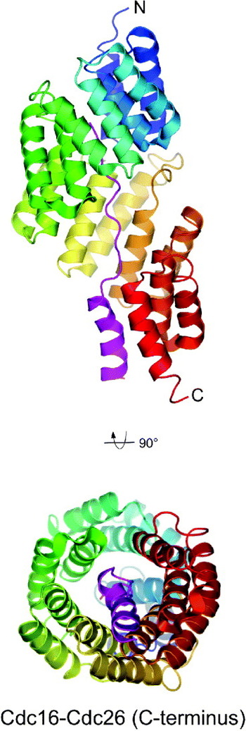
The recent crystal structure determinations of an Apc6/Cdc16-Cdc26 complex and the N-terminal domain of Apc3/Cdc27 reveal considerable insights into the molecular organisation and architecture of the APC/C's TPR-containing subunits (Zhang et al. Reference Zhang, Roe, Diogon, Kong, El Alaoui and Barford2010a) (Fig. 7a). Both Apc6/Cdc16 and Apc3/Cdc27 were found to form homo-dimers, with the dimer interface mediated by the self-association of their respective N-terminal TPR domains. A TPR superhelix created by seven successive TPR motifs (each TPR motif folds into a pair of anti-parallel α-helices) forms an interlocking dimer interface whereby the two subunits of the dimer associate in an N-terminal-to-N-terminal configuration (Fig. 7b) (Zhang et al. Reference Zhang, Roe, Diogon, Kong, El Alaoui and Barford2010a). An intimate homotypic interface is generated as the inner concave surface of each subunit's TPR superhelix encircles its dimer counterpart in a clasp-like arrangement. Apc6/Cdc16 associates with Cdc26 and in the Apc6/Cdc16-Cdc26 hetero-tetramer, the C-terminal seven TPR motifs of Apc6/Cdc16 are arranged into a TPR superhelix contiguous with the TPR superhelix of the N-terminal dimerisation domain (Zhang et al. Reference Zhang, Kulkarni, Hanrahan, Thompson and Barford2010b). Thus, each Apc6/Cdc16 subunit adopts a rod-like structure of a contiguous TPR superhelix of 14 TPR motifs. Dimerisation of Apc6/Cdc16 via its N-terminal domain generates a shallow ‘V’-shaped molecule measuring some 155 Å in length. Cdc26 stabilises Apc6/Cdc16 through an unusual TPR–protein interaction. The N-terminal 12 residues of Cdc26, which are unstructured in isolation (Wang et al. Reference Wang, Dye, Rajashankar, Kurinov and Schulman2009), adopt an extended conformation that interacts with the inner concave groove of the Apc6/Cdc16 superhelix (Figs 7b and 8). Residues 13–25 (human numbering) of Cdc26 on the other hand form an α-helix that packs against the C-terminal non-consensus TPR α-helix of the super-helix, mimicking a TPR motif, although with both helices arranged in parallel. The intimate interactions between Apc6/Cdc16 and Cdc26 explain the stabilising influence exerted by Cdc26 on Apc6/Cdc16 (Wang et al. Reference Wang, Dye, Rajashankar, Kurinov and Schulman2009).
Apc6/Cdc16 and Apc3/Cdc27 share a related architecture and mode of homo-dimerisation. Both homo-dimerise through an N-terminal domain composed of seven TPR units forming a TPR superhelix (Zhang et al. Reference Zhang, Roe, Diogon, Kong, El Alaoui and Barford2010a). (a) Cdc27 homo-dimer. (b) Cdc16-Cdc26 heterodimer (Zhang et al. Reference Zhang, Kulkarni, Hanrahan, Thompson and Barford2010b).

Crystal structure of the C-terminal eight TPR motifs of human Apc6/Cdc16 in complex with N-terminal 25 residues of Cdc26 (pink) (Wang et al. Reference Wang, Dye, Rajashankar, Kurinov and Schulman2009).

The C-terminal seven TPR units of Apc3/Cdc27, missing from the crystallised N-terminal domain of Apc3/Cdc27 (Apc3/Cdc27Nterm), were predicted to form a continuous TPR superhelix contiguous with the Apc3/Cdc27Nterm TPR superhelix. Thus, similar to Apc6/Cdc16, the Apc3/Cdc27 homo-dimer will adopt a ‘V’-shaped molecule, with the C-terminal TPR super-helix projecting away from the apex of the ‘V’ formed by the Apc3/Cdc27Nterm domain interface (Zhang et al. Reference Zhang, Roe, Diogon, Kong, El Alaoui and Barford2010a). Structural similarity of Apc6/Cdc16 and Apc3/Cdc27 architecture and the mode of dimerisation suggests evolutionary divergence from a common ancestor. Cdh1 interactions with Apc3/Cdc27, mediated via its C-terminal IR motif, involve the highly conserved TPR8 and TPR9 motifs of the C-terminal domain of Apc3/Cdc27 (Matyskiela & Morgan, Reference Matyskiela and Morgan2009). Residues implicated in engaging the Cdh1-IR motif line, the inner groove of the extended TPR superhelix, distal from the Apc3/Cdc27 dimerisation domain.
Findings that Apc6/Cdc16 and Apc3/Cdc27 are homo-dimers are consistent with studies showing that TPR subunits are present at supra-stoichiometric abundance within the APC/C (Dube et al. Reference Dube, Herzog, Gieffers, Sander, Riedel, Muller, Engel, Peters and Stark2005; Ohi et al. Reference Ohi, Feoktistova, Ren, Yip, Cheng, Chen, Yoon, Wall, Huang, Penczek, Gould and Walz2007; Passmore et al. Reference Passmore, Booth, Venien-Bryan, Ludtke, Fioretto, Johnson, Chiu and Barford2005) and early studies demonstrating Apc6/Cdc16, Apc8/Cdc23 and Apc3/Cdc27 self-association (Lamb et al. Reference Lamb, Michaud, Sikorski and Hieter1994). Dimerisation of Cdc27 has implications for the number of co-activator molecules bound simultaneously to APC/C, and the abundance of Apc10/Doc1 within the complex. Since both the co-activator and Apc10/Doc1 are required for optimal APC/C catalytic activity and substrate recognition (Passmore et al. Reference Passmore, Mccormack, Au, Paul, Willison, Harper and Barford2003), and both interact with Apc3/Cdc27 via their C-terminal IR motifs (Kraft et al. Reference Kraft, Vodermaier, Maurer-Stroh, Eisenhaber and Peters2005; Matyskiela & Morgan, Reference Matyskiela and Morgan2009; Passmore et al. Reference Passmore, Mccormack, Au, Paul, Willison, Harper and Barford2003; Thornton et al. Reference Thornton, Ng, Matyskiela, Carroll, Morgan and Toczyski2006; Vodermaier et al. Reference Vodermaier, Gieffers, Maurer-Stroh, Eisenhaber and Peters2003; Vodermaier & Peters, Reference Vodermaier and Peters2004; Wendt et al. Reference Wendt, Vodermaier, Jacob, Gieffers, Gmachl, Peters, Huber and Sondermann2001), the minimum condition of APC/C activity would be one co-activator molecule and one Apc10/Doc1 molecule per Apc3/Cdc27 dimer. Presumably, since APC/C lacks global twofold symmetry (Dube et al. Reference Dube, Herzog, Gieffers, Sander, Riedel, Muller, Engel, Peters and Stark2005; Herzog et al. Reference Herzog, Primorac, Dube, Lenart, Sander, Mechtler, Stark and Peters2009; Ohi et al. Reference Ohi, Feoktistova, Ren, Yip, Cheng, Chen, Yoon, Wall, Huang, Penczek, Gould and Walz2007; Passmore et al. Reference Passmore, Booth, Venien-Bryan, Ludtke, Fioretto, Johnson, Chiu and Barford2005), asymmetry imposed on Apc3/Cdc27 within the context of the APC/C, and/or the requirement for additional APC/C subunits to confer high affinity for the APC/C (Thornton et al. Reference Thornton, Ng, Matyskiela, Carroll, Morgan and Toczyski2006), would allow for both the co-activator and Apc10/Doc1 to bind a single Apc3/Cdc27 homo-dimer within the complex.
The crystal structure of the N-terminal domain of human Apc7 (residues 21–166) revealed an N-terminal dimerisation domain differing from Apc6/Cdc16 and Apc3/Cdc27 (Fig. 6b) (Han et al. Reference Han, Kim, Kim, Kang, Lee and Kim2009).
3. Substrate recognition and role of co-activators
3.1 Canonical APC/C recognition motifs: D box and KEN box
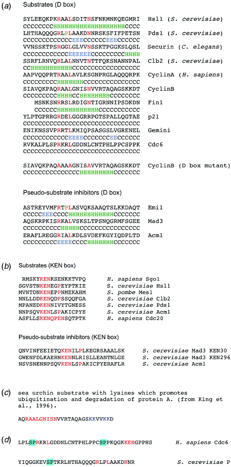
The molecular basis for targeting proteins substrates for ubiquitylation by the APC/C is complex and only partially understood. The classical APC/C degron is the destruction box or D box, a nine-residue motif (RxxLxxI/VxN) first characterised in B-type cyclins as being necessary and sufficient for APC/C-mediated ubiquitylation (Glotzer et al. Reference Glotzer, Murray and Kirschner1991; King et al. Reference King, Glotzer and Kirschner1996; Yamano et al. Reference Yamano, Gannon and Hunt1996) (Fig. 9a). Another APC/C degron, the KEN motif (KENxxxN/D), is commonly present in APC/C substrates usually in addition to the D box (Pfleger & Kirschner, Reference Pfleger and Kirschner2000) (Fig. 9b). Efficient ubiquitylation by either APC/CCdc20 or APC/CCdh1 of substrates harbouring both D and KEN boxes is dependent on both degrons; see for example (Burton & Solomon, Reference Burton and Solomon2001; Passmore et al. Reference Passmore, Mccormack, Au, Paul, Willison, Harper and Barford2003). However, some substrates contain only either a D box or KEN box, in one or more copies. The use of two distinct degrons imposes a degree of substrate specificity on the APC/C with APC/CCdc20 being more dependent on the D box and APC/CCdh1 more dependent on the KEN box (Passmore et al. Reference Passmore, Booth, Venien-Bryan, Ludtke, Fioretto, Johnson, Chiu and Barford2005; Zur & Brandeis, Reference Zur and Brandeis2002). Importantly, APC/CCdh1 is capable of ubiquitylating the KEN box only containing substrates (or artificial substrates; Zur & Brandeis, Reference Zur and Brandeis2002), for example Cdc20 (Pfleger & Kirschner, Reference Pfleger and Kirschner2000), Tome-1 (Ayad et al. Reference Ayad, Rankin, Murakami, Jebanathirajah, Gygi and Kirschner2003) and Sororin (Rankin et al. Reference Rankin, Ayad and Kirschner2005). Cin8p is recognised by APC/CCdh1 in combination with a second recognition element proximal to the KEN box together with a nuclear localisation signal (Hildebrandt & Hoyt, Reference Hildebrandt and Hoyt2001). Substrate recognition by APC/CCdc20 in contrast is dependent on the D box (for example, Hendrickson et al. Reference Hendrickson, Meyn, Morabito and Holloway2001). Additionally, as discussed below, more rarely used APC/C degrons have also been characterised. An analysis of the relative positions and orientations of D boxes and KEN boxes reveals striking variation with no clear common structural theme. Recognition motifs may occur in either order, be separated by between 30 and 200 amino acids and be present at either the N- or C-termini of APC/C substrates. There are little data addressing the conformation of the D box and KEN motifs. Predictions of secondary structure do not provide a consistent view of a defined structural motif. For example, the D box and flanking residues of cyclin B and Hsl1 are predicted to form an α-helix, whereas the D box of other APC/C substrates, for example, Cdc6 and Geminin are predicted to be either unfolded or to adopt an extended β-strand conformation, respectively (Fig. 9a). Consistently, the KEN box secondary structure predictions are indicative of an unstructured motif. A circular dichroism spectrum of a 20-residue peptide modelled on the cyclin B D box at 4 and 20°C revealed an unstructured conformation (D. Barford, unpublished data).
Substrate recognition motifs of the APC/C. (a) Multiple sequence alignments of D box (destruction box) motifs of substrates and pseudo-substrate inhibitors. Predicted secondary structure indicated. In the consensus RxxLxx(I/V)xN motif, the Arg and Leu residues are invariant, whereas the Asn residue is less well conserved. The Asn is not conserved in the D box pseudo-substrate motifs. Consensus residues indicated in red. There is a weak tendency for the D box to be predicted as an α-helix. (b) Multiple sequence alignments of KEN box motifs of substrates and pseudo-substrate inhibitors. (c) Minimal 27-residue sequence of sea urchin cyclin B that when fused to its N-terminus directs degradation of the heterologous protein A (King et al. Reference King, Glotzer and Kirschner1996). Substrate lysines shown in blue, D box in red. (d) Cdk-dependent phosphorylation sites shown highlighted in the vicinity of the D box of Cdc6 (Mailand & Diffley, Reference Mailand and Diffley2005) and securin (Holt et al. Reference Holt, Krutchinsky and Morgan2008) that block APC/C-mediated ubiquitylation.

Little is known concerning the optimal position of target lysine residues relative to the D box and KEN box motifs. In their study of signals responsible for cyclin destruction, King et al. (Reference King, Glotzer and Kirschner1996) found that a fusion of 40–66 residues of sea urchin cyclin B to protein A was sufficient to promote degradation of the fusion protein. With the D box between residues 42 and 50, four Lys residues are positioned within 15 residues (Fig. 9c). Therefore, this study revealed that Lys residues in quite close proximity to the D box serve as targets of ubiquitylation, consistent with findings that a Lys residue immediately C-terminal to the D box can function as a ubiquitin acceptor (Kraft et al. Reference Kraft, Vodermaier, Maurer-Stroh, Eisenhaber and Peters2005).
3.2 Mechanism of D box and KEN box recognition
Mechanistic insights into the role of the D box in substrate recognition and ubiquitylation were provided by findings showing that transferring the cyclin B D box to a heterologous protein, for example, protein A or Gal4BD, conferred similar cell-cycle-dependent instability as observed for cyclin B (Glotzer et al. Reference Glotzer, Murray and Kirschner1991; King et al. Reference King, Glotzer and Kirschner1996; Yamano et al. Reference Yamano, Gannon and Hunt1996). Mutation of any of the three most highly conserved residues, Arg, Leu or Asn, ablated the destruction signal (Glotzer et al. Reference Glotzer, Murray and Kirschner1991); however, although the Arg and Leu residues appear as an invariant feature of a D box motif, the C-terminal Asn residue is less well conserved (Fig. 9a). Short 20–25 residue peptides comprising the D box inhibit APC/C-dependent ubiquitylation of cyclin B at sub-mM concentrations with some 1000-fold lower affinity than full-length natively folded proteins (Kraft et al. Reference Kraft, Vodermaier, Maurer-Stroh, Eisenhaber and Peters2005; Yamano et al. Reference Yamano, Tsurumi, Gannon and Hunt1998). Similar findings have been found for peptides comprising the KEN box (Burton et al. Reference Burton, Tsakraklides and Solomon2005). Importantly, these studies demonstrate that APC/C–co-activator complexes harbour receptor(s) that directly recognise the D box and KEN box motifs.
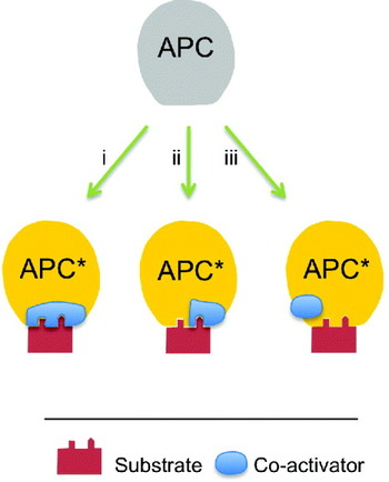
Without the co-activator, the core APC/C is inactive as an E3 ubiquitin ligase and is unable to bind most APC/C substrates (exceptions include recognition of Nek2A, cyclin A and a tandem D-box in vertebrates). It has therefore been proposed that the role of the co-activator is to act as a substrate recognition subunit, analogous to the F-box protein receptors of the SCF, and recruit substrate to the APC/C. This notion is consistent with observations that co-activators promote the interaction of substrate to the APC/C to form a stoichiometric ternary complex of APC/C – co-activator-substrate, dependent on the D box and KEN box degrons (Passmore & Barford, Reference Passmore and Barford2005; Passmore et al. Reference Passmore, Mccormack, Au, Paul, Willison, Harper and Barford2003). In support of the adaptor model, numerous studies demonstrate direct and D box and KEN box-dependent binding of APC/C substrate proteins to co-activators (Amador et al. Reference Amador, Ge, Santamaria, Guardavaccaro and Pagano2007; Burton & Solomon, Reference Burton and Solomon2001; Burton et al. Reference Burton, Tsakraklides and Solomon2005; Hilioti et al. Reference Hilioti, Chung, Mochizuki, Hardy and Cohen-Fix2001; Pfleger et al. Reference Pfleger, Lee and Kirschner2001; Schwab et al. Reference Schwab, Neutzner, Mocker and Seufert2001). Chemical cross-linking of substrates to the WD40 domains of Cdc20 and Cdh1 in a D box and KEN box-dependent manner confirms a role of the co-activators to contribute towards the recruitment of the substrate to the core APC/C (Hilioti et al. Reference Hilioti, Chung, Mochizuki, Hardy and Cohen-Fix2001; Kraft et al. Reference Kraft, Vodermaier, Maurer-Stroh, Eisenhaber and Peters2005). Mutation of conserved (predicted) surface residues of the WD40 domain abolished substrate interactions (Kraft et al. Reference Kraft, Vodermaier, Maurer-Stroh, Eisenhaber and Peters2005). However, given the immeasurably low affinity of co-activator–substrate interactions, it is unlikely that the co-activators alone are sufficient to confer high-affinity substrate binding to the APC/C–co-activator binary complex, and there is increasing evidence that core APC/C subunits play important roles in contributing towards substrate association, especially D box-containing substrates. For example, other studies suggested that APC/C alone could bind tandem D boxes (Yamano et al. Reference Yamano, Gannon, Mahbubani and Hunt2004), and the Apc10/Doc1 subunit of APC/C was shown to be necessary for optimal co-activator-dependent substrate recognition (Passmore et al. Reference Passmore, Mccormack, Au, Paul, Willison, Harper and Barford2003), and for contributing towards substrate processivity (Carroll & Morgan, Reference Carroll and Morgan2002). These findings can be rationalised from two non-mutually exclusive models (Fig. 10). In one, the bi-partite model, the binding of substrates to the APC/C–co-activator complex is formed from a bi-partite binding site created from co-activator and APC/C subunits. In the alternative allosteric model, the co-activator induces a conformational change within the APC/C to generate substrate-binding sites from APC/C subunits (discussed in Eytan et al. Reference Eytan, Moshe, Braunstein and Hershko2006; Passmore & Barford, Reference Passmore and Barford2005). Since co-activators have been shown to bind the substrate directly, this model is less likely; however, a combination of a bipartite-binding model coupled to co-activator-induced conformational change is plausible. Other studies also support the notion of a composite substrate-binding site shared between the co-activator and a core APC/C subunit(s), dependent on Apc10/Doc1. First, Burton et al. discovered that the substrate enhanced the binding of Cdh1 to the APC/C in a D box-dependent manner (Burton et al. Reference Burton, Tsakraklides and Solomon2005), a finding supported by a study showing that substrate compensates for loss of affinity of Cdh1 to APC/C due to mutations in the putative Cdh1-binding region of Apc3/Cdc27 (Matyskiela & Morgan, Reference Matyskiela and Morgan2009), and a finding that cyclin B (D box) peptides interact more efficiently with Cdc20 when it is bound to APC/C (Kraft et al. Reference Kraft, Vodermaier, Maurer-Stroh, Eisenhaber and Peters2005). Significantly, the study by Matyskiela & Morgan (Reference Matyskiela and Morgan2009) showed that mutations of Apc10/Doc1 that impair processivity in a D box-dependent manner (Carroll et al. Reference Carroll, Enquist-Newman and Morgan2005), did not prolong co-activator binding in the presence of high concentrations of a cyclin fragment (containing only a D box). These results support the model that substrate promotes co-activator binding by providing a bivalent bridge between a site on the co-activator and an Apc10/Doc1-dependent site on the APC/C core. A plausible model for D box-dependent substrate recognition, which appears to reconcile data that (i) D box binds to the co-activator, (ii) D box binds core APC/C, (iii) D box is required for substrate–APC/C interactions in a Apc10/Doc1-dependent manner and (iv) that a functional D box stabilises Cdh1 binding to APC/C dependent on Apc10/Doc1, is one that invokes a composite-binding site for the D box involving both the co-activator and Apc10/Doc1. Interestingly, as KEN box motifs also bind directly to the co-activator, but that KEN box-dependent association with the APC/C–co-activator complex does not require Apc10/Doc1 (Carroll et al. Reference Carroll, Enquist-Newman and Morgan2005), and that association of Cdh1 with APC/C is not KEN box dependent (Burton et al. Reference Burton, Tsakraklides and Solomon2005), it is most likely that the KEN box binds predominantly to the co-activator alone, or at least via a mechanism that does not involve Apc10/Doc1.
Schematic indicating different models of co-activator activation on the APC/C, modified from Passmore & Barford (Reference Passmore and Barford2005) . (i) The co-activator functions to bind substrate alone. (ii) The co-activator functions to create a bi-partite-binding site with a core APC/C subunit (possibly Apc10/Doc1). (iii) The co-activator causes an allosteric conformational change allowing core APC/C subunits to bind the substrate.

3.3 Non-canonical APC/C recognition motifs
Ubiquitylation of certain APC/C substrates is not entirely dependent on either the D box or KEN box, and some substrates lack both destruction motifs entirely. Thus, a class of non-canonical APC/C recognition motifs contributes towards APC/C-dependent substrate ubiquitylation. Significantly, as discussed below, although co-activators (Cdc20 and Cdh1) are implicated in activating the APC/C towards these substrates, at least in instances where substrate recognition is not dependent on either a D box or KEN box, it is likely that co-activators play no role in direct substrate recognition. Furthermore, non-canonical recognition motifs in substrates containing D box and KEN box motifs are likely recognised by core APC/C subunits. The capacity of the APC/C to recognise a wide range of structurally varied substrates may underlie its structural complexity and size.
Cyclin A and Nek2A evade the SAC mediated by the MCC, being Cdc20-dependent substrates of the APC/C, even though the MCC inhibits ubiquitylation of the APC/CCdc20-dependent targets securin and cyclin B (Dawson et al. Reference Dawson, Roth and Artavanis-Tsakonas1995; den Elzen & Pines, Reference Den Elzen and Pines2001; Geley et al. Reference Geley, Kramer, Gieffers, Gannon, Peters and Hunt2001; Hames et al. Reference Hames, Wattam, Yamano, Bacchieri and Fry2001; Hayes et al. Reference Hayes, Kimata, Wattam, Lindon, Mao, Yamano and Fry2006; Sigrist et al. Reference Sigrist, Jacobs, Stratmann and Lehner1995). Interestingly, both cyclin A and Nek2A possess modified D boxes (cyclin A-type D boxes), located close to the N-terminus in cyclin A and towards the C-terminus in Nek2A. However, for both cyclin A and Nek2A, mutation or deletion of the D box (and KEN box in Nek2A) does not stabilise the respective protein at prometaphase, questioning the importance of these motifs in APC/C-dependent recognition, and suggesting a different mode of substrate recognition from that occurring at other phases of the cell cycle (den Elzen & Pines, Reference Den Elzen and Pines2001; Geley et al. Reference Geley, Kramer, Gieffers, Gannon, Peters and Hunt2001; Hayes et al. Reference Hayes, Kimata, Wattam, Lindon, Mao, Yamano and Fry2006; Jacobs et al. Reference Jacobs, Keidel and Lehner2001). In the instance of cyclin A, both a region immediately C-terminus of the D box, and interactions with Cdk1 (Geley et al. Reference Geley, Kramer, Gieffers, Gannon, Peters and Hunt2001; Stewart et al. Reference Stewart, Kobayashi, Harrison and Hunt1994), are required for APC/C recognition, whereas a C-terminal Met-Arg motif is responsible for associating Nek2A with the APC/C (Hayes et al. Reference Hayes, Kimata, Wattam, Lindon, Mao, Yamano and Fry2006). In recent work, it was shown that Cdc20 and the Cdk-cyclin A binding partner Cks1, direct spindle checkpoint-independent destruction of cyclin A (Wolthuis et al. Reference Wolthuis, Clay-Farrace, Van Zon, Yekezare, Koop, Ogink, Medema and Pines2008). Although not required for prometaphase-dependent degradation of Nek2A, its KEN box is a required degradation signal after the onset of metaphase, consistent with altered modes of substrate recognition at prometaphase (Hayes et al. Reference Hayes, Kimata, Wattam, Lindon, Mao, Yamano and Fry2006).
Other examples of substrates that incorporate non-canonical destruction motifs include the A box of Aurora A kinase (Castro et al. Reference Castro, Vigneron, Bernis, Labbe, Prigent and Lorca2002; Littlepage & Ruderman, Reference Littlepage and Ruderman2002), an N-terminal 34 residue segment of Cik1L (a splice variant of Cik1) (Benanti et al. Reference Benanti, Matyskiela, Morgan and Toczyski2009), a C-terminal element of Cin8p (Hildebrandt & Hoyt, Reference Hildebrandt and Hoyt2001), elements within cyclin A (Geley et al. Reference Geley, Kramer, Gieffers, Gannon, Peters and Hunt2001; Jacobs et al. Reference Jacobs, Keidel and Lehner2001), a 43-residue segment at the C-terminus of the kinesin Kip1p (Gordon & Roof, Reference Gordon and Roof2001), a GxEN motif in Xenopus chromokinesin XKid (Castro et al. Reference Castro, Vigneron, Bernis, Labbe and Lorca2003), a LxExxxxN motif in S. cerevisiae Spo13 (Sullivan & Morgan, Reference Sullivan and Morgan2007a), and a Cry box in Cdc20 (Reis et al. Reference Reis, Levasseur, Chang, Elliott and Jones2006). Curiously, both the KEN box-dependent degradation of Cdc20, and the GxEN motif-dependent degradation of XKid by APC/CCdh1, are blocked by a D box-containing peptide (Castro et al. Reference Castro, Vigneron, Bernis, Labbe and Lorca2003; Pfleger & Kirschner, Reference Pfleger and Kirschner2000). This could imply partial overlap of receptor sites for D, KEN and GxEN boxes, or alternatively, if the D box-containing peptides were ubiquitylated, could simply compete for cellular ubiquitin.
The discovery of new APC/C substrates will be facilitated by high-throughput-automated approaches based on protein micro-arrays such as the extract-based functional assay of Merbl & Kirschner (Reference Merbl and Kirschner2009) .
3.4 Co-activators as catalytic activators
Substantial evidence now exists for a role for co-activators in contributing to substrate recognition mediated by D box and KEN box motifs. However, as mentioned previously, some substrates, which lack such canonical destruction motifs, are known to be dependent on the co-activator for APC/C-mediated ubiquitylation. Strikingly, cyclin A and Nek2A are targets of the APC/C, dependent on Cdc20 (Hames et al. Reference Hames, Wattam, Yamano, Bacchieri and Fry2001; Hayes et al. Reference Hayes, Kimata, Wattam, Lindon, Mao, Yamano and Fry2006; Wolthuis et al. Reference Wolthuis, Clay-Farrace, Van Zon, Yekezare, Koop, Ogink, Medema and Pines2008), thus implicating co-activators in other roles in APC/C-catalysed substrate ubiquitylation. Fry and Yamano recently obtained substantial new insights into the function and mechanism of co-activators following their analysis of the mechanism of Nek2A ubiquitylation by APC/CCdc20 (Kimata et al. Reference Kimata, Baxter, Fry and Yamano2008a). Although Nek2A binds to the APC/C independent of Cdc20 via its C-terminal Met-Arg motif, resembling the C-terminal Ile-Arg motif of co-activators and Apc10/Doc1, APC/C-mediated ubiquitylation of Nek2A relies on Cdc20 (Hames et al. Reference Hames, Wattam, Yamano, Bacchieri and Fry2001; Hayes et al. Reference Hayes, Kimata, Wattam, Lindon, Mao, Yamano and Fry2006). Kimato and co-workers discovered that the N-terminal C box motif of Cdc20, known to be required for interactions of the co-activator with APC/C (Kraft et al. Reference Kraft, Vodermaier, Maurer-Stroh, Eisenhaber and Peters2005; Schwab et al. Reference Schwab, Neutzner, Mocker and Seufert2001; Thornton et al. Reference Thornton, Ng, Matyskiela, Carroll, Morgan and Toczyski2006), is also required for the APC/C to ubiquitylate Nek2A bound to the APC/C via its Met-Arg motif (Kimata et al. Reference Kimata, Baxter, Fry and Yamano2008a). Thus, the N-terminus of Cdc20 allosterically stimulates the catalytic activity of the APC/C. Fusing the N-terminus of Cdc20 to a canonical substrate (Mes1), thereby targeting the substrate to the APC/C, promoted its ubiquitylation. Significantly, mutation of both Mes1 D box and KEN box motifs did not prevent the ubiquitylation of the Cdc20–Mes1 fusion protein, indicating that the APC/C receptors for these motifs were not required for the catalytic reaction. These data therefore suggest a model whereby the WD40 domain of co-activator functions in substrate recognition (Burton & Solomon, Reference Burton and Solomon2001; Hilioti et al. Reference Hilioti, Chung, Mochizuki, Hardy and Cohen-Fix2001; Kraft et al. Reference Kraft, Vodermaier, Maurer-Stroh, Eisenhaber and Peters2005; Pfleger et al. Reference Pfleger, Lee and Kirschner2001; Schwab et al. Reference Schwab, Neutzner, Mocker and Seufert2001), whereas the N-terminal C box and C-terminal IR motif binds to and activates the APC/C, with the C box functioning to stimulate APC/C catalytic activity.
4. Molecular enzymology of the APC/C
4.1 RING E3 ubiquitin ligases
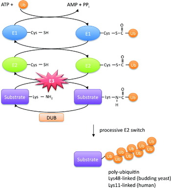
Protein ubiquitylation proceeds via a three-enzyme cascade (Capili & Lima, Reference Capili and Lima2007; Hershko & Ciechanover, Reference Hershko and Ciechanover1998; Passmore & Barford, Reference Passmore and Barford2004; Pickart, Reference Pickart2001) (Fig. 11). In the first step, ubiquitin is activated in an ATP-dependent reaction, resulting in a thioester covalent bond between its C-terminal carboxyl group and the catalytic cysteine of an E1 protein (ubiquitin-activating enzyme). The activated ubiquitin is then transferred, via attachment through its carboxyl terminus, to the catalytic cysteine of an E2 protein (ubiquitin-conjugating enzyme) forming an E2–ubiquitin species. In the final step, an E3 protein ubiquitin ligase catalyses the transfer of activated ubiquitin from E2–Ub to a lysine N∊-amino group (or more rarely an N-terminal amino group) of the target protein substrate forming an isopeptide linkage. E3s function to confer substrate selectivity and biological regulation on the ubiquitin system, and couple a specific protein substrate to one or more cognate E2s. Determination of the type of ubiquitin linkage within a polyubiquitin chain is likely conferred by both E2 and E3. While there are only one or two E1 enzymes in most organisms, and approximately 50 E2s, there are ~300 E3s, reflecting their regulatory and specificity functions and the importance of ubiquitylation as a biological control process.
The three-step ubiquitylation cascade of protein ubiquitylation involving E1 (ubiquitin activating enzyme), E2 (ubiquitin conjugating enzyme) and E3 ubiquitin ligase. Modified from Passmore & Barford (Reference Passmore and Barford2004) .

The largest structural class of E3 ligases are the RING (really interesting new gene) domain containing proteins, members of the cross-brace Zn-binding family (Deshaies & Joazeiro, Reference Deshaies and Joazeiro2009; Petroski & Deshaies, Reference Petroski and Deshaies2005). Within the human genome ~1000 RING genes are encoded. Although it is not known whether all RING proteins function as E3 ligases, the large number of RING proteins suggests enormous potential for structural and functional diversity. Closely structurally related to the RING E3 ligases are the U box proteins, which adopt the same overall fold as RING proteins but lack Zn-binding sites. RING and U box domain E3 ligases function within the context of scaffolding molecules containing protein-substrate recognition sites. A distinct class of E3 ligases is the HECT (homology to E6 C-terminus) domain family. These ligases differ mechanistically and structurally from RING and U box domain E3 ligases as they form an enzyme-ubiquitin intermediate (via a thioester bond with the ubiquitin C-terminus) prior to substrate ligation (Rotin & Kumar, Reference Rotin and Kumar2009).
4.2 Catalytic mechanisms
Unresolved questions remain regarding the catalytic mechanisms of E3 ligases and the role of the RING domain in the ubiquitylation reaction (Deshaies & Joazeiro, Reference Deshaies and Joazeiro2009; Passmore & Barford, Reference Passmore and Barford2004). The RING domain does not participate directly in the catalytic reaction (unlike the HECT domain E3s), and an explanation for their role is to act as scaffolding molecules to coordinate the interaction between the ubiquitin-conjugated E2 and the appropriate protein substrate bound to a protein-interaction domain or subunit attached to the RING domain. The catalytic centre of such an E3–E2–Ub–substrate complex would therefore be mainly generated by the E2 and there is strong evidence that the catalytic site of the E2 is critical for lowering the pK a of the attacking lysine N∊ group to enable nucleophilic attack onto the E2–Ub-thiol linkage (Yunus & Lima, Reference Yunus and Lima2006), reviewed by Ye & Rape (Reference Ye and Rape2009) . However, although the E2–Ub thioester conjugate is inherently reactive and is capable of allowing discharge of ubiquitin to lysine donors in the absence of a RING E3 (Petroski & Deshaies, Reference Petroski and Deshaies2005), RING E3s such as Rbx1 increase the rate of ubiquitin discharge some five-fold (Petroski & Deshaies, Reference Petroski and Deshaies2005). This demonstrates that RING E3s manipulate the catalytic reaction, although structural data indicate that the structure of the ubiquitin E2 (UbcH7) does not alter in complex with the RING E3 (Zheng et al. Reference Zheng, Wang, Jeffrey and Pavletich2000). Two possible explanations emerge from the structure of the SUMO E3 ligase–Ubc9–substrate complex (Reverter & Lima, Reference Reverter and Lima2005). One is that the RING domain promotes the optimal orientation of the attacking nucleophilic lysine residue onto the E2–Ub thioester linkage. Secondly, in this substrate complex, an Asn side chain, conserved in all E2 structures (Wu et al. Reference Wu, Hanlon, Eddins, Tsui, Rogers, Jensen, Matunis, Weissman, Wolberger and Pickart2003), rotates towards the catalytic site approaching the SUMO C-terminal Gly97 carbonyl oxygen, generating an oxyanion hole to stabilise the oxyanion transition state intermediate that develops during the formation of an isopeptide bond.
4.3 Mechanisms of processivity
Proteasome-dependent degradation of ubiquitylated protein substrates is determined by a ubiquitin polymer composed of at least four ubiquitin moieties (Chau et al. Reference Chau, Tobias, Bachmair, Marriott, Ecker, Gonda and Varshavsky1989; Thrower et al. Reference Thrower, Hoffman, Rechsteiner and Pickart2000). Although early studies indicated that proteasomal targeting was dependent on Lys48-linked polymers, recent data have revealed that proteins modified by ubiquitin polymers of other linkages, including Lys11, Lys29 and Lys63, and even single ubiquitin moieties, are recognised for degradation by the proteasome (Jin et al. Reference Jin, Williamson, Banerjee, Philipp and Rape2008; Kirkpatrick et al. Reference Kirkpatrick, Hathaway, Hanna, Elsasser, Rush, Finley, King and Gygi2006; Kravtsova-Ivantsiv et al. Reference Kravtsova-Ivantsiv, Cohen and Ciechanover2009; Saeki et al. Reference Saeki, Kudo, Sone, Kikuchi, Yokosawa, Toh-E and Tanaka2009a; Xu et al. Reference Xu, Duong, Seyfried, Cheng, Xie, Robert, Rush, Hochstrasser, Finley and Peng2009). Human APC/C was shown to catalyse formation of Lys11-linked polyubiquitin (Jin et al. Reference Jin, Williamson, Banerjee, Philipp and Rape2008), unlike S. cerevisiae APC/C that generates Lys48 linkages (Rodrigo-Brenni & Morgan, Reference Rodrigo-Brenni and Morgan2007). How different linked ubiquitin polymers specify distinct biological outcomes is the subject of much research. Structures of Lys48 and Lys63 linked ubiquitin polymers are radically different, explaining their potential to stimulate diverse signalling outcomes (Komander et al. Reference Komander, Reyes-Turcu, Licchesi, Odenwaelder, Wilkinson and Barford2009).
4.4 E2s define polyubiquitin linkage
An assembly of a polyubiquitin chain of defined linkage onto a specific substrate involves complex molecular mechanisms. The process requires that the E3 ubiquitin ligase attaches a priming ubiquitin molecule onto the protein substrate, followed by the generation of an ubiquitin polymer of a specific linkage onto a continuously varying substrate – the growing ubiquitin polymer. Two models for how E3 ligases catalyse processive reactions, required for generation of polyubiquitin chains, have been proposed (Hochstrasser, Reference Hochstrasser2006). In the sequential addition model, ubiquitin donated by the E2–Ub is added successively to the lysine residue of the distal monomer of the conjugated ubiquitin polymer. In this model, the geometry and position of the substrate lysine to which the ubiquitin is to be conjugated continuously varies. In an alternative seesaw model, the ubiquitin polymer is first assembled on the E2 prior to being transferred en bloc to the lysine residue of the substrate. The latter model requires that at least two E2 molecules participate in catalysis, a prediction supported by oligomerisation of numerous E2 and E3s (Passmore et al. Reference Passmore, Booth, Venien-Bryan, Ludtke, Fioretto, Johnson, Chiu and Barford2005), and is consistent with the assembly of ubiquitin polymers on individual E2s (Gazdoiu et al. Reference Gazdoiu, Yamoah, Wu, Escalante, Tappin, Bermudez, Aggarwal, Hurwitz and Pan2005; Li et al. Reference Li, Tu, Brunger and Ye2007; Ravid & Hochstrasser, Reference Ravid and Hochstrasser2007). However, recent time-resolved and thermodynamic studies of the SCF and its cognate E2 Cdc34, provide compelling evidence that at least for the SCF-Cdc34 E3–E2 system, formation of a polyubiquitin chain occurs through a sequential mechanism (Kleiger et al. Reference Kleiger, Saha, Lewis, Kuhlman and Deshaies2009; Pierce et al. Reference Pierce, Kleiger, Shan and Deshaies2009). A sequential reaction requires that Cdc34 is recharged with ubiquitin before dissociation of the modified substrate from the SCF, a process that involves rapid association and dissociation of Cdc34 from the SCF. Kinetic data indicated that the association rate constant of Cdc34 for the SCF exceeds the diffusion-limited rate, in a process facilitated by electrostatic steering of the acidic tail of Cdc34 to engage with a conserved and positively charged groove on the cullin domain of Cul1 (Kleiger et al. Reference Kleiger, Saha, Lewis, Kuhlman and Deshaies2009).
In S. cerevisiae, two E2s are responsible for generating a polyubiquitin chain through a sequential mechanism. Ubc4 catalyses the rapid monoubiquitylation of multiple lysines on APC/C substrates, whereas Ubc1 catalyses Lys48-linked polyubiquitin chain assembly onto a pre-attached ubiquitin moiety (Rodrigo-Brenni & Morgan, Reference Rodrigo-Brenni and Morgan2007). The capacity of Ubc1 to elongate an existing ubiquitin modification is conferred by its unusual C-terminal UBA domain. Deletion of the UBA domain results in shorter reaction products than those produced with wild-type Ubc1, indicating that the UBA domain confers processivity on the APC/C–E2 complex. Notably, specificity for Lys48 linkages is retained in the absence of the UBA domain. The human homolog of Ubc1, E2-25K (UbcH1) can also elongate monoubiquitylated APC/C substrates modified by UbcH10, although the role of the UBA domain in conferring the processivity was not reported; however, this may not represent a physiologically relevant E2 (Summers et al. Reference Summers, Pan, Mukhyala and Jackson2008), as discussed below. In contrast to S. cerevisiae, higher eukaryotes were until recently thought to utilise a single E2, UbcH10, an E2-C family member, for both the priming and elongation reactions, generating a polyubiquitin chain predominantly linked via Lys11 (Aristarkhov et al. Reference Aristarkhov, Eytan, Moghe, Admon, Hershko and Ruderman1996; Jin et al. Reference Jin, Williamson, Banerjee, Philipp and Rape2008; Yu et al. Reference Yu, King, Peters and Kirschner1996). Genetic evidence for S. pombe and Drosophila supports the notion that the E2-C family members, UbcH10 and Vihar, are critical for APC/C function (Mathe et al. Reference Mathe, Kraft, Giet, Deak, Peters and Glover2004; Osaka et al. Reference Osaka, Seino, Seno and Yamao1997). The mechanism of Lys11-linked ubiquitin chain formation catalysed by human APC/C has recently been elucidated through independent studies that identified the E2 Ube2S as the E2 responsible for extending APC/C substrates primed by UbcH10 or UbcH5 (Garnett et al. Reference Garnett, Mansfeld, Godwin, Matsusaka, Wu, Russell, Pines and Venkitaraman2009; Williamson et al. Reference Williamson, Wickliffe, Mellone, Song, Karpen and Rape2009; Wu et al. Reference Wu, Merbl, Huo, Gallop, Tzur and Kirschner2010). Thus, analogous to S. cerevisiae APC/C that utilises separate priming and elongating E2s to generate Lys48-linked ubiquitin polymers – Ubc4 and Ubc1, respectively (Rodrigo-Brenni & Morgan, Reference Rodrigo-Brenni and Morgan2007), in humans, Lys11-linked ubiquitin polymers are generated by UbcH10/UbcH5 and Ube2S. Somewhat different from budding yeast APC/C; however, human APC/C binds both UbcH10 and Ube2S simultaneously, and Ube2S (Williamson et al. Reference Williamson, Wickliffe, Mellone, Song, Karpen and Rape2009) only functions in the presence of UbcH10/UbcH5 (Garnett et al. Reference Garnett, Mansfeld, Godwin, Matsusaka, Wu, Russell, Pines and Venkitaraman2009).
Another factor determining processivity is the affinity of substrate for the APC/C (Rape et al. Reference Rape, Reddy and Kirschner2006). Substrates with high affinity, and therefore low rates of dissociation, remain bound to the APC/C for sufficient length of time to allow extension of the ubiquitin polymer. The shorter residency times of lower affinity substrates, which dissociate more rapidly, allow for only short extensions on each binding event, and therefore, generation of a longer ubiquitin polymer necessary for proteasomal degradation occurs following multiple binding events. Further limiting the chain length of such distributive substrates, once dissociated from the APC/C, deubiquitinases (DUBs) depolymerise the ubiquitin chain. During the cell cycle, the processivity of multi-ubiquitylation by the APC determines the order of substrate degradation.
5. APC/C regulation I: control by phosphorylation
APC/C is activated in early mitosis by Cdk- and Plk1-dependent phosphorylation (Golan et al. Reference Golan, Yudkovsky and Hershko2002; Kraft et al. Reference Kraft, Herzog, Gieffers, Mechtler, Hagting, Pines and Peters2003; Kramer et al. Reference Kramer, Scheuringer, Podtelejnikov, Mann and Peters2000; Lahav-Baratz et al. Reference Lahav-Baratz, Sudakin, Ruderman and Hershko1995; Rudner & Murray, Reference Rudner and Murray2000; Shteinberg et al. Reference Shteinberg, Protopopov, Listovsky, Brandeis and Hershko1999; Yamada et al. Reference Yamada, Kumada and Yanagida1997). The principle consequence being to stimulate Cdc20 binding, possibly mediated via the CCT chaperonin (Camasses et al. Reference Camasses, Bogdanova, Shevchenko and Zachariae2003). However, unlike CRLs that are activated by nedd8ylation (Duda et al. Reference Duda, Borg, Scott, Hunt, Hammel and Schulman2008; Saha & Deshaies, Reference Saha and Deshaies2008), there is no evidence that APC/C subunits are modified with ubiquitin-like proteins. Mutational analysis and mass-spectrometry-based identification of phosphorylation sites has indicated that the major phosphorylated subunits are Apc6/Cdc16, Apc8/Cdc23, Apc3/Cdc27 and Apc1 (Kraft et al. Reference Kraft, Herzog, Gieffers, Mechtler, Hagting, Pines and Peters2003; Rudner & Murray, Reference Rudner and Murray2000; Steen et al. Reference Steen, Steen, Georgi, Parker, Springer, Kirchner, Hamprecht and Kirschner2008).
Reversible phosphorylation of co-activators provides a second phosphorylation-dependent mechanism to control APC/C activity. Whereas the activity of APC/CCdc20 is activated by APC/C phosphorylation, Cdk-mediated-phosphorylation of Cdh1 inactivates Cdh1 by preventing APC/C binding (Jaspersen et al. Reference Jaspersen, Charles and Morgan1999; Visintin et al. Reference Visintin, Craig, Hwang, Prinz, Tyers and Amon1998; Zachariae et al. Reference Zachariae, Schwab, Nasmyth and Seufert1998a). In S. cerevisiae, dephosphorylation of Cdh1 by the protein phosphatase Cdc14, activated by the mitotic exit network (MEN) and FEAR (Cdc14 early anaphase release) networks, stimulates APC/CCdh1 by reversing the effects of the M-phase kinases (Jaspersen et al. Reference Jaspersen, Charles and Morgan1999; Shou et al. Reference Shou, Seol, Shevchenko, Baskerville, Moazed, Chen, Jang, Shevchenko, Charbonneau and Deshaies1999; Visintin et al. Reference Visintin, Hwang and Amon1999). However, the mammalian homologs of Cdc14 (Cdc14A and Cdc14B) appear to play distinct roles (Berdougo et al. Reference Berdougo, Nachury, Jackson and Jallepalli2008). At the mitotic/spindle checkpoint, Cdc20 is negatively regulated by phosphorylation mediated by Cdks, MAP kinases and Bub1 (Chung & Chen, Reference Chung and Chen2003; Tang et al. Reference Tang, Shu, Oncel, Chen and Yu2004; Yudkovsky et al. Reference Yudkovsky, Shteinberg, Listovsky, Brandeis and Hershko2000).
Direct phosphorylation of substrates provides a third level of APC/C control by protein phosphorylation. At S-phase, Cdk-dependent phosphorylation of amino acids in the immediate vicinity of the D box of Cdc6 blocks binding to the APC/C, thereby protecting Cdc6 from ubiquitylation, and promoting DNA replication origin licensing (Mailand & Diffley, Reference Mailand and Diffley2005) (Fig. 9d). Through a related process, Cdk1-mediated phosphorylation of securin at N-terminal sites in close proximity to the KEN box and D box reduce the rate of APC/C-dependent securin ubiquitylation some 5–10-fold (Holt et al. Reference Holt, Krutchinsky and Morgan2008) (Fig. 9d). Dephosphorylation of these sites by Cdc14 therefore promotes securin degradation. Interestingly, since active separase (produced as a result of securin degradation) stimulates Cdc14, a positive feedback loop is generated involving Cdc14-mediated dephosphorylation of securin, which in addition to the partial inactivation of Cdks at metaphase due to APC/CCdc20-mediated destruction of mitotic cyclins, thereby increases the rate of securin degradation and the abruptness of anaphase onset (Holt et al. Reference Holt, Krutchinsky and Morgan2008).
6. APC/C regulation II: APC/C pseudo-substrate-based inhibitors
6.1 Early mitotic inhibitor 1 (Emi1)
In vertebrates, Emi1 is an inhibitor of the APC/C, initially discovered due to its role in preventing premature APC/C activation in early mitosis in the presence of high Cdk1 activity (Reimann et al. Reference Reimann, Freed, Hsu, Kramer, Peters and Jackson2001a), but now recognised to primarily function as an antagonist of APC/CCdh1 during G2 (Di Fiore & Pines, Reference Di Fiore and Pines2008; Grosskortenhaus & Sprenger, Reference Grosskortenhaus and Sprenger2002). The protein contains an F box that targets it for degradation via the SCFβ−TrCP E3 ligase following Plk1-mediated phosphorylation later in mitosis (Hansen et al. Reference Hansen, Loktev, Ban and Jackson2004). In addition, the binding partner of Emi1, the Evi5 oncoprotein, blocks Plk1-dependent phosphorylation of Emi1, and binding to the SCFβ−TrCP ligase, thereby antagonising APC/C activation (Eldridge et al. Reference Eldridge, Loktev, Hansen, Verschuren, Reimann and Jackson2006). Emi1 also functions at the G1-S transition when E2F-induced transcription of Emi1 stabilises APC/CCdh1 targets such as cyclin A, thereby promoting S phase entry (Hsu et al. Reference Hsu, Reimann, Sorensen, Lukas and Jackson2002). The ability of Emi1 to inhibit the APC/C is conferred from a combination of its conserved D box, which provides strong APC/C-binding affinity and a block to substrate – APC/C – co-activator binding, together with a conserved zinc-binding region (ZBR), which provides an APC/C E3 ligase antagonising activity (Miller et al. Reference Miller, Summers, Hansen, Nachury, Lehman, Loktev and Jackson2006; Reimann et al. Reference Reimann, Gardner, Margottin-Goguet and Jackson2001b). Interestingly, disruption of the ZBR converts Emi1 into an APC/C substrate, consistent with the notion that Emi1 functions as a pseudo-substrate-based inhibitor. In interphase, Emi1 is associated with a complex of APC/C and Cdh1, however, importantly, Emi1 can bind to APC/C independent of a co-activator (Miller et al. Reference Miller, Summers, Hansen, Nachury, Lehman, Loktev and Jackson2006). Emi1 interactions with Cdh1 were strongly dependent on an intact D box. Significantly, this study showed that the interactions of Emi1 with either core APC/C (depleted of Cdc20 and Cdh1) or purified Cdh1 were disrupted by mutations within the D box (substituting Ala for the conserved D box residues Arg and Leu). Therefore, in agreement with Yamano et al. (Reference Yamano, Gannon, Mahbubani and Hunt2004), the data of Miller et al. (Reference Miller, Summers, Hansen, Nachury, Lehman, Loktev and Jackson2006) provide support for the notion of a medium-affinity D box receptor on the co-activator, combined with a low-affinity D box receptor present on the core APC/C, consistent with the model of a composite D box receptor provided by APC/C and a co-activator (Fig. 10). Presumably, the ZBR of Emi1 blocks the catalytic activity of the APC/C, possibly by either preventing access of substrates to the catalytic site or interfering with interactions with the E2 enzyme.
In addition to controlling the APC/C during mitosis, an Emi1 homolog Emi2/Erp1 contributes to the cytostatic factor (CSF) (Masui & Markert, Reference Masui and Markert1971; Tung et al. Reference Tung, Hansen, Ban, Loktev, Summers, Adler and Jackson2005) to mediate meiotic arrest at metaphase II in unfertilised eggs by inhibiting APC/C (Rauh et al. Reference Rauh, Schmidt, Bormann, Nigg and Mayer2005; Tung et al. Reference Tung, Hansen, Ban, Loktev, Summers, Adler and Jackson2005). Fertilization and continued embryonic development follows from sperm-induced increases of intracellular Ca2+. To overcome meiotic arrest, eggs must destroy cyclin B thereby inactivating M-phase promoting factor (MPF). Meiotic arrest is exerted via a kinase network involving Mos, MEK, MAPK and p90Rsk (Sagata et al. Reference Sagata, Watanabe, Vande Woude and Ikawa1989). Phosphorylation of Emi2 by p90Rsk leads to meiotic arrest (Inoue et al. Reference Inoue, Ohe, Kanemori, Nobui and Sagata2007; Nishiyama et al. Reference Nishiyama, Ohsumi and Kishimoto2007a). Ca2+ relieves arrest by stimulating a Ca2+-dependent-protein kinase – CamKII (Liu & Maller, Reference Liu and Maller2005; Rauh et al. Reference Rauh, Schmidt, Bormann, Nigg and Mayer2005) and Ca2+-dependent-phosphatase – calcineurin (Mochida & Hunt, Reference Mochida and Hunt2007; Nishiyama et al. Reference Nishiyama, Yoshizaki, Kishimoto and Ohsumi2007b). CamKII-mediated phosphorylation of Emi2 promotes its phosphorylation by Plx1, targeting it for degradation via SCFβ−TrCP. Calcineurin also acts to repress the Mos-p90Rsk-Emi2-induced inactivation of APC/C, but the exact target of calcineurin-mediated dephosphorylation has not been established.
6.2 APCCdh1 modulator 1 (Acm1)
The concept of inhibiting APC/C–co-activator complexes via a pseudo-substrate mechanism has recently been found to also apply to two other APC/C regulatory systems, the MCC and the S. cerevisiae inhibitor of Cdh1, Acm1 (APC/CCdh1 modulator 1). Phosphorylated Acm1 protein functions with the 14-3-3 proteins Bmh1 and Bmh2 forming ternary complexes to inhibit APC/CCdh1 (Dial et al. Reference Dial, Petrotchenko and Borchers2007; Martinez et al. Reference Martinez, Jeong, Choi, Billings and Hall2006). Acm1 is cell cycle regulated, appearing in late G1 and disappearing in late M. Assembly of the ternary complex inhibits ubiquitylation of Clb2. Acm1 contains two D box motifs and a single KEN box motif. Within the central region of Acm1 (residues 89–122), conserved D box and KEN box motifs are required for the inhibitory properties of Acm1 (Choi et al. Reference Choi, Dial, Jeong and Hall2008; Enquist-Newman et al. Reference Enquist-Newman, Sullivan and Morgan2008; Ostapenko et al. Reference Ostapenko, Burton, Wang and Solomon2008). These two motifs mediate interactions of Acm1 with the WD40 domain of Cdh1 (Choi et al. Reference Choi, Dial, Jeong and Hall2008), indicating that they function as a pseudo-substrate motif, blocking the interaction of substrate with Cdh1. High-affinity binding of Acm1 to Cdh1 requires the central region of Acm1 and in addition a conserved N-terminal region (Enquist-Newman et al. Reference Enquist-Newman, Sullivan and Morgan2008). Acm1 is degraded by APC/CCdc20 during anaphase, in a process that requires the N-terminal D box, highly specific for Cdc20 (Enquist-Newman et al. Reference Enquist-Newman, Sullivan and Morgan2008). An Acm1 fragment lacking the central inhibitory region containing the D box and KEN box, but retaining the N-terminal portion was a poor Cdh1 inhibitor but an efficient substrate of APC/CCdh1.
6.3 The SAC complex
The SAC is a cell cycle mitotic checkpoint mechanism that detects inappropriate attachment and tension between chromosomal kinetochores and microtubules and prevents anaphase onset (Rieder et al. Reference Rieder, Schultz, Cole and Sluder1994, Reference Rieder, Cole, Khodjakov and Sluder1995). The SAC is exerted through inhibiting APC/CCdc20-mediated ubiquitylation of securin and cyclin B1, both inhibitors of separase (reviewed by Musacchio & Salmon, Reference Musacchio and Salmon2007; Yu, Reference Yu2007). Suppressing the activation of separase prevents loss of cohesion between sister chromatids which are not correctly aligned on the bi-polar spindle. The SAC therefore performs a crucial function to ensure correct segregation of sister chromosomes during mitosis, delaying anaphase onset until all chromosomes have achieved bipolar kinetochore-microtubule attachment. The assembly checkpoint is mediated by the MCC comprising the checkpoint proteins Mad2, Mad3/BubR1, Bub3 and Cdc20 (Braunstein et al. Reference Braunstein, Miniowitz, Moshe and Hershko2007; Hardwick et al. Reference Hardwick, Johnston, Smith and Murray2000; Sudakin et al. Reference Sudakin, Chan and Yen2001). Mad2 and Mad3/BubR1 bind directly to Cdc20 and can inhibit APC/CCdc20 activity in vitro (Fang et al. Reference Fang, Yu and Kirschner1998b; Hwang et al. Reference Hwang, Lau, Smith, Mistrot, Hardwick, Hwang, Amon and Murray1998; Hwang & Murray, Reference Hwang and Murray1997; Kim et al. Reference Kim, Lin, Matsumoto, Kitazono and Matsumoto1998; Sudakin et al. Reference Sudakin, Chan and Yen2001; Tang et al. Reference Tang, Bharadwaj, Li and Yu2001a). Mad3/BubR1 and Bub3 interact directly (Hardwick et al. Reference Hardwick, Johnston, Smith and Murray2000) via a WD40 domain in Bub3 and the GLEBS motif in Mad3/BubR1 (Fraschini et al. Reference Fraschini, Beretta, Sironi, Musacchio, Lucchini and Piatti2001; Larsen et al. Reference Larsen, Al-Bassam, Wei and Harrison2007). Functional MCC complexes can assemble in vitro from recombinant Mad2, Mad3/BubR1, Bub3 and Cdc20 proteins in the absence of kinetochores, although at a much slower rate (Kulukian et al. Reference Kulukian, Han and Cleveland2009). Interestingly, a single unattached kinetochore is sufficient to inhibit all APC/CCdc20 activity, due to a catalytic amplification effect of the kinetochore-induced APC/CCdc20 inhibitor. Unattached kinetochores catalytically generate a diffusible Cdc20 inhibitor or inhibit Cdc20 already bound to APC/C. The chromosome-produced inhibitor requires both recruitment of Mad2 by Mad1 that is stably bound at unattached kinetochores, and dimerisation-competent Mad2. Immobilised Mad1–Mad2 at kinetochores provides a template for initial assembly of Mad2 bound to Cdc20 that is then converted to a final mitotic checkpoint inhibitor with Cdc20 bound to Mad3/BubR1 (Kulukian et al. Reference Kulukian, Han and Cleveland2009; Luo et al. Reference Luo, Tang, Rizo and Yu2002; Luo & Yu, Reference Luo and Yu2008; Mapelli et al. Reference Mapelli, Massimiliano, Santaguida and Musacchio2007; Sironi et al. Reference Sironi, Mapelli, Knapp, De Antoni, Jeang and Musacchio2002; Yang et al. Reference Yang, Li, Liu, Tomchick, Machius, Rizo, Yu and Luo2008). Although the MCC binds to APC/C to inhibit APC/C activity towards securin and cyclin B, Nek2A and cyclin A escape MCC-mediated inhibition, being targeted for ubiquitin-mediated proteolysis at prometaphase. Additionally, the checkpoint may act via promoting Cdc20 degradation since checkpoint proteins including Mad3/BubR1 are required for APC/C-mediated degradation of Cdc20 (Nilsson et al. Reference Nilsson, Yekezare, Minshull and Pines2008; Pan & Chen, Reference Pan and Chen2004).
The molecular mechanisms underlying MCC-mediated inhibition of APC/C activity towards securin and cyclin B are related to the pseudo-substrate-based inhibition of APC/C by Acm1 and Emi1, notions consistent with findings that the presence of MCC components on the APC/C leads to strongly reduced substrate binding (Herzog et al. Reference Herzog, Primorac, Dube, Lenart, Sander, Mechtler, Stark and Peters2009). The N-terminus of Mad3/BubR1 functions as a pseudo-substrate inhibitor binding to Cdc20 and blocking substrate interactions (Burton & Solomon, Reference Burton and Solomon2007; King et al. Reference King, van Der Sar and Hardwick2007; Malureanu et al. Reference Malureanu, Jeganathan, Hamada, Wasilewski, Davenport and Van Deursen2009; Sczaniecka et al. Reference Sczaniecka, Feoktistova, May, Chen, Blyth, Gould and Hardwick2008). Two KEN boxes of Mad3/BubR1 are required for the SAC function, whereas the D box (present in S. cerevisiae) and both KEN boxes are required for Cdc20 binding. Supporting the notion that these motifs mediate binding of Mad3/BubR1 to Cdc20 via a mechanism that is reminiscent of Cdc20–substrate interactions, binding to common overlapping sites, Mad3/BubR1 binding to Cdc20 is mutually competitive with that of the substrate Hsl1, and peptides corresponding to the D box and KEN box of Hsl1 compete with Mad3/BubR1 for binding to Cdc20 (Burton & Solomon, Reference Burton and Solomon2007). In an independent study, Hardwick and co-workers found that the conserved KEN box motifs of Mad3/BubR1 are necessary for spindle checkpoint function and for MCC formation. Interestingly, mutation of the N-terminal KEN box stabilised Cdc20 in mitosis, findings also reported by Burton & Solomon (Reference Burton and Solomon2007), and Mad3/BubR1 in G1 (King et al. Reference King, van Der Sar and Hardwick2007). Recent studies extend these findings by showing that the Cdc20-binding domain within the N-terminal region of Mad3/BubR1, which adopts a TPR-like structure (Bolanos-Garcia et al. Reference Bolanos-Garcia, Kiyomitsu, D'ARCY, Chirgadze, Grossmann, Matak-Vinkovic, Venkitaraman, Yanagida, Robinson and Blundell2009), acts as a soluble pseudo-substrate inhibitor of Cdc20 during interphase to inhibit cyclin B ubiquitylation, allowing cyclin B accumulation prior to mitosis onset (Malureanu et al. Reference Malureanu, Jeganathan, Hamada, Wasilewski, Davenport and Van Deursen2009).
A key question for understanding regulation by the SAC is the mechanism of its inactivation. In one scenario, it is possible that appropriate attachment of all kinetochores to the mitotic spindle terminates the conformational activation of Mad2. However, other models have recently been proposed based on observations that Cdc20 undergoes auto-ubiquitylation catalysed by the APC/C in conjunction with UbcH10, which is reversed by the DUB, Usp44 (Reddy et al. Reference Reddy, Rape, Margansky and Kirschner2007; Stegmeier et al. Reference Stegmeier, Rape, Draviam, Nalepa, Sowa, Ang, Mcdonald, Li, Hannon, Sorger, Kirschner, Harper and Elledge2007). These studies showed that Cdc20 ubiquitylation releases APC/CCdc20 from the SAC by promoting the disassembly of Cdc20 and Mad2, in a process counteracted by Usp44. The study of Pines and co-workers showing that Cdc20 ubiquitylation leads to its degradation, thereby imposing the SAC (Nilsson et al. Reference Nilsson, Yekezare, Minshull and Pines2008), clearly contradicts that of Reddy et al. (Reference Reddy, Rape, Margansky and Kirschner2007) and Stegmeier et al. (Reference Stegmeier, Rape, Draviam, Nalepa, Sowa, Ang, Mcdonald, Li, Hannon, Sorger, Kirschner, Harper and Elledge2007), and also questions the relevance of Mad3-mediated pseudo-substrate inhibition, reviewed by Fry & Yamano (Reference Fry and Yamano2008) .
6.4 Other APC/C inhibitors
In a study that further supports the notion that negative regulators may act as pseudo-substrate inhibitors of the APC/C, Yamano and co-workers found that the APC/C substrate Mes1, required for meiotic progression in fission yeast, functions as both a substrate and a competitive inhibitor dependent on its D box and KEN box motifs. In support of the notion that the WD40 domain of co-activators functions to bind D box and KEN box motifs (Kraft et al. Reference Kraft, Vodermaier, Maurer-Stroh, Eisenhaber and Peters2005), Mes1 interactions with the Cdc20 WD40 domain were disrupted by mutations of these motifs (Kimata et al. Reference Kimata, Trickey, Izawa, Gannon, Yamamoto and Yamano2008b). Mes1-mediated inhibition of APC/CCdc20 sequesters the co-activator to slow down cyclin B proteolysis. Although Mes1 is capable of binding Cdc20 alone, it was also observed to form a ternary complex with APC/CCdc20 via a related mechanism of substrate inhibition dependent on D box and KEN box motifs. In prophase of meiosis, securin competitively inhibits cyclin B degradation, allowing increased Cdk activity necessary for M-phase entry (Marangos & Carroll, Reference Marangos and Carroll2008; Solomon & Burton, Reference Solomon and Burton2008).
In a recent work, Hershko and co-workers have identified a novel human mitotic checkpoint inhibitor, distinct from the MCC, termed mitotic checkpoint factor 2 (MCF2). MCF2 is associated with APC/C only in the checkpoint-arrested state (Eytan et al. Reference Eytan, Braunstein, Ganoth, Teichner, Hittle, Yen and Hershko2008).
7. APC/C regulation III: control exerted via E2s
Finally, coupling of APC/C activity to an E2 provides another mechanism of APC/C regulation. UbcH10, an E2 for human APC/C, is itself a target of APC/CCdh1-mediated ubiquitylation. Degradation of UbcH10 inactivates the APC/C; however, this occurs only once high-affinity substrates such as cyclins have been ubiquitylated. Because APC/C substrates inhibit the auto-ubiquitylation of UbcH10, but not its E2 function, APC/C activity is maintained as long as G1 substrates are present. Through UbcH10 degradation, APC/C autonomously down-regulates its activity (Rape & Kirschner, Reference Rape and Kirschner2004). This model that APC/C activity is controlled through UbcH10 auto-ubiquitylation has been questioned in the work of Pines and co-workers showing that UbcH10 has a rate-limiting role in G1, but might not act as an autonomous oscillator (Walker et al. Reference Walker, Acquaviva, Matsusaka, Koop and Pines2008).
The N-terminus of UbcH10 controls the threshold for APC/C activation, and contributes to substrate specificity and sensitivity to the inhibitors Emi1 and BubR1/Mad3, whereas its catalytic ubiquitin conjugating enzyme (E2) domain confers specificity for pairing with APC/C, involving subunits in addition to the RING subunit Apc11 (Summers et al. Reference Summers, Pan, Mukhyala and Jackson2008).
8. Conclusions and future perspectives
Fifteen years since the discovery of the APC/C have witnessed important and exciting discoveries revealing APC/C mechanisms, function and regulation including the discovery of non-cell cycle APC/C functions, and much progress has been made towards elucidating the molecular mechanisms of the APC/C, and other related E3 ligases. However, quantitative mechanistic insights concerning the assembly of this large multi-subunit complex, basis for substrate recognition, role of co-activators in substrate binding and catalysis, and regulation conferred by phosphorylation and inhibitory subunits and complexes, can only be addressed through high-resolution structural studies, specifically X-ray crystallography and single-particle cryo-electron microscopy. Crystallisation of the APC/C requires milligram quantities of the complex, and it is likely that such quantities will be obtained through the application of recent advances in multi-gene expression in the insect cell system (Fitzgerald et al. Reference Fitzgerald, Schaffitzel, Berger, Wellinger, Bieniossek, Richmond and Berger2007). On the other hand, within a few years structural analysis of endogenous APC/C using cryo-EM techniques would be expected to achieve at least 6 Å resolution, sufficient for definition of secondary structure. Before an atomic-resolution structure of the APC/C is available (either by crystallography or EM), we can expect that a quasi-atomic model of the APC/C, generated through a hybrid approach of cryo-EM at sub-nm resolution combined with docking individual APC/C structures (from X-ray crystallography or homology modelling) into the APC/C molecular envelop, will provide important insights concerning the overall architecture and organisation of the complex.