Introduction
The thyroid gland develops as a diverticulum from the floor of the primitive foregut and descends along the thyroglossal duct to its final midline position. Aberrancy of this process results in ectopic thyroid tissue anywhere along or adjacent to its path of descent, including within the wall of branchial cleft cysts.Reference Noussios, Anagnostis, Goulis, Lappas and Natsis1 While papillary thyroid carcinoma outside the thyroid gland is typically due to metastases, it rarely can be a primary malignant transformation of ectopic thyroid tissue. However, to distinguish these two aetiologies can be a diagnostic challenge.
We present a case of papillary thyroid carcinoma found within the wall of a resected lateral neck cyst, and we discuss the diagnostic considerations in determining a primary versus metastatic aetiology.
Case report
A 75-year-old woman presented with a recurrent, right-sided neck cyst that had been aspirated 6 times over the previous 5 years. Physical examination revealed an impressively large (10-cm), non-tender, mobile, cystic mass occupying her entire right anterior triangle. Her trachea was significantly deviated to the left, and the ipsilateral vocal fold could not be visualised on laryngoscopy due to bulging of the lateral pharyngeal wall. Interestingly, the patient did not experience any dyspnoea, dysphagia or dysphonia. She was a non-smoker and non-drinker with no history of radiation exposure or significant family history.
Approximately 700 ml of fluid was aspirated and sent for cytology, which showed foamy inflammatory cells consistent with benign cystic contents. The patient consented to surgical resection with the presumed diagnosis of a branchial cleft cyst.
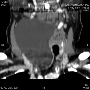
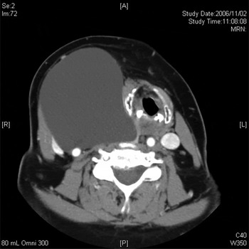
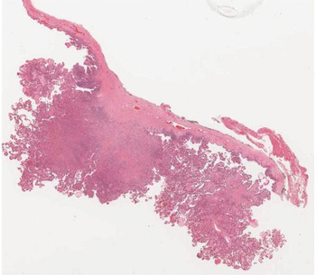
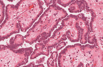
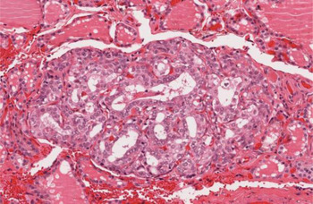
On previous computed tomography imaging, the cyst had been characterised as being 10 × 8.2 × 6.6 cm and extending from the inferior aspect of the parotid gland to the thoracic inlet (Figures 1 and 2). Intra-operatively, it was found to be 15 cm in longest diameter; it reached superiorly as far as the skull base and extended posteriorly to the paravertebral fascia. There was no anatomical connection to the thyroid gland. The cyst was removed en bloc. No tract arising from the cyst was identified. Histologically, the wall of the cyst was composed of thick fibroconnective tissue containing thyroid papillae and follicles (Figure 3). The papillae and follicles were composed of follicular cells exhibiting nuclear features diagnostic for papillary carcinoma (Figure 4). The neoplastic cells stained positive for thyroid transcription factor 1 and thyroglobulin, confirming thyroid origin. The cells were also positive for papillary thyroid carcinoma markers, such as galectin-3, keratin-19 and Hector Battifora mesothelial epitope. No lymph node parenchyma was seen on the specimen. There was no evidence of extracystic extension, vascular invasion or lymphatic invasion.
Coronal, contrast-enhanced computed tomography scan showing a cystic mass extending from the inferior aspect of the parotid to the thoracic inlet.

Axial, contrast-enhanced computed tomography scan showing a cystic mass occupying the right anterior triangle and measuring 10 × 8.2 cm.

Low-power photomicrograph of the resected cyst wall showing thyroid papillae and follicles. (H&E; ×2)

High-power photomicrograph of follicular cells showing nuclear features of papillary thyroid carcinoma. (H&E; ×20)

At this point, the possibility of an occult primary could not be ruled out, so the patient was treated as though she had an isolated cervical lymphatic metastasis. She was taken back to the operating theatre for a total thyroidectomy and right central neck dissection, followed by post-operative radioiodine treatment. Initial histological examination of the thyroid gland, however, found only normal tissue and was negative for carcinoma. Eight out of eight central compartment lymph nodes were also benign. At this point, the working diagnosis was changed to primary papillary carcinoma arising from ectopic thyroid tissue in the wall of a branchial cleft cyst. However, on a later review, a 0.5-mm, confined papillary thyroid microcarcinoma was identified within the thyroid gland (Figure 5). The differential diagnosis now included metastatic papillary thyroid carcinoma, although whether the identified papillary thyroid microcarcinoma represented the primary lesion could not be conclusively determined.
High-power photomicrograph of the total thyroidectomy specimen showing a 0.5-mm focus of papillary carcinoma. (H&E; ×20)

At the time of writing, the patient remained well after four years of follow up, with no clinical evidence of recurrence, a negative ultrasound and an undetectable serum thyroglobulin level.
Discussion
Papillary thyroid carcinoma is the most common malignant thyroid neoplasm accounting for 80 per cent of thyroid cancers.Reference Cramer, Fu, Harth, Margevicius and Wilhelm2 Our case highlights the finding of papillary thyroid carcinoma in the wall of a resected lateral neck cyst, and the diagnostic dilemma of determining whether this represents cystic degeneration of a metastatic lymph node, or primary carcinoma of ectopic tissue in a branchial cleft cyst. The former situation is well described, as up to 11 per cent of lateral neck cysts presenting in adulthood harbour occult papillary thyroid carcinoma.Reference Seven, Gurkan, Cinar, Vural and Turgut3 The latter possibility, of a primary ectopic papillary thyroid carcinoma within a branchial cyst, is very rare, with only nine cases reported to date.Reference Balasubramaniam, Stillwell and Kennedy4–Reference Fumarola, Trimboli, Cavaliere, Coletta, Veltri and Di Fiore12
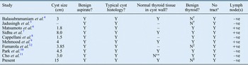
Sidhu et al. suggested the following diagnostic features of primary branchial papillary thyroid carcinoma: (1) benign contents of the cyst aspirate; (2) typical histology of a branchial cyst consisting of an epithelial layer and a subepithelial collection of lymphoid tissue; (3) normal thyroid tissue within the cyst wall with adjacent focus of papillary carcinoma; (4) no evidence of papillary carcinoma within the thyroid gland; and (5) lack of a tract travelling from the cyst upwards to exclude the possibility of a laterally placed thyroglossal cyst.Reference Sidhu, Lioe and Clements7 A review of the nine previously published cases with regards to these criteria is presented in Table I. Interestingly, only three out of nine cases definitively satisfy all five criteria.Reference Sidhu, Lioe and Clements7, Reference Mehmood, Basha and Ghareeb9, Reference Park, Kwon, Kim, Baik and Choi10
Summary of previously reported cases

*No study other than Sidhu et al. explicitly stated a lack of tract travelling from the cyst; however, this was presumed or deduced based on specimen photographs. †Thyroidectomy not performed. ‡Multifocal lesions with extracapsular invasion, largest 4 mm. **No normal thyroid tissue in cyst. §0.5 mm papillary thyroid microcarcinoma. Y = yes, criteria satisfied; N = no, criteria not satisfied; –ve = negative; +ve = positive; – = not described
Our case fulfils four of Sidhu and colleagues' criteria but fails to meet criterion four, as a 0.5-mm papillary thyroid microcarcinoma was found in the thyroid and presumably represents the primary. However, papillary thyroid microcarcinomas – defined as lesions smaller than 1 cm – are typically considered benign and are found in 10–30 per cent of autopsies for unrelated causes of death.Reference Harach, Franssila and Wasenius13–Reference Yamamoto, Maeda, Izumi and Otsuka15 The thyroid lesion in our case was likewise potentially an unrelated incidental finding. The small size (0.5 mm), lack of extracapsular invasion, absence of lymph node parenchyma in the cyst specimen, negative lymph nodes with no evidence of metastasis, and complete anatomical and vascular separation of the cyst to the thyroid are all features that would support a primary ectopic aetiology. Metastatic papillary thyroid microcarcinoma, on the other hand, usually involves the central compartment while only 3 per cent develop lateral lymph node metastasis.Reference Kim16 Our case lacked factors predictive of lateral nodal metastasis, i.e. male sex, larger tumour size, tumour stage four and central nodal metastasis.Reference Kim16 Nevertheless, the possibility of metastatic degeneration of a lateral lymph node cannot be ruled out. A similar diagnostic dilemma was presented by Briggs et al. for branchial cleft squamous cell carcinoma.Reference Briggs, Pou and Schnadig17
• Papillary thyroid carcinoma arising from ectopic thyroid tissue in branchial cleft cyst walls is rare
• Diagnosis is based on histological criteria and exclusion of occult thyroid gland primary carcinoma
• Papillary thyroid microcarcinomas (i.e. <1 cm) are incidentally found in 10–30 per cent of autopsies
• Suspected primary ectopic papillary thyroid carcinoma is not disproved by a solitary thyroid gland microcarcinoma as the latter may be incidental
How thyroid tissue can come to exist in the wall of a branchial cyst is unclear. During the fourth week of embryogenesis, the second branchial arch grows over the third and fourth arches, creating a space between them to form the cervical sinus that will later become obliterated. Branchial cysts are classically thought of as vestigial remnants from incomplete regression of this process.Reference Ozolek18 Meanwhile, anlages from the fourth branchial pouches fuse with the lateral aspects of the descending thyroid to give rise to additional follicular progenitors that will account for up to 30 per cent of the mature thyroid.Reference Fagman and Nilsson19 In this model, ectopic thyroid tissue may form in branchial cysts if thyroid progenitors from the fourth pouch become aberrantly incorporated into the nearby cervical sinus. An alternative theory challenges this view and proposes that branchial cysts arise from early inflammatory degeneration of lymph nodes secondary to epithelial entrapment.Reference Glosser, Pires and Feinberg20 Benign thyroid tissue metastasis to cervical lymph nodes has been described and estimated to have an incidence of 0.03 per cent; such an event may act to trigger the formation of a branchial cyst with entrapped ectopic thyroid tissue.Reference Sidhu, Lioe and Clements7
Conclusion
Primary papillary carcinoma can arise from ectopic thyroid tissue. One diagnostic feature of this condition is the lack of occult disease in the thyroid gland. Papillary thyroid microcarcinoma is an incidental finding in up to 30 per cent of normal thyroids on autopsy.Reference Harach, Franssila and Wasenius13 In a case of suspected primary ectopic papillary thyroid carcinoma, a solitary papillary thyroid microcarcinoma in the thyroid gland may be an unrelated incidental finding and should not automatically preclude the diagnosis.