Introduction
Sialendoscopy is a modern, minimally invasive method for diagnosing and treating diseases of the salivary gland duct system. The ducts of the major salivary glands can be visualised using semi-flexible mini-endoscopes with an outer diameter of 0.8–1.6 mm. The insertion of additional tools into the working channel facilitates the treatment of pathologies such as intraductal salivary stones or strictures.Reference Iro, Dlugaiczyk and Zenk1, Reference Luers, Grosheva, Stenner and Beutner2 In general, sialendoscopy is considered a low-risk procedure that can often be performed under local anaesthesia.Reference Luers, Damm, Klussmann and Beutner3, Reference Luers, Stenner, Schinke, Helmstaedter and Beutner4 The risk and complication spectrum for sialendoscopy includes injuries to the salivary duct system and the development of duct stenoses. Analysis of learning curves has shown that the more experienced a surgeon is in sialendoscopy, the fewer complications arise.Reference Luers, Damm, Klussmann and Beutner3, Reference Kroll, Finkensieper, Hauk, Guntinas-Lichius and Wittekindt5 One of the risks associated with sialendoscopy is that the rinsing solution applied throughout endoscopy can cause severe swelling of the salivary gland. The swelling usually completely disappears within 48 hours after endoscopy.Reference Koch, Zenk, Bozzato, Bumm and Iro6, Reference Chu, Chow, Lim and Kwok7 However, in up to 5 per cent of patients the swelling can result in severe inflammation or formation of an abscess, which must then be treated with antibiotics or surgery.Reference Marchal, Dulguerov, Becker, Barki, Disant and Lehmann8–Reference Konstantinidis, Chatziavramidis, Tsakiropoulou, Malliari and Constantinidis11 In the submandibular gland, sialendoscopy can be followed infrequently by severe dyspnoea resulting from submucosal swelling at the base of the tongue.Reference Marchal and Dulguerov12
A possible pathomechanism for sialadenitis after sialendoscopy is injury of the salivary duct system followed by bacterial invasion into the tissue. Direct injury to duct epithelium may be caused by the endoscope itself or by tools used inside the salivary duct system. Indirect injuries may result from the high intraluminal pressure generated in the duct system by the irrigation fluid. In particular, damage to the smaller and deeper duct sections, i.e. striated ducts, intercalated ducts or the acinar area, which cannot be visualised during sialendoscopy, could play a pathophysiological role. Physiologically, the epithelial cells of salivary ducts are connected by different kinds of tight junctions.Reference Baker13 With rising intraductal pressure, these tight junctions might disintegrate, allowing penetration of the intraductal fluid. The resulting extracellular oedema might then lead to severe inflammation of the salivary gland. Another possible pathomechanism could be rupture of the basal lamina itself and destruction of the entire epithelium.
Our aim was to investigate the intraductal pressure generated during sialendoscopy and explore whether high pressure can damage the salivary duct system.
Materials and methods
First, physiological intra-operative intraluminal pressure was analysed during six sialendoscopies (four of the submandibular gland and two of the parotid gland). Indications for all sialendoscopies were recurrent episodes of glandular swelling and inflammation. The final outcome was sialolithiasis in three glands with salivary stones of 1–4 mm, chronic duct inflammation in two glands and ductal papilla stenosis in one gland.
We used all-in-one sialendoscopes with outer diameters of 1.1 and 1.6 mm (11573 A and 11574 A, Karl Storz, Tuttlingen, Germany). The irrigation solution comprised a 10:1 ratio of 0.9 per cent NaCl and 1 per cent Xylocaine®. On the surgeon's instruction, the assisting nurse performed intermittent flushing by manual pressure on a 10 ml syringe. A three-way stopcock (Discofix 3, B. Braun, Melsungen, Germany) was inserted between the syringe and the endoscope, and a device for continuous pressure monitoring (IntelliVue MP2, Philips Medizin Systeme Boeblingen, Boeblingen, Germany) was connected to the assembly.
Sialendoscopy is always started by flushing the salivary duct system and filling it with approximately 2 ml irrigation solution to unfold the ducts. During sialendoscopy, irrigation fluid continuously drained in a retrograde manner from the duct system via the ostium into the oral cavity, so that irrigation fluid had to be replenished regularly throughout the procedure. For this, a small volume was applied whenever the ducts collapsed. This strategy resulted in an average application rate of about 0.5 ml irrigation fluid per minute. Stronger and longer-lasting bursts of irrigation may be necessary during endoscopy to flush out plaques and microsialoliths from the salivary duct system. The duct orifice was not obstructed with any instruments, so fluid could escape from the duct system.
The effects of different irrigation pressures on the integrity of the salivary duct system were investigated ex vivo in 10 freshly excised human salivary glands (8 submandibular glands and 2 parotid glands). Specimens were obtained from neck dissections or parotidectomies or submandibulectomies of patients with suspected tumours. Informed consent was obtained from each patient and the study was approved by the local institutional ethics committee. Histological examination showed all glands to be unaffected by metastatic disease.
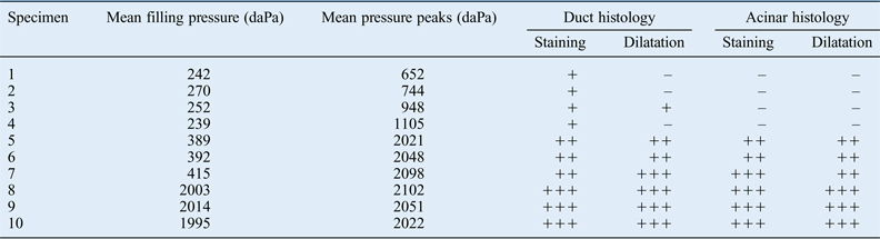
These experiments closely reproduced the intra-operative setting. A green histological marker (WAK-HM-G-1, WAK Chemie, Steinbach, Germany), which remains stable and does not diffuse during histological processing, was used as irrigation fluid. The green colour was chosen to provide a visible contrast to the reddish cells of the salivary gland. Specimens were irrigated at variable pressures, in terms of filling pressure and pressure peaks, adjusted to intra-operative measurements (Table I). For each pressure value, irrigation lasted for 30 seconds, after which the tissue was immediately fixed in formalin. After embedding in paraffin wax, 5 µm sections were cut and stained with haematoxylin and eosin for histological analysis.
Overview of in vitro experiments*

* The duct systems of 10 salivary glands were flushed for 30 seconds with a histological stain at increasing pressures. – = no staining; + = weak staining; ++ = moderate staining; +++ = strong staining
Results
At the beginning of each sialendoscopy, i.e. before any irrigation fluid was applied, the salivary duct system was under normal pressure (0–40 daPa). After the initial filling of the salivary duct system, the intraductal pressure was approximately 100 daPa (1 kPa). The filling pressure was dependent on the extent of swelling and thus on the quantity of irrigation fluid present in the duct system. On average, the filling pressure was 100–250 daPa during the course of sialendoscopy. In rare cases of a long sialendoscopy period and a heavily swollen gland, a maximum filling pressure of 500 daPa was observed. There was no significant difference in the filling pressures of the submandibular and parotid glands.
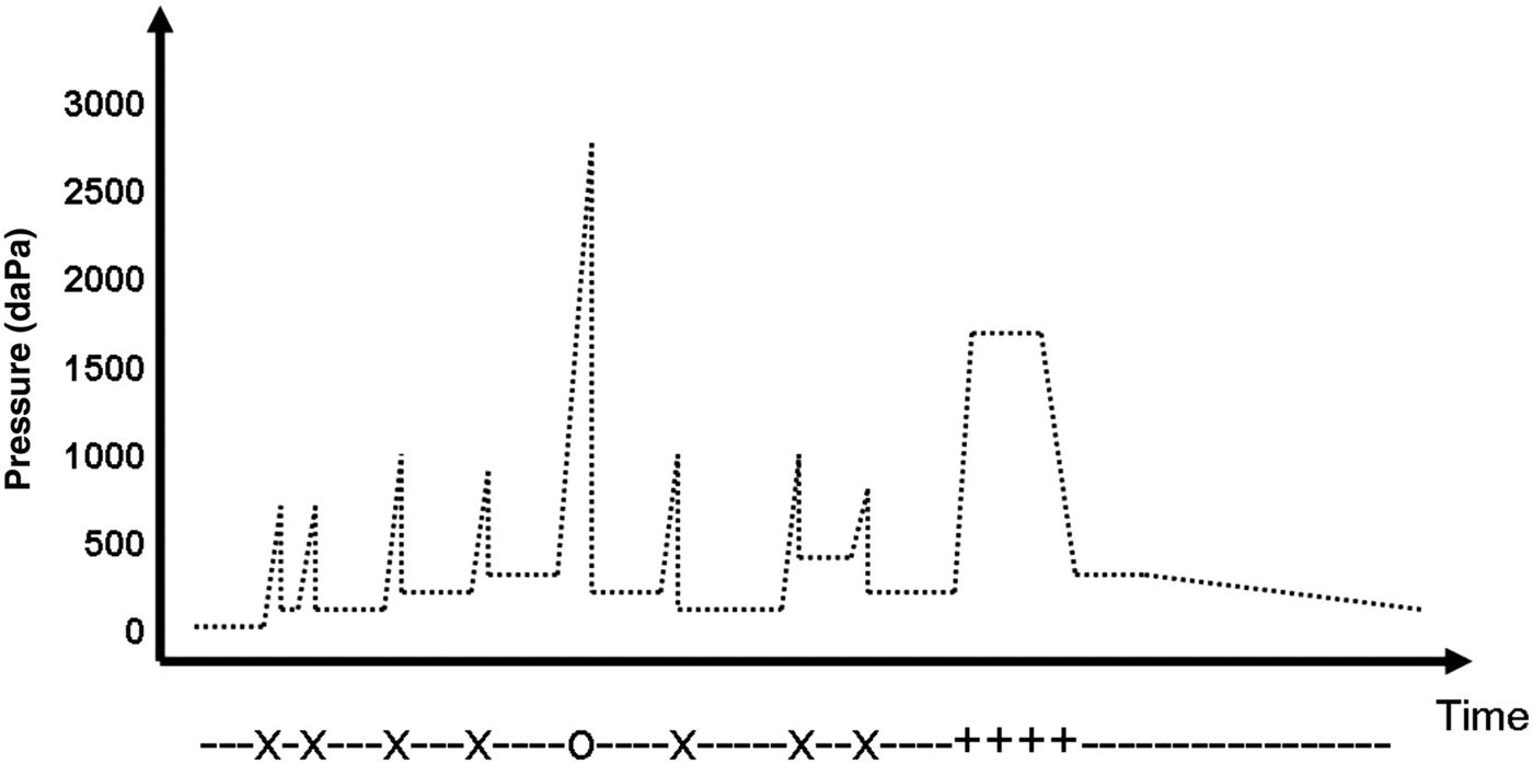
When the duct lumen is visible during sialendoscopy, the application of short flushes of irrigation fluid can result in pressure peaks of 650–1100 daPa. When the duct lumen is not endoscopically visible during the application of irrigation fluid, vigorous flushing can result in higher short-term pressure values. For example, pressure peaks of up to 2600 daPa when a 10 ml syringe was used, and 4000–4500 daPa for a 20 ml syringe were observed. Vigorous flushing of the salivary duct system in the event of plaques showed pressure peaks of up to about 2000 daPa. Figure 1 shows an example of a time course of pressure changes during sialendoscopy.
Schematic time course showing exemplar pressure conditions during a sialendoscopy. X = irrigation when the duct lumen is visible; O = irrigation when the duct lumen is not visible; += irrigation when flushing out plaques (+). The filling pressure (–) does not exceed 400 daPa
In ex vivo experiments measurements, the pressure conditions applied were equivalent to those observed during a normal sialendoscopy (filling pressure of approximately 250 daPa, pressure peaks of 650–1100 daPa every 5 seconds). This resulted in minor and major salivary ducts being marked with tissue colour (Table I, specimen 1–4). The duct lumen in the acinar area was not dilatated and was hardly visible (Figure 2). When the filling pressure was raised to about 400 daPa and pressure peaks were increased to about 2000 daPa, increased dilatation of the ducts was observed. In addition, the duct lumen was visible in acinar areas and showed slight staining (Table I, specimen 5–7). Epithelial cells showed no signs of damage (Figure 3). When the salivary duct system was irrigated at maximum pressure, all major and minor ducts down to the level of the acini were highly enlarged and strongly stained (Table I, specimen 8–10). Furthermore, weak interstitial staining was also visible in some areas, but there were no histological signs of duct rupture (Figure 4). An overview of the experimental setup and histology results is given in Table I.
Photomicrograph of the salivary ducts and the acinar area of a submandibular gland after irrigation at an intraductal filling pressure of approximately 250 daPa and pressure peaks of 650–1100 daPa, showing salivary ducts with no dilatation but filled with stain, and no dilatation or stain in the acinar area. (H&E; ×10)
Photomicrograph of the salivary ducts and duct lumen of a submandibular gland after irrigation at an intraductal filling pressure of approximately 400 daPa and pressure peaks of approximately 2000 daPa, showing staining but no dilatation of the salivary ducts and the duct lumen of the acinar areas. (H&E; ×10)
Photomicrograph of the salivary ducts and duct lumen of a submandibular gland after irrigation with maximum manual pressure for about 30 seconds, showing strong dilatation and staining of all salivary ducts and highly enlarged acini. In the lower right corner, a microsialolith is visible in a salivary duct (*). (H&E; ×20)
Discussion
During sialendoscopy, salivary duct flushing is performed manually with a syringe, and there is usually no direct control of the pressure inside the salivary duct system. Most sialendoscopes currently feature an irrigation channel with a diameter of 0.25 mm and a length of 10 cm. If laminar flow is assumed, the Hagen-Poiseuille formula predicts that a manual pressure of 869.6 daPa (1 daPa ≈ 1 mmH2O) is required to empty a 10 ml syringe within 20 minutes. During sialendoscopy, this value is directly related to the intraductal filling pressure. In our experience of over 300 sialendoscopies, about 10–40 ml irrigation fluid is used during a single procedure, which is consistent with the figures shown above (10 ml in 20 minutes). The present study demonstrates the intraductal pressures that can result from this irrigation strategy during sialendoscopy. We observed the pressure values to vary depending on the extent of swelling and the endoscopic visibility of the duct. Normally, only a relatively low filling pressure of 100–250 daPa is needed during sialendoscopy. To prevent the intraductal filling pressure increasing unnecessarily during sialendoscopy, it is essential not to obstruct the duct orifice so that irrigation fluid can escape into the oral cavity. Only in the case of papillotomy does additional partial obstruction with forceps appear reasonable. Otherwise, the salivary duct system might not be able to hold the fluid, leading to its collapse and making endoscopy more difficult.
During sialendoscopy, regular short flushes are followed by transient high pressures of usually 650–1100 daPa. If only the epithelial wall is visible (and not the duct lumen itself), then extremely high maximum pressures of up to 5500 daPa (approximately 0.5 atm) can occur during vigorous flushing. However, because the epithelium probably obstructs the irrigation channel to a large extent in these cases, such very high pressure peaks are only likely to develop in the tube system of the measuring unit (i.e. in front of the sialendoscope) and not in the salivary duct system (i.e. behind the sialendoscope). Nevertheless, it seems advisable to flush or irrigate the duct system only when the duct lumen is endoscopically visible. For this, the surgeon should always properly instruct the assisting nurse who often only has a limited view of the intraductal area. Furthermore, we recommend that prior to sialendoscopy the assisting nurse should test how much pressure is manually generated with a 10 ml syringe using the anaesthesiological pressure measuring unit.
During sialendoscopy, pressure in the salivary duct system becomes higher if manually applied using a syringe with a rather low volume (e.g. 5 ml) than one with a higher volume (e.g. 10 or 20 ml). This is a hydraulic effect in which the pressure in a dependent small-tube system (salivary duct) decreases with increasing diameter of the syringe, providing that the manually applied force remains constant. Consequently, high-volume rather than low-volume syringes should be used for flushing during sialendoscopy to maintain a moderate intraductal pressure. However, in our experiments the assisting nurse applied much more manual force when working with a bigger syringe. As a result, intraductal pressures with a 20 ml syringe were even higher than those obtained with a 10 ml syringe. We therefore recommend using a 10 ml syringe for the manual application of irrigation fluid because this provides a compromise between small (2–5 ml; higher pressure resulting from the hydraulic factor) and large (20 ml; risk that manual force is unintentionally increased) syringes. Ideally, a mechanical pressure limiter (i.e. a safety valve) should be integrated into the irrigation assembly.
In our experiments, the major and minor salivary ducts became more dilatated with increasing pressure. At high intraductal pressure, duct dilatation occurred down to the level of the acini (Figure 4). Interestingly, no salivary duct damage was visible histologically. Although the intercellular junctional complexes could not be directly visualised, epithelial cells appeared to be intact, with no signs of rupture. However, weak staining of the interstitial areas was evident after flushing at the maximum pressure. A possible explanation is that rupture of a duct in fact did occur during the experiment but was not seen in tissue sections. However, a thorough analysis of tissue sections showed all apparent interstitial staining to be artefacts resulting not from intraductal staining but from staining of the gland surface. Such unintentional marking of the tissue is not completely avoidable when flushing at maximum pressure because it is very difficult to keep the salivary gland capsule intact during excision. Thus, during irrigation, stain released from the salivary duct system marked parenchymal areas of the gland. Alternatively, retrograde flow of stain from the main duct during flushing may have marked the gland surface.
Elastic fibres located along the salivary ducts, around the acini and in the gland capsuleReference Seifert14 may play a key role in preventing duct rupture, even at maximal intraductal pressure. These fibres might cause the gland to be highly extensible; however, they may also allow a continuous outflow of fluid through the main excretory duct, thus limiting the maximum intraductal pressure. Recovery after severe duct dilatation might also depend on the function of myoepithelial cells located around the acini and salivary ducts. This might be a fruitful area of future research.
A limitation of our experiments is that no living tissue was observed. Hence, our observations may not replicate the situation in vivo. Furthermore, long-term effects of irrigation at high intraductal pressure on the acini and the epithelial cells could not be studied in this ex vivo system.
In our opinion, during sialendoscopy irrigation of the duct system should not be continuous, but should be done in response to changes in intraductal dilatation. Irrigation fluid should only be applied if the ductal walls collapse slightly and hinder the surgeon's view. Intermittent flushing enables the volume of instilled irrigation fluid to be greatly reduced,Reference Marchal, Dulguerov, Becker, Barki, Disant and Lehmann8 and thus gland swelling is decreased. Our experiments showed that very high intraductal pressure causes strong dilatation of the acinar area of salivary glands. As this dilatation might initiate pathological processes such as oedema and inflammation of the gland, intraductal pressures higher than 400 daPa (≈40 cmH2O) should be avoided during sialendoscopy. A pressure limiter in the form of a safety valve should therefore be integrated into the irrigation assembly or the assisting nurse should test the manual pressure applied to a 10 ml syringe using an anaesthesiological pressure measuring device.
• During sialendoscopy, normal intraductal filling pressure is 100–250 daPa, with pressure peaks of up to 2000 daPa during flushing
• High filling pressures and pressure peaks result in dilatation of salivary ducts, including the acinar area
• Intermittent irrigation fluid administration reduces the risk of post-operative oedema and inflammation