Diabetes mellitus is a diverse group of metabolic disorders with varied clinical complications and undoubtedly one of the leading causes of mortality and morbidity throughout the world. Even after various medical breakthroughs and technological intervention for diabetes treatment, the number of diabetic patients is constantly on the rise. According to the annual report of International Diabetes Federation, estimated number of adults living with diabetes had soared to 366 million and health care spending on diabetes has reached US$465 billion. New data have projected that the number of people with diabetes will rise to 552 million by 2030 (Whiting et al. Reference Whiting, Guariguanta, Weil and Shaw2011).
Dietary intervention with non-digestible fibre is one of the main therapies proposed in the case of diabetic patients (Luo et al. Reference Luo, Van Yperselle, Rizkalla, Rossi, Bornet and Slama2000). Prebiotics are emerging functional products that are generally useful for the improvement of the nutritional quality of foods. Prebiotics possess interesting functional and physiological attributes such as low sweetness, non-cariogenicity, low energetic value, and hypolipidaemic and hypocholesterolemic properties. Most of the research on prebiotics mainly focused on inulin and fructooligosaccharides (FOS). Another oligosaccharide, GOS has also emerged as one of the most potent prebiotic and is known to modulate both digestive and immune health. Because of its solubility and stability over a range of pH and temperature, GOS is an ideal choice for formulating a wide variety of foods and beverages. One of the most interesting characteristic of GOS is its similarity to human milk oligosaccharides (HMO) because of which it became an ideal choice for addition in infant foods and also attracted global attention from research and clinical professionals. GOS modulate the intestinal microbiota in two different ways, along with the selective enhancement of health promoting bacteria or probiotics, its molecules mimic the structure present on epithelial cells acting as a decoy receptor to inhibit/reduce the adhesion of pathogenic microbes to the intestinal cells thereby protecting the host from various health complications (Sangwan et al. Reference Sangwan, Tomar, Singh and Ali2011). GOS are commercially produced from lactose in a reaction catalysed by β-galactosidase, termed transglycosylation. GOS are able to modulate intestinal microbiota and to improve the intestinal barrier functions. Other reported beneficial health effects include the promotion of mineral absorption, weight management, prevention of carcinogenesis and the alleviation of allergies (Tzortzis & Vulevic, Reference Tzortzis, Vulevic, Charalampopoulos and Rastall2009; Ohr, Reference Ohr2010; Figueroa-Gonzalez et al. Reference Figueroa-Gonzalez, Quijano, Ramirez and Cruz-Guerrero2011). Potential use of GOS in food products has been thoroughly described (Wang, Reference Wang2009; Sangwan et al. Reference Sangwan, Tomar, Singh and Ali2011).
Today, prebiotics are not only examined for their antipathogenic effects, but they are also developed to decrease faecal transit time, lower cholesterol and the glycaemic response, improve bone health, lower daily energy (fat) intake, relieve symptoms of inflammatory bowel disease, and attempt to lower colon cancer rates. There have been a number of studies carried out on effectiveness of prebiotics like FOS and xylooligosaccharides (XOS) in reduction or prevention of diabetes. Feeding of FOS and XOS to diabetic rats significantly improved the body weight and reduced glucose level, cholesterol and other related complications like glucosuria, diabetic nephropathy, blood creatinine and urea concentrations (Gobinath et al. Reference Gobinath, Madhu, Prashant, Srinivasan and Prapulla2010). XOS was reported as a sweetener capable of improving diabetic symptoms like growth retardation, polydipsia, hyperphagia, elevation of serum glucose, cholesterol and triglycerides in streptozotocin-induced diabetic rats (Imaizumi et al. Reference Imaizumi, Nakatsu, Sato, Sedarnawati and Sugano1991). Because of its soluble fibre effect, FOS, was reported to alleviate diabetic-related metabolic complications to a certain degree (Mabel et al. Reference Mabel, Sangeetha, Platel, Srinivasan and Prapulla2008). However, there are few reports available for GOS in this regard. The present study, therefore, was carried out to evaluate the hypoglycaemic effect of GOS on diabetes (induced by administration of alloxan) using rats as animal model. Alloxan (2,4,5,6-pyrimidinetetrone) is a toxic glucose analogue, which selectively destroys insulin-producing cells in the pancreas (that is β-cells) causing an insulin-dependent diabetes mellitus (called ‘Alloxan Diabetes’). After the induction of diabetes in male Sprague-Dawley (SD) rats, effect of GOS supplementation was studied on various metabolic parameters like fasting blood glucose, lipid profile, urea, creatinine and on oxidative enzymes. As prebiotics are known to increase the lactobacilli count in the host and lactobacilli are reported to aid in the reduction of diabetes and population of pathogens, counts of lactobacilli and coliforms were also monitored in this study. As there is a dearth of information on the hypoglycaemic effect of GOS, this study could provide valuable information regarding the role of this novel oligosaccharide in improvement of diabetes and related metabolic parameters.
Methods and materials
Chemicals
All the components of rat diet except casein were procured from Hi Media Laboratories Pvt Ltd, (Mumbai, India). Casein was locally procured from Modern Dairy plant, Karnal. Alloxan was procured from Sigma-Aldrich (CA, USA). GOS used in the present study (prepared using lactose as substrate by the action of enzyme β-galactosidase) were supplied by FrieslandCampina Domo, Great Ocean Ingredients Plant, Allansford, Australia on complementary basis. All other chemicals and growth media used in the study were procured from Hi Media Laboratories Pvt. Ltd.
Animals
The study was approved (Approval No. 19/11) by the Ethical Committee for Animal Experiments of National Dairy Research Institute (Registration No. 382/01/ab/CPCSEA), Karnal, Haryana, India. Male SD rats were procured from Central Animal Facility, National Institute of Pharmaceutical Education & Research, S.A.S. Nagar, Punjab, India. At the time of procurement rats were about 6 weeks old and approximately of similar body weight (~90 g). Rats were housed under standard conditions of temperature (24±1 °C) and relative humidity (56±5%) with a 12-h light/dark cycle.
Induction of diabetes
A total of 60 rats were handled and adapted to their new environment with a basal diet (standard rodent diet AIN-93) for 14 d prior to 42 d study. All rats were fed ad libitum on basal diet and water. Forty five rats were administered with alloxan for induction of diabetes. Freshly prepared solution of alloxan (dissolved in water) was injected at an intraperitoneal dose of 100 mg/kg body weight for this purpose. Since alloxan could evoke fatal hypoglycaemia as a result of massive insulin release, glucose (5%) was administered (prepared by mixing 5% glucose in drinking water) for 48 h following the injection to prevent drug-induced hypoglycaemic mortality. Blood drawn from the tail vein 10 d after alloxan administration was used for the determination of fasting blood glucose.
Grouping of animals and feeding schedule
The animals were allocated into four treatment groups of six rats each.
Group 1: Normal control group (NCG) – Non diabetic group-fed with basal diet
Group 2: Prebiotic control group (PCG) – Non diabetic group-fed with basal diet supplemented with GOS (100 g/kg of diet)
Group 3: Diabetic control group (DCG) – Injected with Alloxan (100 mg/kg body weight) – fed with basal diet
Group 4: Diabetic prebiotic group (DPG) – Injected with Alloxan (100 mg/kg body weight) – fed with basal diet supplemented with GOS (100 g/kg diet)
The oligosaccharide diets contained GOS at the 10% level by replacing an equivalent amount of starch in the basal diet (w/w).
Collection and processing of samples
On day 0 (10 d after alloxan administration) blood samples were collected from the tail vein for the assessment of various biochemical parameters. At the end of the trial all the animals were sacrificed (on day 42). Blood samples were collected in ethylenediaminetetraacetic acid (EDTA) coated vacutainers by heart puncture (in right ventricle). A small part of whole blood samples was also collected in EDTA coated 1·5 ml Eppendorf tubes for haemoglobin (Hb) determination. Vacutainers containing blood were centrifuged at 4 °C at 4000 g for 15 min. The separated plasma was stored at −20 °C until processed. Liver was removed, washed with phosphate buffer saline, blotted dry and processed for biochemical measurements.
Analysis of triglycerides, cholesterol, urea and creatinine in plasma
Triglycerides, Cholesterol, and Urea in plasma were analysed using Autospan® kits, (Span Diagnostic India Pvt. Ltd., Baroda, India). These lipid fractions were analysed as per the instructions of manufacturer. Creatinine was analysed by using a creatinine kit (Coral Clinical Systems, Goa, India). The kits were based on enzymatic colorimetric methods.
Haemoglobin and glycosylated haemoglobin estimation
Blood samples collected as above were used for Hb estimation by Drabkin's Cyanmethaemoglobin method (Drabkin, Reference Drabkin1950). HbA1c was estimated by using Cation-Exchange Resin kit method (Monozyme Laboratories Pvt Ltd.). In brief, whole blood (with heparin) was mixed with lysing reagent to prepare hemolysate and mixed with cation-exchange resin. The non-glycosylated haemoglobin (Hb) binds to the resin, leaving HbA1c free in the supernatant. The HbA1c level (%) was determined by measuring the absorbance of the HbA1c fraction and total Hb at 415 nm by spectrophotometer. Percent Hb1Ac of the samples were calculated as follows:
 $${\rm \% HbA1C = (AHbA1C/AHb)} \times {\rm 4} {\cdot} {\rm 61}\,{\rm assay}\,{\rm factor}$$
Measurement of antioxidative enzymes
Activity of catalase (CAT), glutathione peroxidase (GPx) and superoxide dismutase (SOD) was measured in liver and blood using the methods of Aebi (Reference Aebi, Colowick and Kaplan1984), Lawrence & Burk (Reference Lawrence and Burk1976) and Marklund & Marklund (Reference Marklund and Marklund1974) respectively.
Collection of faeces for lactobacilli and coliforms count
The ennumeration of faecal lactobacilli and coliform was performed weekly. The faecal samples were collected by gently squeezing the rectal part into sterile test tubes and were processed within 60 min of collection. Samples obtained were homogenised with sterile phosphate buffer saline (pH 7·2) and serially diluted followed by plating of appropriate dilutions (10−6–10−8) on MRS (de Man, Rogosa and Sharpe) agar for total lactobacilli counts. The plates were incubated at 37 °C for 48 h. The number of faecal coliforms was determined by pour plating diluted samples on violet red bile agar after incubation at 37 °C for 24 h.
Statistical analysis
Values were expressed as means and se of six rats per group. Statistical analysis was carried out using Data was analysed using Statistical Analysis System (SAS, V. 6, SAS Institute Inc.). Results were analysed and the significance level was calculated using the Duncan multiple range test and results are considered significant at P<0·05.
Results and discussion
Diabetes was induced by single intraperitoneal administration of alloxan (100 mg/kg). Initially after alloxan administration a hypoglycaemic phase (lasts maximum of 30 min) starts because of stimulation of insulin secretion followed by an increase in blood glucose (lasts for 2–4 h) due to inhibition of insulin secretion from the pancreatic beta cells. Thereafter, another hypoglycaemic phase begins typically 4–8 h after alloxan administration, leading to fatal convulsions (West et al. Reference West, Simon and Morrison1996). To prevent the rats from this hypoglycaemic mortality, they were given 5% solution of glucose. After administration of alloxan, rats with fasting blood glucose level more than 200 mg/dl were considered as diabetic. From the 45 rats administered with alloxan, 28 were found to be diabetic and out of these 12 with approximately same blood glucose levels were finally included in the study. The change in body weight, fasting blood glucose level and Hb levels of all the rats are given in Table 1. Body weight of all the animals was recorded at day 0 and 42. Initial body weight of all the groups was approximately same. A significant increase in body weight was observed in NCG and PCG while body weight of DCG and DPG showed a significant reduction. Compared with DCG, DPG showed a less pronounced reduction in the body weight during the feeding period. Our results are in agreement with the results obtained by Byung-sun (Reference Byung-Sung2011) and Gobinath et al. (Reference Gobinath, Madhu, Prashant, Srinivasan and Prapulla2010), who reported a marked increase in body weight of diabetic rats fed on oligosaccharide diet. However, Sheu et al. (Reference Sheu, Lee, Chen and Chan2008) observed no significant effect of XOS feeding on body weight.
Effect of GOS supplementation on body weight and blood glucose levels in control and alloxan-induced diabetic rats

GOS, Galactooligosaccharides; NCG, Normal control group; PCG, Prebiotic control group; DCG, Diabetic control group; DPG, Diabetic prebiotic group
A,B (Mean±se of 6 replicates) values with different superscript in a row are significantly different (P⩽0·05)
a,b,c,d (Mean±se of 6 replicates) values with different superscript in a column are significantly different (P⩽0·05)
As evident from Table 1, consumption of GOS for a period of 42 d, decreased blood glucose level in both PCG and DPG. Diabetes mellitus leads to constant elevated blood sugar levels which damage the body leading to the multiple health problems. The present study has shown that the substitution of sugars in foods by prebiotics has been suggested to decrease the amount of available glucose for absorption into the bloodstream. Prebiotics also help in reduction in the time of gastric emptying along with shortening of small intestine transit time preventing excessive blood glucose elevations after a meal (Swennen et al. Reference Swennen, Courtin and Delcour2006). Production of short chain fatty acids (SCFA) like propionate and butyrate is another mechanism by which prebiotics can modulate blood glucose and even insulin level. Propionate has been shown to reduce hepatic gluconeogenesis along with an enhancement in hepatic glycolysis. Butyrate is believed to be responsible for increasing a potent antihyperglycaemic hormone, glucagon-like peptide-1 (GLP-1) which was demonstrated to promote beta cell proliferation and inhibit apoptosis (Brubake & Drucker, Reference Brubake and Drucker2004). Intake of FOS and XOS was reported to ameliorate the hyperglycaemic condition in diabetic subjects (Mabel et al. Reference Mabel, Sangeetha, Platel, Srinivasan and Prapulla2008; Sheu et al. Reference Sheu, Lee, Chen and Chan2008; Gobinath et al. Reference Gobinath, Madhu, Prashant, Srinivasan and Prapulla2010).
Effect of GOS supplementation on triglyceride, total cholesterol, high density lipoproteins (HDL) and low density lipoproteins (LDL) was assessed. In our body, carbohydrates consumed in the diet are converted to glucose which in turn, if in excess in blood, is converted to triglycerides (fats). In DCG, a significant increase in levels of triglycerides, total cholesterol and LDL was observed with a concomitant decrease in HDL. Feeding of diet supplemented GOS to rats of DPG group significantly improved the lipid profile of rats. Table 2 shows the lipid levels in different groups. Diabetic groups were found to have significantly higher concentration of triglycerides compared with non-diabetic groups. DPG had significantly lower triglycerides level compared with DCG. Production of SCFA by fermentation of prebiotics leads to a decline in liver lipogenesis is the principle mechanism of action behind the hypotriglyceridemic activity of prebiotics. Reduction in activity of lipogenic enzymes may also result from a prebiotic mediated decrease in postprandial glucose and insulin concentrations (Swennen et al. Reference Swennen, Courtin and Delcour2006). Metabolism of lipids was found to be modulated by inulin type fructans (ITF) leading to a decrease in both fasting and postprandial plasma triglyceride levels (Fiordaliso et al. Reference Fiordaliso, Kok, Desager, Goethals, Deboyser, Roberfroid and Delzenne1995). Consumption of FOS and Oligofructose has also been shown to prevent hypertriglyceridaemia in rats (Busserolles et al. Reference Busserolles, Gueux, Rock, Demigne, Mazur and Rayssiguier2003; Moroti et al. Reference Moroti, Magri, de Rezende Costa, Cavallini and Sivieri2012).
Effect of GOS supplementation on triglycerides, cholesterol, low density lipoproteins and high density lipoproteins levels in control and alloxan-induced diabetic rats

GOS, Galactooligosaccharides; NCG, Normal control group; PCG, Prebiotic control group; DCG, Diabetic control group; DPG, Diabetic prebiotic group
A,B (Mean±se of 6 replicates) values with different superscript in a row are significantly different (P⩽0·05)
a,b,c,d (Mean±se of 6 replicates) values with different superscript in a column are significantly different (P⩽0·05)
Diabetes comes with various other complications including higher risk of heart disease and stroke because of an increase in LDL concentration. One of the main problems in case of LDL is that in diabetics levels of LDL may appear in normal range even after being at higher levels. This is because of the fact that diabetics actually have a more dangerous form of smaller and denser LDL particles which are more atherogenic and are more readily oxidised and glycated. In this study, the concentration of total cholesterol and LDL showed a significant decline in rats fed with a GOS supplemented diet. Several studies have shown that intake of GOS, FOS and XOS led to significant reduction in cholesterol and LDL levels in diabetic subjects (Hsu et al. Reference Hsu, Liao, Chung, Hsieh and Chan2004; Liong et al. Reference Liong, Dunshea and Shah2007; Sheu et al. Reference Sheu, Lee, Chen and Chan2008; Gobinath et al. Reference Gobinath, Madhu, Prashant, Srinivasan and Prapulla2010). This decline in blood cholesterol may in part be due to the reduction in its absorption and an increase in its excretion via faeces or because of reduction in the levels of enzyme involved in fatty acid synthesis (Van Bennekum et al. Reference Van Bennekum, Nguyen, Schulthess, Hauser and Phillips2005).
Unlike LDL, higher concentration of HDL (possess multiple antiatherogenic activities) reduce the risk of cardiovascular diseases. In the present study a significant decrease in HDL levels was observed in case of diabetic rats compared with normal rats and feeding of GOS was found to be positively associated with significant improvement in HDL levels as depicted by the improved HDL concentration in DPG and PCG (Table 2). Inulin, oligofructose and soybean oligosaccharides were reported to increase the levels of HDL-cholesterol (Kim & Shin, Reference Kim and Shin1998; Kiebling et al. Reference Kiebling, Schneider and Jahreis2002; Chen et al. Reference Chen, Li-jun, Jian-jun, Xua and Lib2010).
Kidney disease – known as nephropathy – is a serious complication associated with long-term diabetes. One way to measure the damage in kidney is by the assessment of blood urea concentration. Increment in blood urea level with increasing blood glucose concentration is a clear indication of kidney damage due to elevated blood glucose concentration. In this study higher serum urea and creatinine levels were observed in DCG and DPG compared with NCG and PCG. In DPG a significant improvement in urea and creatinine levels was observed after GOS feeding (Table 3). Reduction in the muscle mass (also called muscle atrophy) during diabetes is indicated by the higher level of creatinine. This muscle atrophy is reduced by the consumption of prebiotic containing diet which may be an explanation to the improvement in body weight in GOS fed rats. Similar results were obtained in earlier studies which show the intake of XOS and FOS reduced the amount of urea and creatinine in diabetic rats (Gobinath et al. Reference Gobinath, Madhu, Prashant, Srinivasan and Prapulla2010). Another study demonstrated that the addition of oligosaccharides and gum arabic gum to the diet decreased blood urea by 20–30% relative to the control (Younes et al. Reference Younes, Garleb, Behr, Remesy and Demigne1995).
Effect of GOS supplementation on urea and creatinine levels in control and alloxan-induced diabetic rats

GOS, Galactooligosaccharides; NCG, Normal control group; PCG, Prebiotic control group; DCG, Diabetic control group; DPG, Diabetic prebiotic group
A,B (Mean±se of 6 replicates) values with different superscript in a row are significantly different (P⩽0·05)
a,b,c,d (Mean±se of 6 replicates) values with different superscript in a column are significantly different (P⩽0·05)
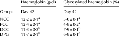
Level of Hb and glycosylated Hb are among the reliable markers for diabetes. Diabetes led to decrease in Hb concentration with a concomitant increase in glycosylated Hb in rats (Table 4). The decreased level of total Hb observed in diabetic rats might be due to the increased formation of glycosylated Hb. Increase in glycosylated Hb was observed in uncontrolled diabetes and the increase is directly proportional to the fasting blood glucose level. Excess glucose present in blood in diabetics reacts with Hb to form glycosylated Hb. As the concentration of glycosylated Hb provide a measure of blood glucose, the reduced level of glycosylated Hb and an increase in the level of Hb in GOS fed diabetic rats showed its hypoglycaemic activity.
Effect of GOS supplementation on haemoglobin and glycosylated haemoglobin levels in control and alloxan-induced diabetic rats
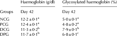
GOS, Galactooligosaccharides; NCG, Normal control group; PCG, Prebiotic control group; DCG, Diabetic control group; DPG, Diabetic prebiotic group
a,b,c,d (Mean±se of 6 replicates) values with different superscript in a column are significantly different (P⩽0·05)
Increasing evidence suggests that oxidative stress (generated because of production of free radicals) plays a major role in the pathogenesis of diabetes. Keeping in mind the importance of oxidative parameters in diabetes, level of oxidative enzymes was measured in the liver and RBC of the animals. A considerable difference was observed in the levels of oxidative enzymes of diabetic groups fed on basal and GOS supplemented diet. Diabetes led to a significant decrease in SOD, CAT and GPx level in liver of rats (Table 5). In diabetic patients, the formation of hydrogen peroxide takes place as a result of the autoxidation of glucose which inactivates SOD leading to its decreased activity. The declined activity of GPx could be directly explained by the low content of GSH found in diabetic subjects, since GSH is a substrate and cofactor of GPx or may be due to the glycation of enzyme governed by prevailing glucose concentration (Domingues et al. Reference Domingues, Ruiz, Gussinye and Carrascosa1998). Various studies have reported the reduction of hepatic SOD, CAT and GPx activities in STZ-induced diabetic rats (Sheu et al. Reference Sheu, Lee, Chen and Chan2008; Nirmala et al. Reference Nirmala, Saroja and Gayathri2011; Shanmugasundaram et al. Reference Shanmugasundaram, Devi, Soris, Maruthupandian and Mohan2011).
Effect of GOS feeding on concentration of antioxidative enzymes in liver and RBC (Red blood cells) in control and alloxan-induced diabetic rats

GOS, Galactooligosaccharides; NCG, Normal control group; PCG, Prebiotic control group; DCG, Diabetic control group; DPG, Diabetic prebiotic group
a,b,c,d (Mean±se of 6 replicates) values with different superscript in a column are significantly different (P⩽0·05)
The activity of these antioxidative enzymes was also assessed in RBC in the present study. Feeding of GOS improved the levels of these marker enzymes. SOD and GPx levels in RBC's were significantly reduced in diabetic groups compared with non-diabetic groups (Table 5). A different pattern was obtained in case of catalase level in RBC as activity of catalase increased in case of diabetic group. Observations regarding catalase activity available in literature are rather inconclusive. The activity of catalase in the liver and kidneys of diabetic animals is generally believed to decrease although there are reports of its increase (Szaleczky et al. Reference Szaleczky, Prechl, Feher and Somogyi1999) as well. A number of researchers have reported an increase in catalase activity in RBC (Stoppa et al. Reference Stoppa, Cesquini, Roman, Ogo and Torsoni2006; Qujeq & Rezvani, Reference Qujeq and Rezvani2007; Shanmugasundaram et al. Reference Shanmugasundaram, Devi, Soris, Maruthupandian and Mohan2011). We found a significantly higher catalase activity in diabetic rats compared with controls. The increase in the erythrocyte antioxidant enzymes such as catalase is related to the oxidative damage of membrane protein and lipid by increased ROS in the body.
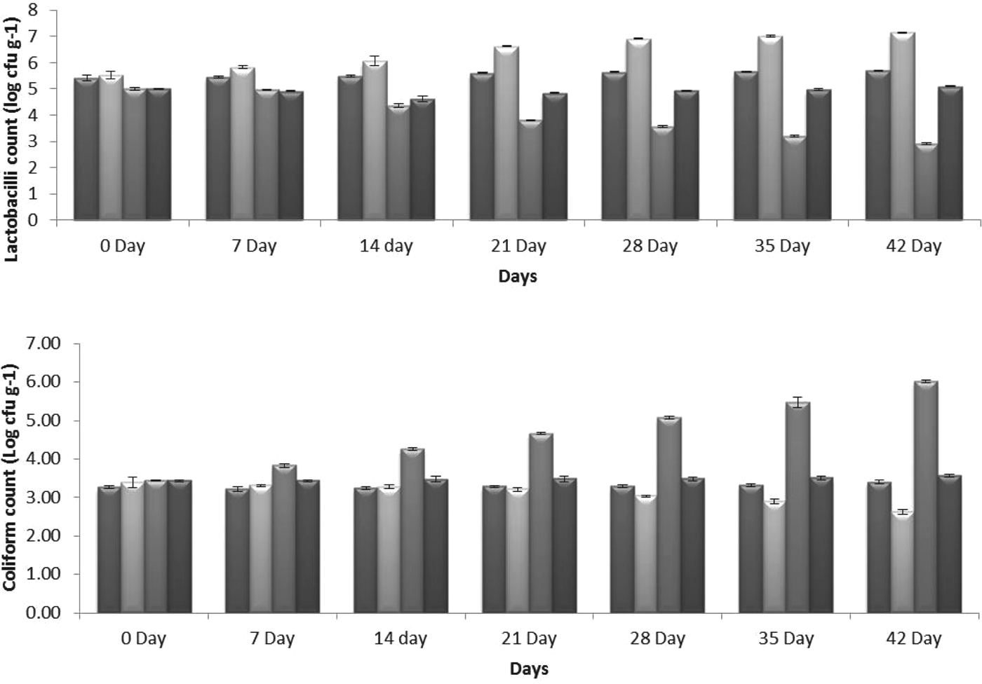
There is good evidence to support the hypothesis that prebiotics can influence gut microbiota composition and, as such, metabolic disturbances. In this study, apart from direct prebiotic effects, the improvement in the overall health of diabetic rats may also be due to the restoration of lactobacilli population in the intestine. As shown in Fig. 1, lactobacilli count in DCG showed a gradual decrease from day 0 to 42 while GOS supplemented diet restored the lactobacilli count. Various reports are available which showed a reduction in the incidence of diabetes on feeding with lactobacillus strains (Yadav et al. Reference Yadav, Jain and Sinha2008; Kang et al. Reference Kang, Yun and Park2010; Valladares et al. Reference Valladares, Sankar, Li, Williams, Lai, Abdelgeliel, Gonzalez, Wasserfall, Larkin, Schatz, Atkinson, Triplett, Neu and Lorca2010; Naito et al. Reference Naito, Yoshida and Makino2011). Along with an increase in lactobacilli count feeding of GOS also reduced the coliform count in diabetic rats. Coliform counts were found to be increased with the progression of diabetes. GOS feeding led to a reduction of coliforms in both DPG and PCG (Fig. 1). The reduction in coliform count may be partially due to less effective absorption of GOS by coliforms and partially due to increase in lactobacilli count which thereby competitively inhibit their adhesion. Prebiotics have been suggested to be directly involved in protecting the gut from infection and inflammation through the inhibition of the attachment and/or invasion of pathogenic bacteria or their toxins to the colonic epithelium (Searle et al. Reference Searle, Best, Nunez, Salguero, Johnson, Weyer, Dugdale, Cooley, Carter, Jones, Tzortzis, Woodward and La Ragione2009).
Lactobacilli and Coliform count in faecal samples of control and alloxan-induced diabetic rats. GOS, Galactooligosaccharides; NCG, Normal control group; PCG, Prebiotic control group; DCG, Diabetic control group; DPG, Diabetic prebiotic group.  , NCG;
 , PCG;
 , DCG;
 , DPG. Values are mean±se (indicated error bars in graph) of 6 animals in each group.
Conclusion
The current research is a preliminary study related to the antidiabetic potential of GOS and indicates that supplementation of GOS at a level of 10% to the diet can reduce severity of diabetes. A GOS supplemented diet modulates the various biochemical markers associated with diabetes like blood glucose, lipid profile, serum urea, serum creatinine and level of antioxidative enzymes in diabetic rats. Besides, GOS supplementation significantly increased the lactobacilli count in the faeces with a concomitant decrease in faecal coliform count. In the light of this study it can be concluded that a GOS containing diet might be used as an adjunct to dietary therapy to derive antidiabetic benefit. Further studies to draw firm conclusions should include molecular studies on the markers associated with diabetes, organised trials on higher animals.
The authors thank National Agriculture Innovative Project, New Delhi, for funding the project and FrieslandCampina Domo for making the GOS available for this study. The authors thank the Director of NDRI for supporting the work. There are no conflicts of interest whatsoever among the authors.