Introduction
The family Longidoridae Thorne, Reference Thorne1935 (order Dorylaimida Pearse, Reference Pearse1942), comprises a wide and diverse group of migratory ectoparasitic nematode species and is considered one of the most economically important nematode families in agriculture. The importance of this group of nematodes lies not only in their polyphagia and cosmopolitan distribution but also in their status as vectors of some plant nepoviruses (Archidona-Yuste et al. Reference Archidona-Yuste, Navas-Cortés, Cantalapiedra-Navarrete, Palomares-Rius and Castillo2016a, Reference Archidona-Yuste, Navas-Cortés, Cantalapiedra-Navarrete, Palomares-Rius and Castillob, Reference Archidona-Yuste, Wiegand, Castillo and Navas-Cortés2019; Coomans Reference Coomans1996; Decraemer and Geraert Reference Decraemer, Geraert, Perry and Moens2013; Decraemer and Robbins Reference Decraemer and Robbins2007; MacFarlane et al. Reference Macfarlane, Neilson and Brown2002, Reference Macfarlane2003; Taylor and Brown Reference Taylor and Brown1997). The genus Xiphinema Cobb, Reference Cobb1913, commonly known as dagger nematodes, is the most diverse genus within Longidoridae (Ahmed and Holovachov Reference Ahmed and Holovachov2021; Coomans Reference Coomans1996; Gutiérrez-Gutiérrez et al. Reference Gutiérrez-Gutiérrez, Bravo, Santos, Vieira and Mota2016; Lamberti et al. Reference Lamberti, Molinari, Moens and Brown2000). According to Andrássy (Reference Andrássy, Csuzdi and Mahunka2009), it is now the largest genus not only within the order Dorylaimida but also within the entire phylum Nematoda Potts, Reference Potts, Borradaile and Potts1932. Despite the genus comprising approximately 330 species (including ~270 in the Xiphinema non-americanum group and around 60 in the Xiphinema americanum group), only nine species have been confirmed to transmit nepoviruses to date (Decraemer and Robbins Reference Decraemer and Robbins2007).
Based on morphological and biological features, members of the genus Xiphinema are currently divided into two subgroups: (i) the Xiphinema americanum group (~60 species), with species additions by Lamberti et al. (Reference Lamberti, Molinari, Moens and Brown2000), Mobasseri et al. (Reference Mobasseri, Hutchinson, Jahanshahi Afshar and Pedram2019), Jahanshahi Afshar et al. (Reference Jahanshahi Afshar, Pedram and Mobasseri2021), Gu et al. (Reference Gu, Ye and Munawar2022), and Naghavi et al. (Reference Naghavi, Niknam and Vazifeh2022), and (ii) the Xiphinema non-americanum group (~270 species), originally listed in Loof and Luc (Reference Loof and Luc1990, Reference Loof and Luc1993) and Loof et al. (Reference Loof, Luc and Baujard1996), with recent species additions by Jahanshahi Afshar et al. (Reference Jahanshahi Afshar, Shahryari, Gharibzadeh, Pourjam and Pedram2019), Mirzaie Fouladvand et al. (Reference Mirzaie Fouladvand, Pourjam, Castillo and Pedram2019), Vazifeh et al. (Reference Vazifeh, Niknam, Jabbari and Naghavi2019), Archidona-Yuste et al. (Reference Archidona-Yuste, Cai, Cantalapiedra-Navarrete, Carreira, Rey, Viñegla, Liébanas, Palomares-Rius and Castillo2020a, Reference Archidona-Yuste, Wiegand, Castillo and Navas-Cortésb), Cai et al. (Reference Cai, Archidona-Yuste, Cantalapiedra-Navarrete, Palomares-Rius and Castillo2020), and Poureskandarian et al. (Reference Poureskandarian, Pourjam and Pedram2023). Historically, Xiphinema species identification relied on morphological traits and diagnostic keys (Lamberti et al. Reference Lamberti, Molinari, Moens and Brown2000, Reference Lamberti, Hockland, Agostinelli, Moens and Brown2004; Loof and Luc Reference Loof and Luc1990; Loof et al. Reference Loof, Luc and Baujard1996). However, with the advent of molecular tools, integrative taxonomy – combining morphology with molecular data – has become the standard procedure in many species’ descriptions/delineations of this group (e.g., Archidona-Yuste et al. Reference Archidona-Yuste, Navas-Cortés, Cantalapiedra-Navarrete, Palomares-Rius and Castillo2016a, Reference Archidona-Yuste, Navas-Cortés, Cantalapiedra-Navarrete, Palomares-Rius and Castilloc, Reference Archidona-Yuste, Navas-Cortés, Cantalapiedra-Navarrete, Palomares-Rius and Castillod, Reference Archidona-Yuste, Wiegand, Castillo and Navas-Cortés2019; Cai et al. Reference Cai, Maria, Barsalote, Subbotin and Zheng2018, Reference Cai, Archidona-Yuste, Cantalapiedra-Navarrete, Palomares-Rius, Zheng and Castillo2019; Gutiérrez-Gutiérrez et al. Reference Gutiérrez-Gutiérrez, Palomares-Rius, Cantalapiedra-Navarrete, Landa, Esmenjaud and Castillo2010, Reference Gutiérrez-Gutiérrez, Palomares-Rius, Cantalapiedra-Navarrete, Landa and Castillo2011, Reference Gutiérrez-Gutiérrez, Cantalapiedra-Navarrete, Remesal, Palomares-Rius, Navas-Cortés and Castillo2013; Mobasseri et al. Reference Mobasseri, Hutchinson, Jahanshahi Afshar and Pedram2019; Naghavi et al. Reference Naghavi, Niknam and Vazifeh2022). This point is very important because of the frequent presence of cryptic species in plant-parasitic nematodes (Palomares-Rius et al. Reference Palomares-Rius, Cantalapiedra-Navarrete and Castillo2014) and the possibility of the use of molecular markers with juveniles (with not many characters for species identification). Analysis of the most promising molecular markers for Longidoridae have been studied in detail by Palomares-Rius et al. (Reference Palomares-Rius, Cantalapiedra-Navarrete, Archidona-Yuste, Subbotin and Castillo2017).
To date, 36 Xiphinema species have been recorded from Iran (Fadakar et al. Reference Fadakar, Pourjam, Barsi and Pedram2021; Fekrat et al. Reference Fekrat, Ghaderi, Pourjam, Castillo and Pedram2024; Jahanshahi Afshar et al. Reference Jahanshahi Afshar, Shahryari, Gharibzadeh, Pourjam and Pedram2019, Reference Jahanshahi Afshar, Shahryari, Mokhtassi-Bidgoli, Castillo, Mirzaie Fouladvand and Pedram2020, Reference Jahanshahi Afshar, Pedram and Mobasseri2021; Mirzaie Fouladvand et al. Reference Mirzaie Fouladvand, Pourjam, Castillo and Pedram2019; Mobasseri et al. Reference Mobasseri, Hutchinson, Jahanshahi Afshar and Pedram2019; Naghavi et al. Reference Naghavi, Niknam and Vazifeh2022; Pedram Reference Pedram, Ghaderi, Kashi and Karegar2018; Poureskandarian et al. Reference Poureskandarian, Pourjam and Pedram2023; Vazifeh et al. Reference Vazifeh, Niknam, Jabbari and Naghavi2019), of which four species viz. X. diversicaudatum (Micoletzky, Reference Micoletzky1927) Thorne, Reference Thorne1939; X. castilloi Roshan-Bakhsh et al., Reference Roshan-Bakhsh, Pourjam, Pedram, Robbins and Decraemer2014; X. ingens Luc & Dalmasso, Reference Luc and Dalmasso1964, and X. hyrcaniense Fadakar et al., Reference Fadakar, Pourjam, Barsi and Pedram2021, belong to the morphospecies group 5 (sensu Loof and Luc Reference Loof and Luc1990) (i.e., have pseudo-Z-organ (globular structures/inclusions) or pseudo-Z-organ plus crystalloids/spines in uteri). During a recent survey of plant-parasitic nematodes in southern Iran (Bushehr province), three dagger nematode populations belonging to the Xiphinema non-americanum group were recovered. Preliminary morphological studies revealed that two populations shared identical morphological features and represent an undescribed species, which is formally described herein, while the third population corresponds with a known species from morphospecies group 5. An integrative taxonomic approach based on morphological, morphometric, and molecular data was used to characterise these populations.
The objectives of this study were (i) to characterise two Xiphinema species from the non-americanum group recovered during this study through integrative methods and (ii) to reconstruct their phylogenetic relationships using partial sequences of the D2-D3 expansion segments of the 28S rDNA (D2-D3/LSU), internal transcribed spacer region separating the 18S rDNA and 5.8S rDNA (ITS1), 18S rDNA (SSU), and cytochrome c oxidase subunit I of the mtDNA (COI).
Materials and methods
Sampling, extraction, and morphological identification
A total number of 35 soil samples were collected during March and April 2024 from the rhizosphere of different plants (including palm trees) in natural habitats from Bushehr province, southern Iran. The samples were taken from a depth of 5–30 cm. Nematodes were extracted using the tray method (Whitehead and Hemming Reference Whitehead and Hemming1965) and a modification of Cobb’s decanting and sieving method (Flegg Reference Flegg1967). Nematodes were handpicked under a stereomicroscope, killed by adding hot FPG (4:1:1, formaldehyde: propionic acid: glycerin) solution, transferred to anhydrous glycerine according to De Grisse (Reference De Grisse1969), and mounted on permanent glass slides to allow handling and observation. In total, three populations of the genus Xiphinema were recovered. For studying the uterine structure, live females were selected, killed by gentle heat in a drop of water, studied, and photographed in temporary slides. Morphometric values and photomicrographs were taken using a Dino-Eye digital eyepiece camera (Model AM7023, bundled with the DinoCapture 2.0 software; AnMo Electronics Corporation; New Taipei City; Taiwan) attached to a Leitz Dialux 22 light microscope. Line drawings were first made using a drawing tube, and then re-drawn and prepared for publication using CorelDRAW software version 16. The morphometric study of each nematode population included classic diagnostic features in Longidoridae (i.e., de Man body ratios, lip region and amphid shape, oral aperture–guiding ring distance, odontostyle and odontophore length). All measurements were expressed in micrometers (μm), unless otherwise indicated in the text. Morphological comparisons were performed using the polytomous identification keys for the identification of Xiphinema species (Loof and Luc Reference Loof and Luc1990) and with the descriptions of all other characterised species up to the present. The position of pharyngeal gland nuclei was calculated according to Loof and Coomans (Reference Loof and Coomans1972), and the juvenile developmental stages were identified according to Robbins et al. (Reference Robbins, Brown, Halbrendt and Vrain1996).
Molecular characterisation
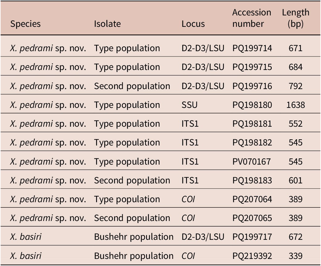
Morphological observations and molecular analyses were conducted in parallel to ensure accurate species identification through an integrative taxonomic approach. A total of seven individuals (four from Xiphinema pedrami sp. nov. [type population], two from X. pedrami sp. nov. [second population], and one from X. basiri Siddiqi, Reference Siddiqi1959) were used for molecular characterisation. Prior to DNA extraction, each individual nematode was examined on a temporary slide, particularly focusing on uterine characteristics, to reconfirm morphological identification. DNA was extracted using the modified Chelex method (Rashidifard et al. Reference Rashidifard, Marais, Daneel, Mienie and Fourie2019). Each nematode was transferred to an Eppendorf tube containing 20 μl of nuclease-free water, 25 μl Chelex (5%, w/v), and 5 μl of proteinase K (20 mg/ml). The microtubes were incubated at 56°C for 2 hr., then at 95°C for 10 min., and the obtained solutions were used as DNA template. 5 μl of each extracted DNA was added to the polymerase chain reaction (PCR) mixture in a 0.2-ml Eppendorf tube containing 15 μl 2X Master mix (Ampliqon, Odense, Denmark), 1 μl of each primer (10 pmol/μl), and 8 μl ddH2O, to a final volume of 30 μl. The D2-D3/LSU region was amplified using forward D2A (5’–ACAAGTACCGTGAGGGAAAGTTG–3’) and reverse D3B (5’–TCGGAAGGAACCAGCTACTA–3’) primers (De Ley et al. Reference De Ley, Felix, Frisse, Nadler, Sternberg and Thomas1999). The SSU portion was amplified as two overlapping fragments, using forward 988F (5′–CTCAAAGATTAAGCCATGC–3′) and reverse 1912R (5′–TTTACGGTCAGAACTAGGG–3′) primers for the first fragment, and forward 1813F (5′–CTGCGTGAGAGGTGAAAT–3′) and reverse 2646R (5′–GCTACCTTGTTACGACTTTT–3′) primers for the second fragment (Holterman et al. Reference Holterman, van derWurff, van den Elsen, van Megen, Bongers, Holovachov, Bakker and Helder2006). The ITS1 portion was amplified using forward primer 18S (5′–TTGATTACGTCCCTGCCCTTT–3′) (Vrain et al. Reference Vrain, Wakarchuk, Lévesque and Hamilton1992) and reverse primer rDNA1 (5′–ACGAGCCGAGTGATCCACCG–3′) (Gutiérrez-Gutiérrez et al. Reference Gutiérrez-Gutiérrez, Palomares-Rius, Cantalapiedra-Navarrete, Landa, Esmenjaud and Castillo2010). The COI region was amplified using forward primer JB3 (5′–TTTTTTGGGCATCCTGAGGTTTAT–3′) and reverse primer JB5 (5′–AGCACCTAAACTTAAAACATAATGAAAATG–3′) (Bowles et al. Reference Bowles, Blair and McManus1992). PCR reactions were carried out in a DNA thermal cycler (Hybaid, Ashford, Middlesex, UK). The PCR cycle conditions were as follows: initial denaturation cycle at 94°C for 15 min., followed by 35 cycles of denaturation at 94°C for 45 sec.; annealing cycle at 56°C (LSU), 55°C (ITS1), 54°C (SSU and COI) for 45 sec.; extension cycle at 72°C for 1 min.; and finally elongation cycle at 72°C for 5 min. After DNA amplification, the quality of PCR was checked by electrophoresis of 4 μl of the PCR reactions in 1% agarose gel containing SYBR Green I. Products were visualised and photographed under ultraviolet light. The length and concentration of each PCR product was measured by comparison with a low DNA mass ladder (Invitrogen, Carlsbad, CA). The PCR products were purified and sequenced directly for both strands using the same primers with an ABI 3730XL sequencer (Bioneer, Seoul, South Korea). The newly obtained sequences were submitted to the GenBank database under these accession numbers (D2-D3/LSU: PQ199714–PQ199716 for X. pedrami sp. nov., PQ199717 for X. basiri, SSU: PQ198180 for X. pedrami sp. nov., ITS1: PQ198181–PQ198183 and PV070167 for X. pedrami sp. nov., COI: PQ207064, PQ207065 for X. pedrami sp. nov., PQ219392 for X. basiri).
Phylogenetic analyses
The newly obtained sequences (D2-D3/LSU, SSU, ITS1, and COI) were compared with those of other Xiphinema species available in GenBank using the BLAST homology search program. For D2-D3/LSU phylogeny, 95 sequences of several Xiphinema non-americanum group species/populations (including three newly generated sequences from the unidentified new species and one from the known species); for SSU phylogeny, 67 sequences of several Xiphinema non-americanum group species/populations (including one newly generated sequence from the unidentified new species); for ITS1 phylogeny, 37 sequences of several Xiphinema non-americanum group species/populations (including four newly generated sequences from the unidentified new species), and for COI phylogeny, 57 sequences of several Xiphinema non-americanum group species/populations (including two newly generated sequences from the unidentified new species and one from the known species) were included. Outgroup taxa for each dataset were chosen following previously published studies (Jahanshahi Afshar et al. Reference Jahanshahi Afshar, Shahryari, Gharibzadeh, Pourjam and Pedram2019; Mirzaie Fouladvand et al. Reference Mirzaie Fouladvand, Pourjam, Castillo and Pedram2019; Poureskandarian et al. Reference Poureskandarian, Pourjam and Pedram2023). Specifically outgroups for each dataset were: i) D2-D3 region: Nevadanema nevadense (JN242245), Tylencholaimus mirabilis (AY593059), Xiphinema brevicollum (HM163209), Xiphinema diffusum (AY601600) and Xiphinema californicum (KX263165); ii) ITS1 region: Xiphinema americanum (AY430189) and Xiphinema pachtaicum (AY430178); iii) SSU region: T. mirabilis (EF207253), Opisthodorylaimus sylphoides (AY284785), Mesodorylaimus centrocercus (EF207248), Xiphinema rivesi (AF036610), Xiphinema cf. americanum (AM086671), Xiphinema citricolum (AM086686), Xiphinema floridae (AM086687), Xiphinema georgianum (AM086688) and X. americanum (KJ934158); and iv) COI region: X. americanum (AM086690 and KX263051) and X. peruvianum (AM086692). Multiple sequence alignments for each gene were made using the FFT-NS-2 algorithm of MAFFT V.7.450 (Katoh et al. Reference Katoh, Rozewicki and Yamada2019, https://mafft.cbrc.jp/alignment/server/). The BioEdit program V.7.2.5 (Hall Reference Hall1999) was used for sequence alignment visualisation and manually editing/trimming of the poorly aligned positions using a light filtering strategy (up to 20% of alignment positions). The best-fit model of DNA evolution, the base frequency, the proportion of invariable sites, the gamma distribution shape parameters, and the substitution rates for each of the four datasets were obtained by JModelTest version 2.1.7 (Darriba et al. Reference Darriba, Taboada, Doallo and Posada2012) using the Akaike information criterion (AIC) and used to infer evolutionary relationships. A general time-reversible model, incorporated with a proportion of invariable sites and a gamma distributed rate variation among sites (GTR+I+G), was used in phylogenetic analyses of the D2-D3/LSU, SSU, and COI datasets, and a general time-reversible model, with the addition of a gamma distributed rate variation among sites (GTR+G), was used in phylogenetic analyses of ITS1 dataset. Phylogenetic analyses of the sequence datasets were performed based on the Bayesian inference (BI) using MrBayes 3.1.2 (Ronquist and Huelsenbeck Reference Ronquist and Huelsenbeck2003) with selecting a random starting tree and running the chains for 10×106 generations (SSU, ITS1, and COI) and 5×10⁶ generations (D2-D3/LSU). The Markov chains were sampled at intervals of 100 generations and 25% of the converged runs being regarded as ‘burn-in’. A 50% majority rule consensus tree was generated for each dataset, and posterior probabilities (PP) were calculated for each clade using Markov chain Monte Carlo (MCMC) method within a Bayesian framework (Larget and Simon Reference Larget and Simon1999). Phylogenetic trees from all analyses were visualised using FigTree software version 1.4.4 (Rambaut Reference Rambaut2018) and were digitally draw in CorelDRAW software version 16.
Results and discussion
By combining nematode morphology with morphometric analysis and molecular data from ribosomal and mitochondrial sequences, we were able to describe a new species of the genus, Xiphinema pedrami sp. nov., as well as the known species X. basiri.
Xiphinema pedrami Footnote 1 sp. nov.
urn:lsid:zoobank.org:pub:30F96817-BF26-40C0-84C8-E19EA1A87035
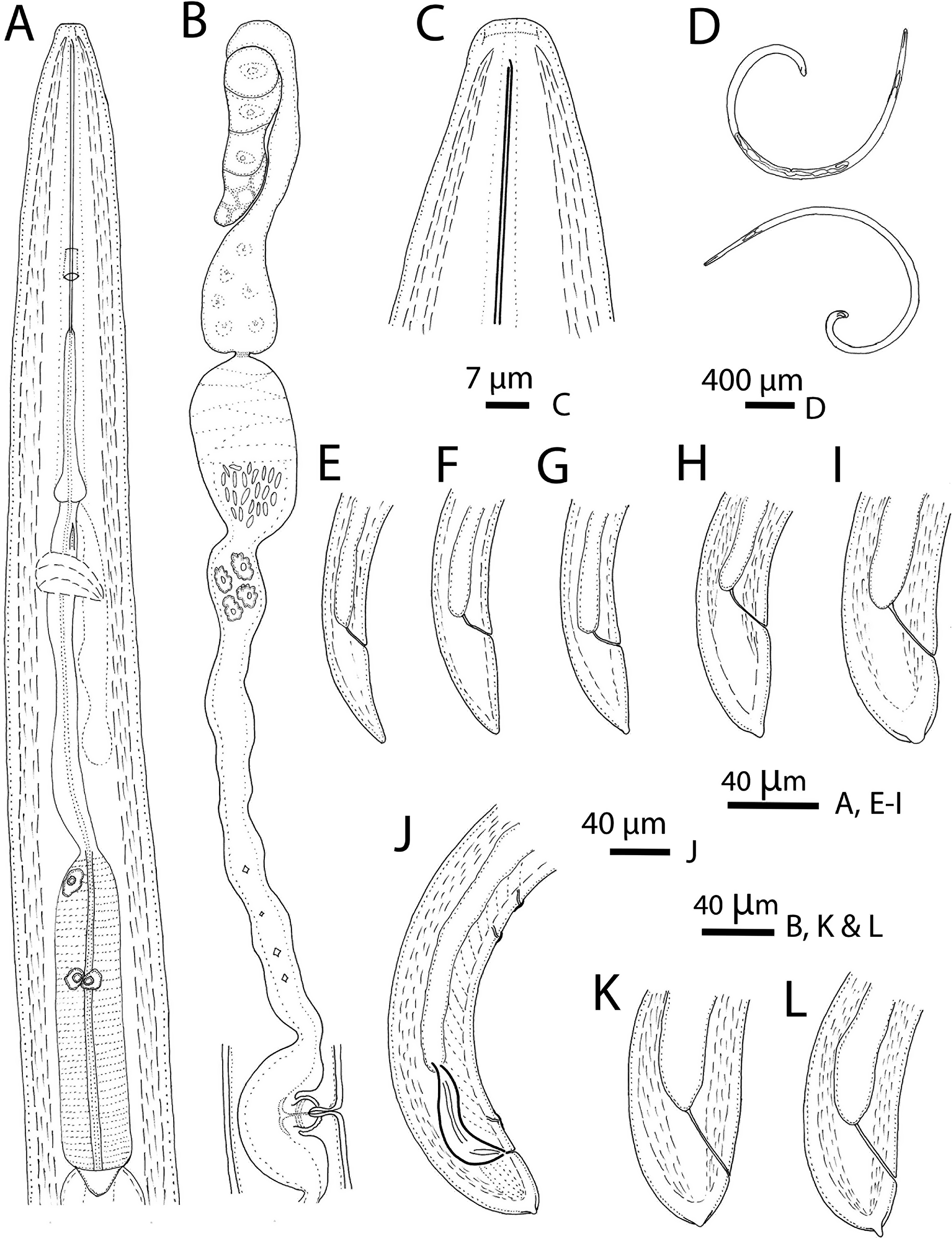
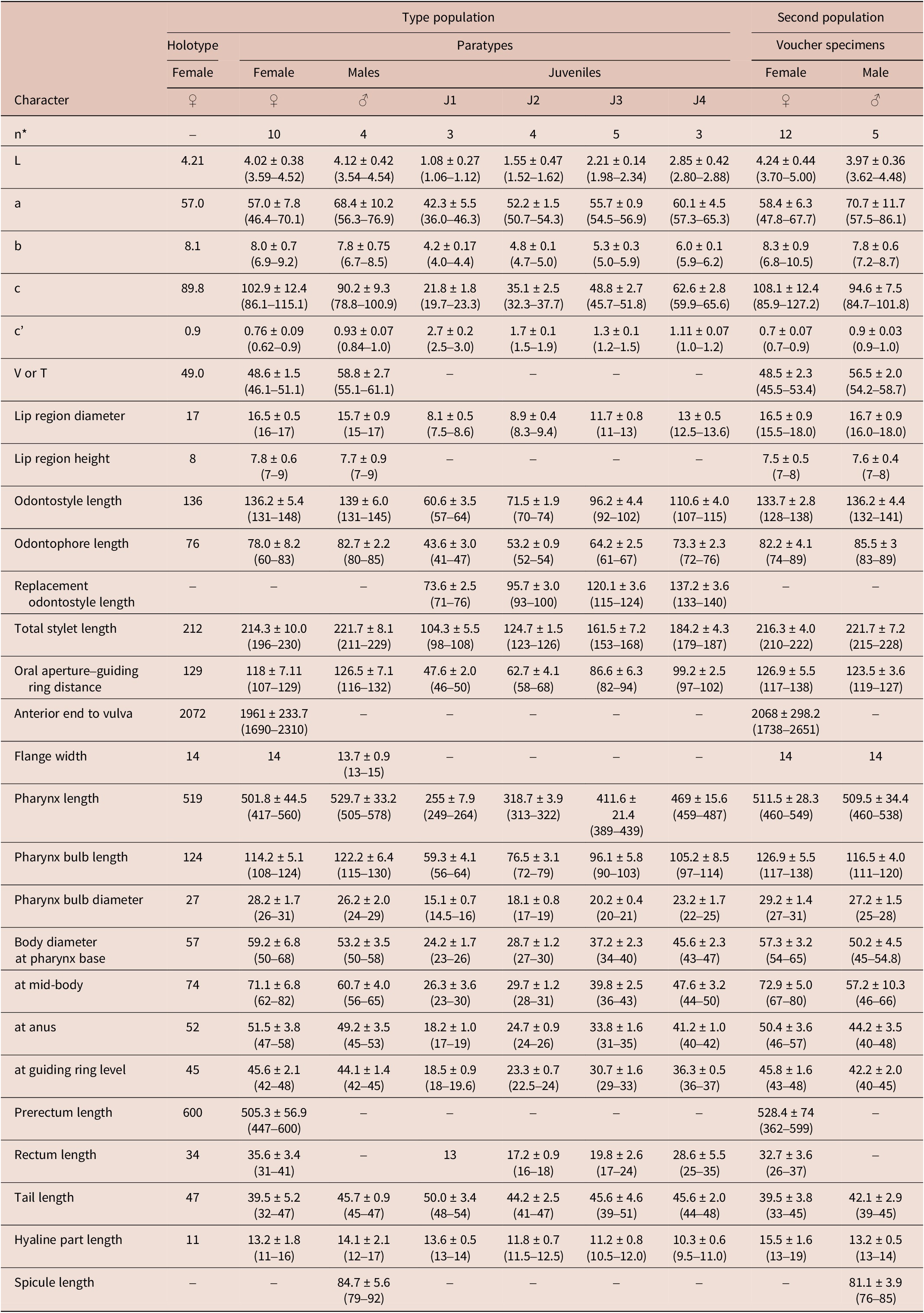
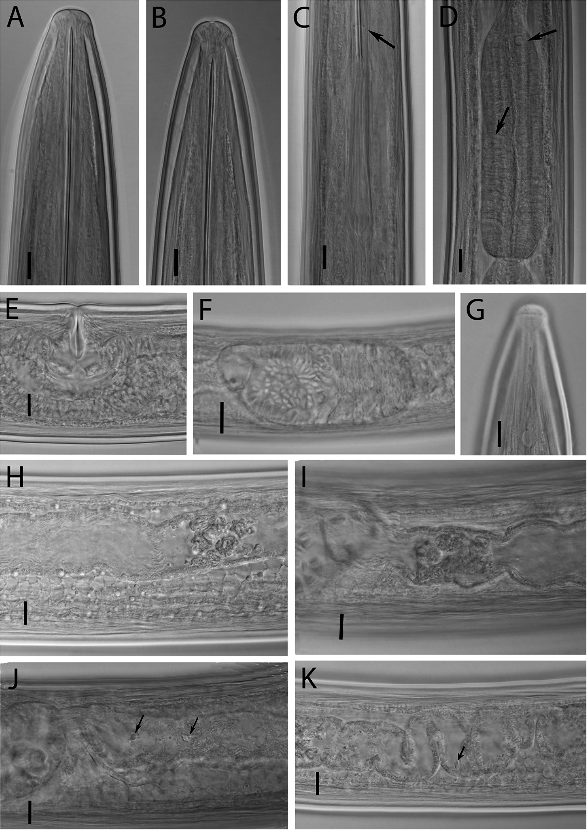
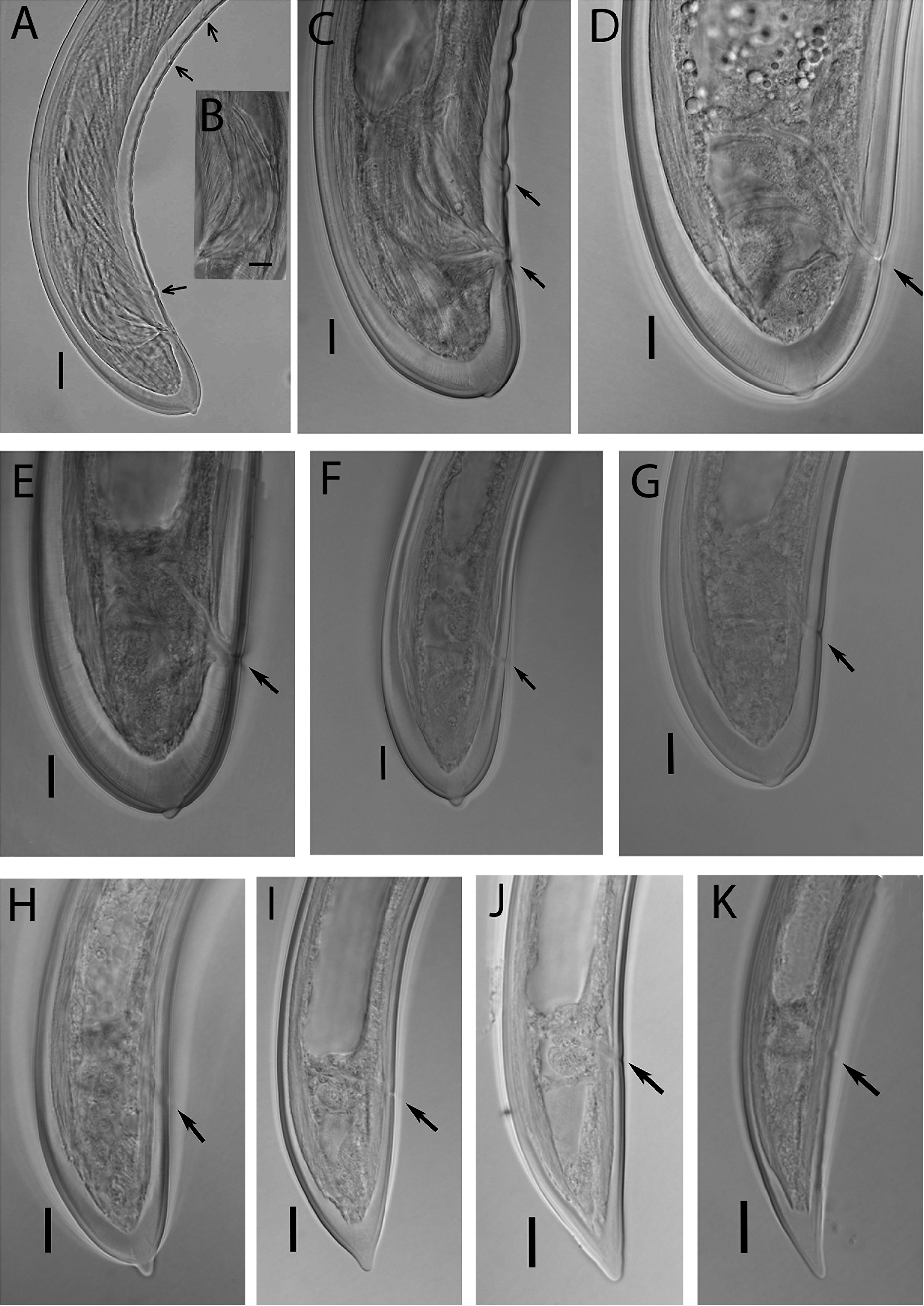
(Figures 1–4, morphometrics in Table 1).
Line drawings of Xiphinema pedrami sp. nov. (A) Female neck region; (B) Female reproductive system; (C) Anterior body region; (D) Female and male entire body; (E) First-stage juvenile tail; (F, G) Second-stage juvenile tail; (H, I) Third- and fourth-stage juvenile tails, respectively; (J) Male posterior body region; (K, L) Female tail. Scale bars: (A, B, E–L) 40 μm; (C) 7 μm; (D) 400 μm.

Morphometrics of Xiphinema pedrami sp. nov. from Bushehr province, southern Iran. All measurements are in μm (except L, in mm), and in the form: mean ± standard deviation (range)

*Abbreviations: n = number of specimens on which measurements are based; L = overall body length; a = body length/greatest body diameter; b = body length/distance from anterior end to pharyngo–intestinal junction; c = body length/tail length; c’ = tail length/tail diameter at anus or cloaca; V = distance from body anterior end to vulva expressed as percentage (%) of the body length; T = distance from cloacal aperture to anterior end of testis expressed as percentage (%) of the body length
Description
Female. Body cylindrical, narrowing very gradually towards anterior end, ventrally curved, assuming an open C to G-shaped when killed by heat. Cuticle two-layered, with fine transverse striae visible in tail region, 3.0–4.0 μm thick at guiding ring level, 4.0–5.0 μm at mid-body, 7.0–8.0 μm at anterior lip of anus and 11.0–16.0 μm at tail end (the hyaline part of tail). Lateral chord 23–35% of corresponding body diameter. Lip region broadly rounded, 1.8−2.2 times as wide as high, separated from the rest of the body by a weak depression. Amphidial fovea cup-shaped, its slit occupies more than half of the lip region diameter and is located just anterior to demarcation line. Body pores present between anterior end and guiding ring, three and four on the dorsal and ventral side, respectively. Odontostyle long and slender, 7.8–8.7 times longer than lip region diameter or 1.5–2.2 times longer than odontophore length, odontophore with well-developed flanges, 13–14 μm wide. Guiding ring double, guiding sheath 12–18 μm long depending on degree of protraction/retraction of the stylet. Nerve ring surrounding the slender portion of the pharynx behind the odontophore base, located at 265–315 μm or 56–63% of the total neck length from the anterior end. Pharynx dorylaimoid, consisting of an anterior slender narrow part, posteriorly extending to a terminal pharyngeal bulb that occupies about 1/4 to 1/5 of the total pharynx length. Pharyngeal bulb 3.1–5.1 times as long as wide and 1.4–2.5 times as long as corresponding body diameter, with three gland nuclei, the larger dorsal gland nucleus (DN) at 8.3–14.1% of pharyngeal bulb length, the two smaller ventro-sublateral nuclei (S1N) at about the same level, at 46–57% of pharyngeal bulb length. Glandularium 96–110 μm long. Cardia conoid-rounded, 5.0–7.0 μm long. Intestine simple, prerectum 7.7–11.5, rectum 0.5–0.8 anal body diameter long. Reproductive system didelphic-amphidelphic with two almost equally-developed branches. Ovary reflexed, 76–146 μm long. Oviduct consisting of a narrow distal part and a well-developed proximal pars dilatata oviductus portion, 110–205 μm long. Sphincter visible between oviducts and uterus. Uterus bipartite, consisting of a distal pars dilatata uteri portion and a tubular proximal part, 210–399 μm long, the pars dilatata uteri portion with ellipsoid-bacilliform sperm cells, the tubular part of uterus devoid of spiniform structures, but with Z-differentiation in the type of irregular-shaped sclerotised bodies of variable size (four to eight in number, varying in size) close to the pars dilatata uteri and small crystalloid bodies (two to four in number) distributed over the entire length of the proximal part (Figure 2, J, K, arrows). Ovejector well-developed. Vagina perpendicular to body axis, 35–40 μm long or extending inwards for 43–62% of corresponding body diameter, pars distalis vaginae 21–28 μm long, pars proximalis vaginae 14–16 μm long and 24–31 μm wide. Vulva a transverse slit. Tail short, rounded, dorsally more convex with a small bulge (2–3 μm) at the end, three or four body pores visible on each side of the tail.
Photomicrographs of Xiphinema pedrami sp. nov. (A, B) Female and male anterior body region; (C) Guiding ring and odontophore flanges; (D) Pharyngeal basal bulb showing dorsal and ventro-sublateral glands nuclei (arrowed); (E) Vulval region; (F) Pars dilatata uteri; (G) Female lip region showing amphidial fovea; (H, I) pseudo-Z-organ; (J, K) Rhomboid-like crystalloids bodies. Scale bars: (A–E, G–K) 10 μm; (F) 20 μm.

Male. (Common, about half as many as females). General morphology similar to that of females, except for reproductive system and more curved posterior part of body, after heat relaxation. Genital system diorchic, with opposed testes, containing multiple rows of different stages of spermatogonia. Spicules paired, massive, ca. two times longer than tail length, lateral guiding pieces more or less straight or with curved proximal end, 16–20 μm long. Copulatory supplements composed one pair of cloacal supplements located at 24–28 μm from cloacal aperture, and three to five single ventromedian supplements. Tail similar to that of females.
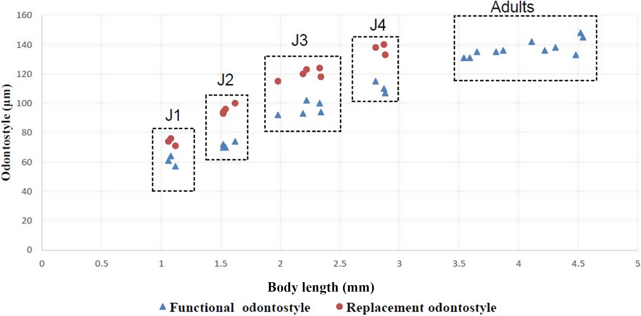
Juveniles. Morphologically similar to adults in most respects except for their smaller body size, having replacement odontostyle, differences in tail shape and development of reproductive system. All juvenile stages were detected and distinguished for the type population using morphological characters and relative lengths of body, functional and replacement odontostyle (Table 1, Figures 3, 4). The first juvenile stage (J1) is characterised by the replacement odontostyle tip close to the base of the functional odontostyle, that is located at the level with the odontophore. In all other stages, the replacement odontostyle is posterior to the flanges of odontophore in its resting position. The first juvenile stage has a conoid tail, dorsally convex, ventrally concave, its tip bluntly rounded; the second juvenile stage (J2) has a dorsally convex, ventrally straight tail with more or less differentiated mucron-like structure at its tip; the third juvenile stage (J3) has a dorsally convex, ventrally convex tail with a subdigitate terminus and the fourth juvenile stage (J4) has a short tail, convex on both sides, ending in a small bulge (Figure 3, F–K).
Photomicrographs of Xiphinema pedrami sp. nov. (A–C) Male tail showing spicules, adanal and ventromedian supplements (arrowed); (D, E) Female tail variations; (F, G) Fourth-stage juvenile tail variations; (H) Third-stage juvenile tail; (I, J) Second-stage juvenile tail variations; (K) First-stage juvenile tail. Scale bars: (B–K) 10 μm; (A) 20 μm.

Correlation of functional and replacement odontostyle length to body length in juvenile stages and adults of Xiphinema pedrami sp. nov.

Diagnosis and relationships
Morphologically, Xiphinema pedrami sp. nov. belongs to the Xiphinema non-americanum group 5 of Loof and Luc (Reference Loof and Luc1990) and can be differentiated from all known species of the genus by a combination of characters. It is characterised by having a medium body length (3.5–5.0 mm), spear length 196–230 μm, composed of a 128–148 μm long odontostyle and 60–89 μm long odontophore, lip region broadly rounded, separated from the rest of the body by a weak depression; vulva position at 45.5–53.4%, two equally developed genital branches having well-developed pseudo-Z-organ, comprising four to eight sclerotised bodies of variable size, located in the vicinity of the pars dilatata uteri, crystalloid bodies (rhomboid shaped) along uterus and spines absent in the uterus; female tail short, rounded, convex on both sides, ending in a small bulge. Males frequent (1:2 ratio), spicules medium-sized (76–92 μm long) and 1 + 3–5 ventromedian supplements. The identification codes for the new species, according to the polytomous key of Loof and Luc (Reference Loof and Luc1990) are A4, B2, C5a5b, D6, E56, F4(5), G23, H2, I34, J5a5b, K3, L2.
Based on using the polytomous key of Loof and Luc (Reference Loof and Luc1990), and character analysis by Coomans et al. (Reference Coomans, Huys, Heyns and Luc2001), including type of female genital system, uterine differentiation, ratio of tail length to anal body diameter and body length, and sorting on matrix codes A (type of female genital system), C (tail shape), D (c´ ratio), E (vulva position), F (body length), and G [total spear length (odontostyle + odontophore)]. Xiphinema pedrami sp. nov. closely resembles to X. andalusiense Archidona-Yuste et al., Reference Archidona-Yuste, Navas-Cortés, Cantalapiedra-Navarrete, Palomares-Rius and Castillo2016a; X. baetica Gutiérrez-Gutiérrez et al., Reference Gutiérrez-Gutiérrez, Cantalapiedra-Navarrete, Remesal, Palomares-Rius, Navas-Cortés and Castillo2013; X. hispanum Lamberti et al., Reference Lamberti, Castillo, Gomez-Barcina and Agostinelli1992; X. hyrcaniense; X. pseudocoxi Sturhan, Reference Sturhan1984 and X. macroacanthum Lamberti et al., Reference Lamberti, Roca and Agostinelli1989; from which it can be differentiated by a combination of characters discussed below.
Xiphinema pedrami sp. nov. differs from:
-
– X. andalusiense, by distant placement in all four phylogenetic trees, having a shorter body and odontostyle (3.5–5.0 and 128–148 vs. 4.0–6.1 mm and 137–151 μm, respectively), wider lip region (15.5–18.0 vs. 11.5–15.5 μm), lower c´ ratio (0.6–0.9 vs. 1.0–1.3), absence vs. presence of spiniform structures in uterus, and presence vs. absence of males.
-
– X. baetica, by distant placement in all four phylogenetic trees, having a shorter body and odontostyle (3.5–5.0 and 128–148 vs. 4.6–6.2 mm and 140–156 μm, respectively), wider lip region (15.5–18.0 vs. 12.5–15.0 μm), longer spicules (76–92 vs. 61.0–70.5 μm), and difference in female tail shape (rounded with a small central bulge vs. convex-conoid with distinctly digitate terminus, 8–14 μm long).
-
– X. hispanum, by distant placement in D2-D3/LSU, SSU and COI phylogenetic trees, having a wider lip region (15.5–18.0 vs. 14.1–15.9 μm), lower a ratio (46.4–70.1 vs. 73–84), absence vs. presence of spiniform structures in uterus, difference in J1 tail shape (conical, dorsally convex, ventrally concave with rounded tip vs. elongate-conoid without terminal swelling), and difference in female tail shape (rounded with a small central bulge vs. conoid with a central bulge.
-
– X. hyrcaniense, by distant placement in D2-D3/LSU and ITS1 phylogenetic trees, having a shorter body (3.5–5.0 vs. 5.4–7.3 mm), more anteriorly located guiding ring (107–138 vs. 123–143 μm), and difference in number of pseudo-Z-organ inclusion bodies (4–8 vs. 3–4).
-
– X. macroacanthum, by distant placement in D2-D3/LSU, ITS1 and COI phylogenetic trees, having a shorter body (3.5–5.0 vs. 4.1–6.1 mm), lower c ratio (85.9–127.1 vs. 122.5–167), more anteriorly located guiding ring (107–138 vs. 112.5–164.5 μm), shorter odontostyle (128–148 vs. 156.5–180 μm), and shorter odontophore (60–89 vs. 83.5–98.5 μm).
-
– X. pseudocoxi (the data of X. pseudocoxi from Archidona-Yuste et al. Reference Archidona-Yuste, Navas-Cortés, Cantalapiedra-Navarrete, Palomares-Rius and Castillo2016a), by distant placement in all four phylogenetic trees, having a longer odontostyle (128–148 vs. 114.5–126.0 μm), lower a ratio (46.4–70.1 vs. 70.3–91.9), wider lip region (15.5–18.0 vs. 11.5–12.5 μm), more posteriorly located guiding ring (107–138 vs. 98.0–113.5 μm), and difference in female tail length (32–47 vs. 39.5–56.0 μm).
In addition, X. pedrami sp. nov. is molecularly related to X. ingens and X. artemisiae Chizhov et al., Reference Chizhov, Tiev and Turkina1986; from which it can be mainly differentiated morphologically as follows: From X. ingens (see Mirzaei et al. Reference Mirzaei, Pourjam and Pedram2015), it differs by having a shorter body and odontostyle (3.5–5.0 and 128–148 vs. 4.1–6.0 mm and 146–167 μm, respectively), more anteriorly situated guiding ring (107–138 vs. 135–165 μm), having vs. lacking males, and difference in J1 tail shape (conical, dorsally convex, ventrally concave with rounded tip vs. conical, dorsally convex, ventrally flat with digitate mucro). From X. artemisiae (see Fekrat et al. Reference Fekrat, Ghaderi, Pourjam, Castillo and Pedram2024), it differs by having a shorter body (3.5–5.0 vs. 5.1–5.6 mm), lower a ratio (46–70 vs. 70–95), longer spicules (76–92 vs. 75–79 μm), and different shape of the tail end of juvenile stages (J2, J3 and J4).
Type locality and habitat
The type population was collected from the rhizosphere of date palm tree (Phoenix dactylifera L.) in Azhdar Khowsh village, Dashti county, Bushehr province, southern Iran, GPS coordinates: 28° 30′ 56″ N, 51° 49′ 38″ E; altitude: 501 m. above sea level. The second population was collected from the rhizosphere of Jujube tree [Ziziphus spina-christi (L.) Desf.] in Kalameh city, Dashtestan county, Bushehr province, southern Iran, GPS coordinates: 28° 56′ 37″ N, 51° 27′ 30″ E; altitude: 389 m. above sea level.
Type material
Female holotype (slide: k23-1), seven female paratypes, two male paratypes, and fifteen juvenile paratypes (slides: k23 1-10) from the type population and ten female vouchers and three male vouchers from the second population were deposited in the nematode collection at Faculty of Agriculture, University of Zanjan, Zanjan, Iran. Three female paratypes and two male paratypes from the type population and two female vouchers and two male vouchers from the Kalameh population were deposited in the nematode collection at Institute for Sustainable Agriculture (IAS), Spanish National Research Council (CSIC), Cordoba, Spain.
Iranian populations of Xiphinema basiri
(Figures 5, 6; morphometrics in Table 2).
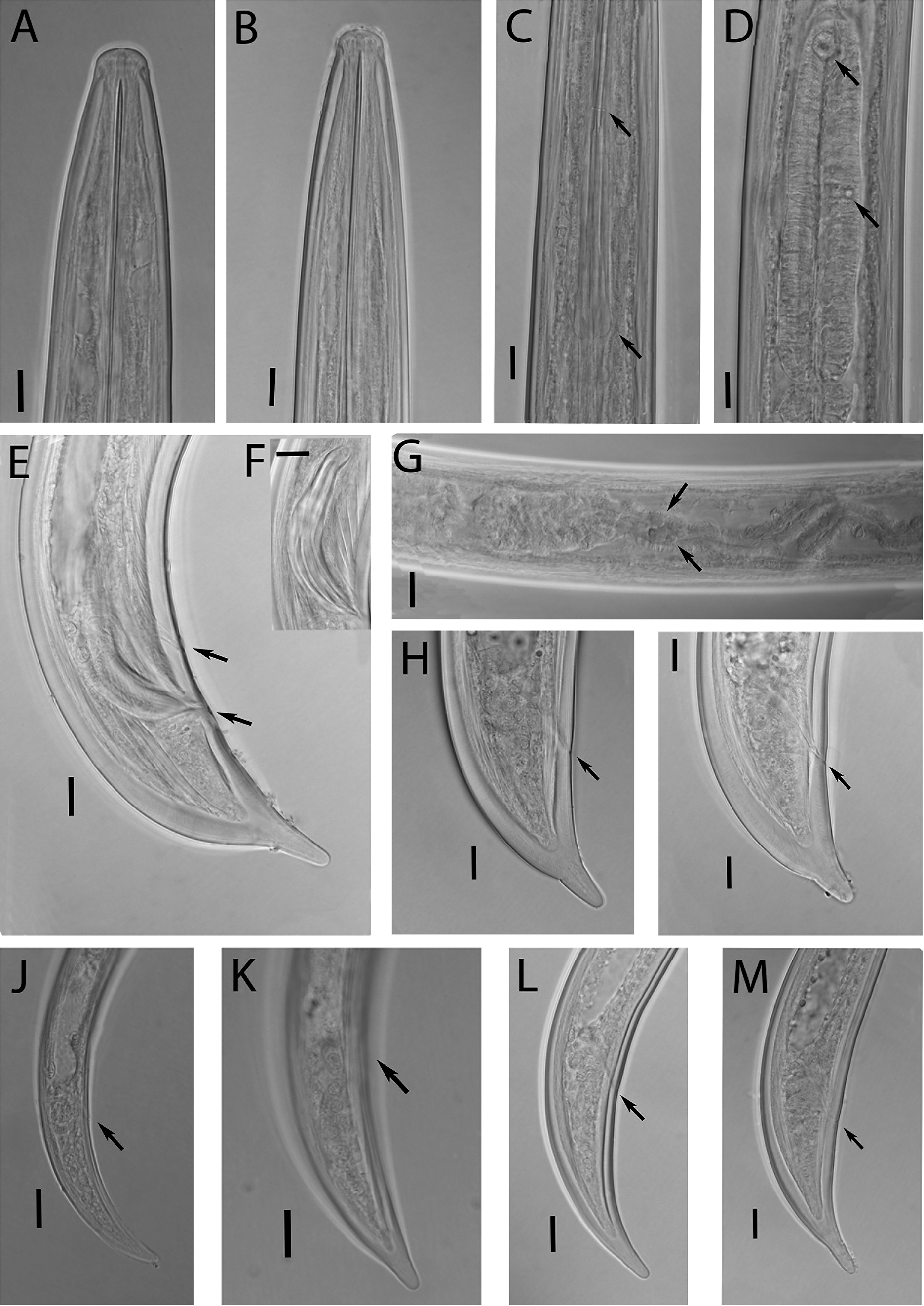
Photomicrographs of Xiphinema basiri Siddiqi, Reference Siddiqi1959. (A, B) Female and male anterior region; (C) Guiding ring and odontophore flanges; (D) Pharyngeal basal bulb showing dorsal and ventro-sublateral glands nuclei (arrowed); (E, F) Male tail showing spicules, adanal and ventromedian supplements (arrowed); (G) pseudo-Z-organ; (H, I) Female tail; (J–M) First-, second-, third-, and fourth-stage juvenile (J1-J4) tails, respectively. Scale bars: (A–M) 10 μm.

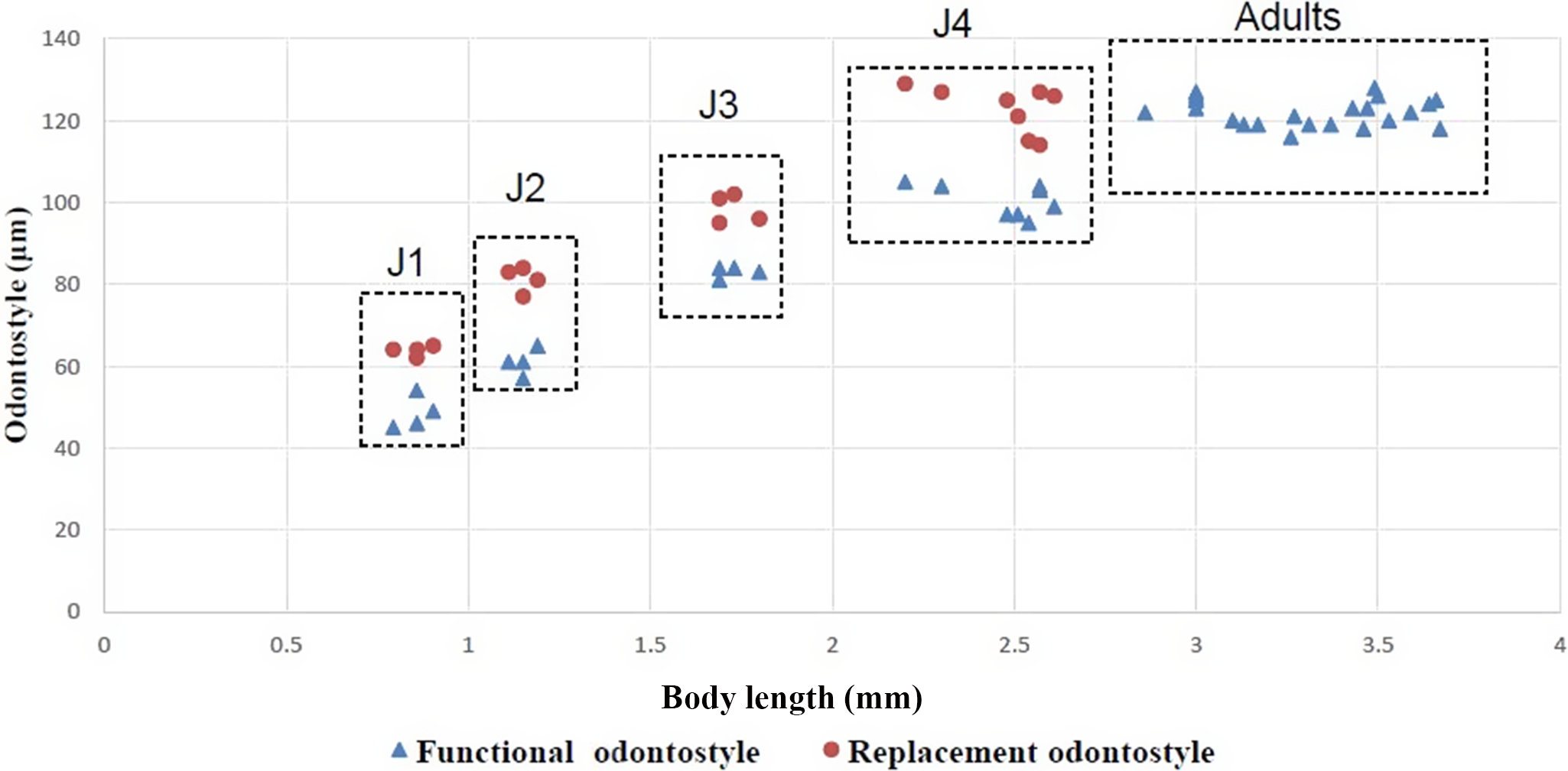
Correlation of functional and replacement odontostyle length to body length in juvenile stages and adults of Xiphinema basiri Siddiqi, Reference Siddiqi1959.

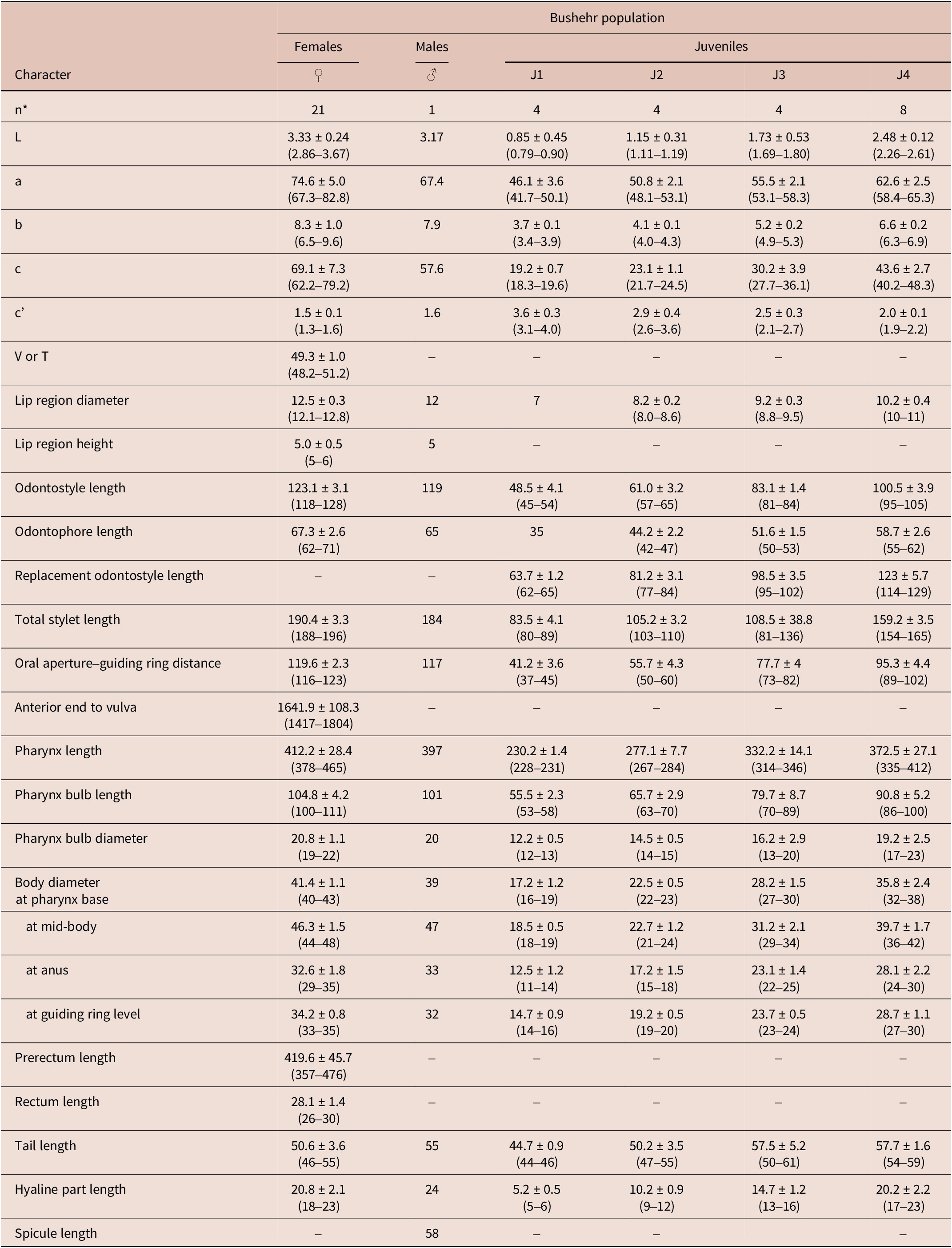
Morphometrics of Xiphinema basiri Siddiqi, Reference Siddiqi1959 from Bushehr province, southern Iran. All measurements are in μm (except L, in mm), and in the form: mean ± standard deviation (range)

*Abbreviations: n = number of specimens on which measurements are based; L = overall body length; a = body length/greatest body diameter; b = body length/distance from anterior end to pharyngo–intestinal junction; c = body length/tail length; c’ = tail length/tail diameter at anus or cloaca; V = distance from body anterior end to vulva expressed as percentage (%) of the body length; T = distance from cloacal aperture to anterior end of testis expressed as percentage (%) of the body length.
Description
Female. Habitus in specimens killed by gentle heat usually open C-shaped, almost straight anteriorly and more curved behind the vulva, occasionally J-shaped; body cylindrical, tapering very gradually towards the anterior extremity. Cuticle apparently smooth, 2–3 μm thick at guiding ring level, 4–5 μm at mid-body, and 5–8 μm at tail hyaline part. Lip region broadly rounded, slightly offset, 2.5−3.2 times as high as width. Amphidial fovea stirrup-shaped, aperture a transverse slit, occupying more than three-fifths of the lip region width. Lateral chord approximately one-fourth of the body diameter. Odontostyle 1.6–2.0 times as long as odontophore, odontophore with well-developed flanges. Guiding ring double and guiding sheath variable in length. Nerve ring at 20–35 μm posterior to base of spear extension. Pharynx typical of the genus, dorsal gland nucleus located at 7.3–12.3% of beginning of the pharyngeal bulb, the two subventral glands almost at the middle. Glandularium 89–94 μm long. Cardia conoid, 6.0–7.0 μm long, well evident within the intestinal tissue. Reproductive system amphidelphic, with both branches equally and fully developed, each branch composed of a 40–65 μm long ovary, a 70–90 μm long reflexed oviduct with pars dilatata oviductus, 20–40 μm long, a sphincter, and an elongated uterus, 150–200 μm long with Z-differentiation near pars dilatata uteri, consisting of three to four globular bodies, neither spiniform structures/crystalloid bodies nor sperm cells observed along uterus. Ovejector well-developed. Vagina perpendicular to body axis, extending for 51–59% of corresponding body diameter, and vulva as a transverse slit. Prerectum variable in length, about 8.0–10.5 times corresponding body diameter, rectum 0.7–1.0 anal body width long. Tail short, conoid, dorsally convex and ventrally concave with a sub digitate peg-like terminus, and bearing three or four caudal pores on each side.
Male. Extremely rare, only one male specimen was found. Genital system diorchic, with testes containing multiple rows of different stages of spermatogonia. Spicules slightly curved ventrally, lateral guiding pieces more or less straight or with curved proximal end, 16 μm long. Copulatory supplements composed one pair of cloacal and three ventromedian supplements. Tail similar to that of female, with terminal peg ventrally located in relation to the body axis and a vague blind terminal canal, three caudal pores are visible on each side of the tail.
Juveniles. All four juvenile stages were identified using morphological characters and relative lengths of body, functional and replacement odontostyle (Table 2, Figures 5, 6). Tail of J1 elongate-conoid, bearing three caudal pores on each side, tail of J2 similar to that of adults, expect that the ventral region of the tail is flat. In other juvenile stages, the tail is similar to that of adults, except that the width of the tail is less (Figure 5, J–M).
Examined material
Twenty-one females, one male and twenty juveniles from four developmental stages, collected from the rhizosphere of Phragmites sp., in Tangestan county, Bushehr province, southern Iran, GPS coordinates: 28° 54’ 14” N, 51° 21’ 52” E; altitude: 369 m. above sea level.
Remarks
Xiphinema basiri was described by Siddiqi (Reference Siddiqi1959) from India. This species was also reported from Cuba, Martinique, Mexico, Nigeria, Pakistan, Puerto Rico, Sri Lanka, Sudan, USA, and Zimbabwe (Almarales Antúnez and Gandarilla Basterrechea Reference Almarales Antúnez and Gandarilla Basterrechea2011; Cohn and Sher Reference Cohn and Sher1972; Gill and Firoza Reference Gill and Firoza2014; Knobloch and Laughlin Reference Knobloch and Laughlin1973; Robbins and Brown Reference Robbins and Brown1991; Roy Reference Roy1973; Scheck Reference Scheck2022; Swart and Quénéhervé Reference Swart and Quénéhervé1998). Based on morphology and morphometry, the presently recovered population belongs to the Xiphinema non-americanum group 5 in Loof and Luc (Reference Loof and Luc1990), which agrees closely with the original description and other descriptions of the species (Cid del Prado-Vera et al. Reference Cid del Prado-Vera, Ferris and Subbotin2024; Cohn and Sher Reference Cohn and Sher1972; Knobloch and Laughlin Reference Knobloch and Laughlin1973; Swart and Quénéhervé Reference Swart and Quénéhervé1998). Up to our knowledge, this is the first report of this species from Iran. According to the polytomous key of Loof and Luc (Reference Loof and Luc1990), the Iranian population of X. basiri has the following specific alphanumeric codes (codes in parentheses are exceptions): A4, B2, C4, D5(4), E5(6), F3(4), G2, H2, I3, J3, K2, L1.
Molecular characterisations and phylogenetic analyses of Xiphinema pedrami sp. nov. and Iranian population of Xiphinema basiri
Molecular markers and accession numbers of the newly sequences determined in the present study are given in Table 3. The amplification of the D2-D3/LSU (~671bp), ITS1 (~552bp), SSU (~1638bp), and COI (~389bp) products of X. pedrami sp. nov. and X. basiri yielded single fragments based on gel electrophoresis. BLAST analyses confirmed that sequences of X. pedrami sp. nov. and X. basiri were most identical to those of the Xiphinema non-americanum group species deposited in GenBank.
Species, isolates names, sequenced loci, and accession numbers of the newly obtained sequences in the present study

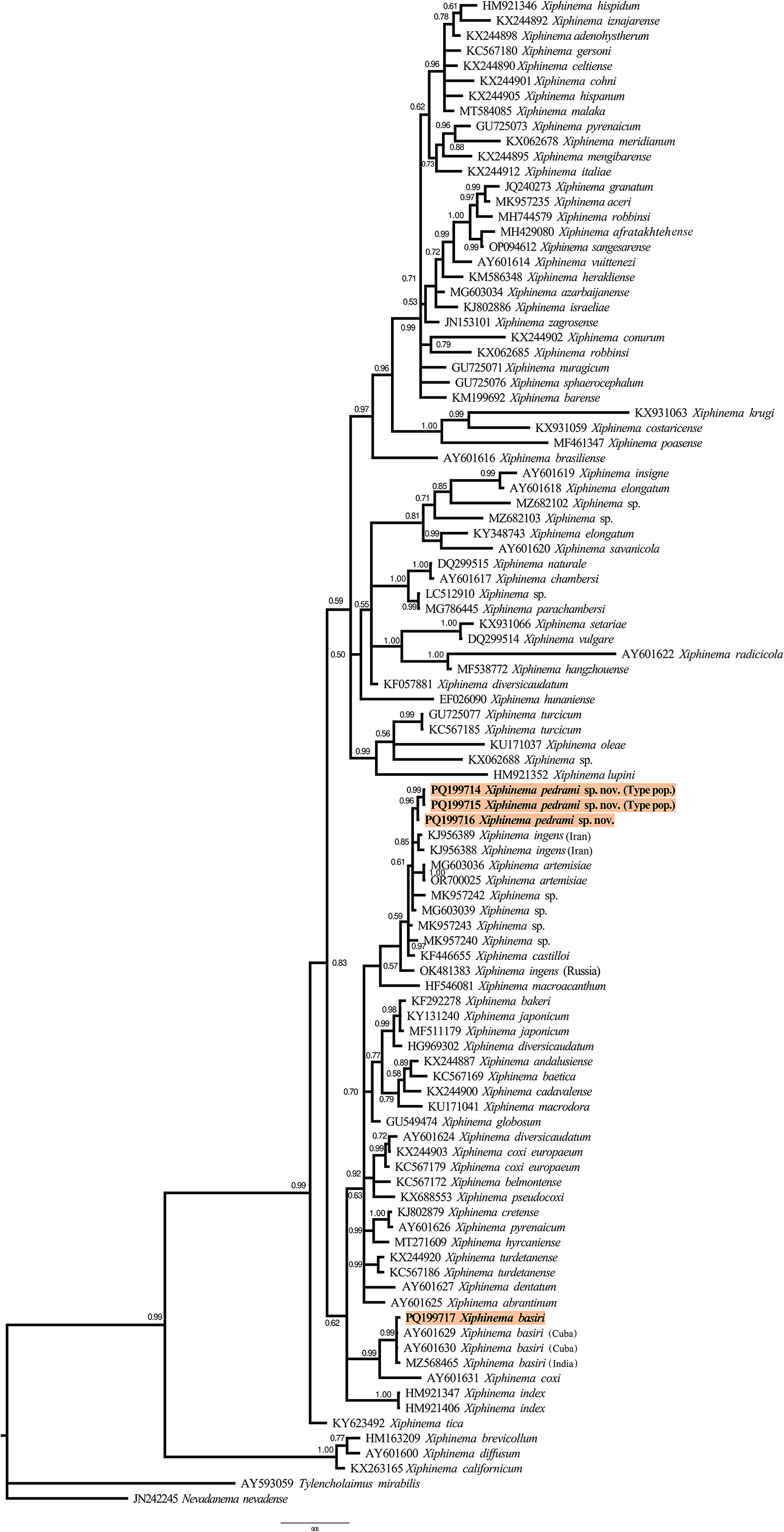
Three new D2-D3/LSU sequences from X. pedrami sp. nov. were obtained in the present study (PQ199714–PQ199716). Intraspecific variation among the D2-D3/LSU sequences of X. pedrami sp. nov. was low; only 2–6 variable positions were found in type and second populations. The D2-D3/LSU sequences of X. pedrami sp. nov. showed 97–98% similarity (11 to 26 different nucleotides and 1 to 2 indels) with X. ingens (KJ956388, KJ956389, OK481383), X. artemisiae (MG603036, OR700025), X. castilloi (KF446655), and several Xiphinema sp. (MG603039, MK957240, MK957242, MK957243). For X. basiri, one new D2-D3/LSU sequence was obtained in the present study (PQ199717). The D2-D3/LSU sequence of X. basiri showed 99% similarity with sequences form Cuba and India (AY601629, AY601630, MZ568465).
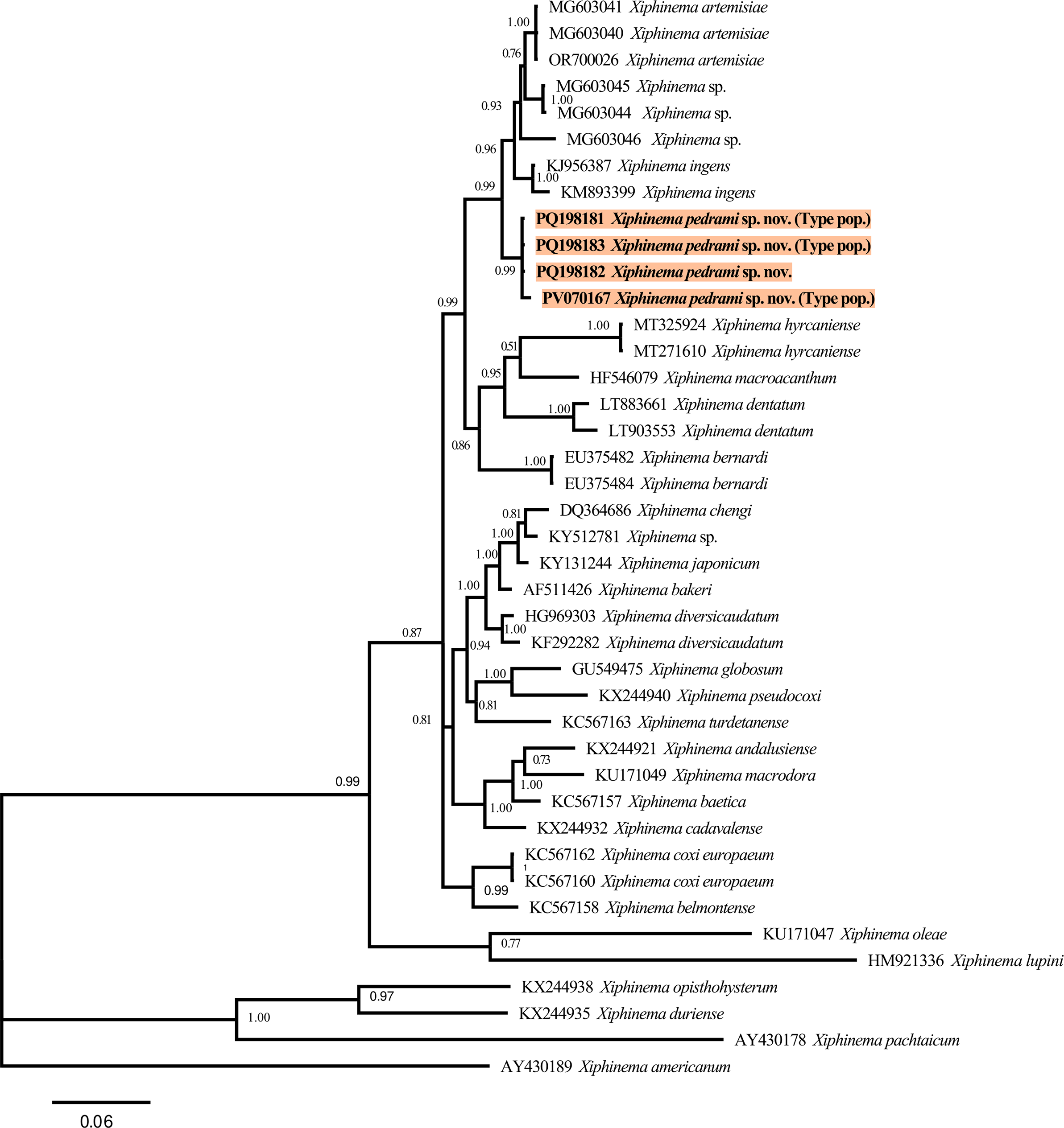
Four new ITS1 sequences from X. pedrami sp. nov. were obtained in the present study (PQ198181–PQ198183, PV070167). ITS1 sequences from both populations of X. pedrami sp. nov. were 98.1–99.8% identical (1–24 different nucleotides), and the closest matches were X. ingens (KJ956387, KM893399), X. artemisiae (OR700026, MG603041), and some Xiphinema sp. (MG603044–MG603046), with 94.4–95.1% similarity, 47 to 51 different nucleotides, and 10 to 12 indels.
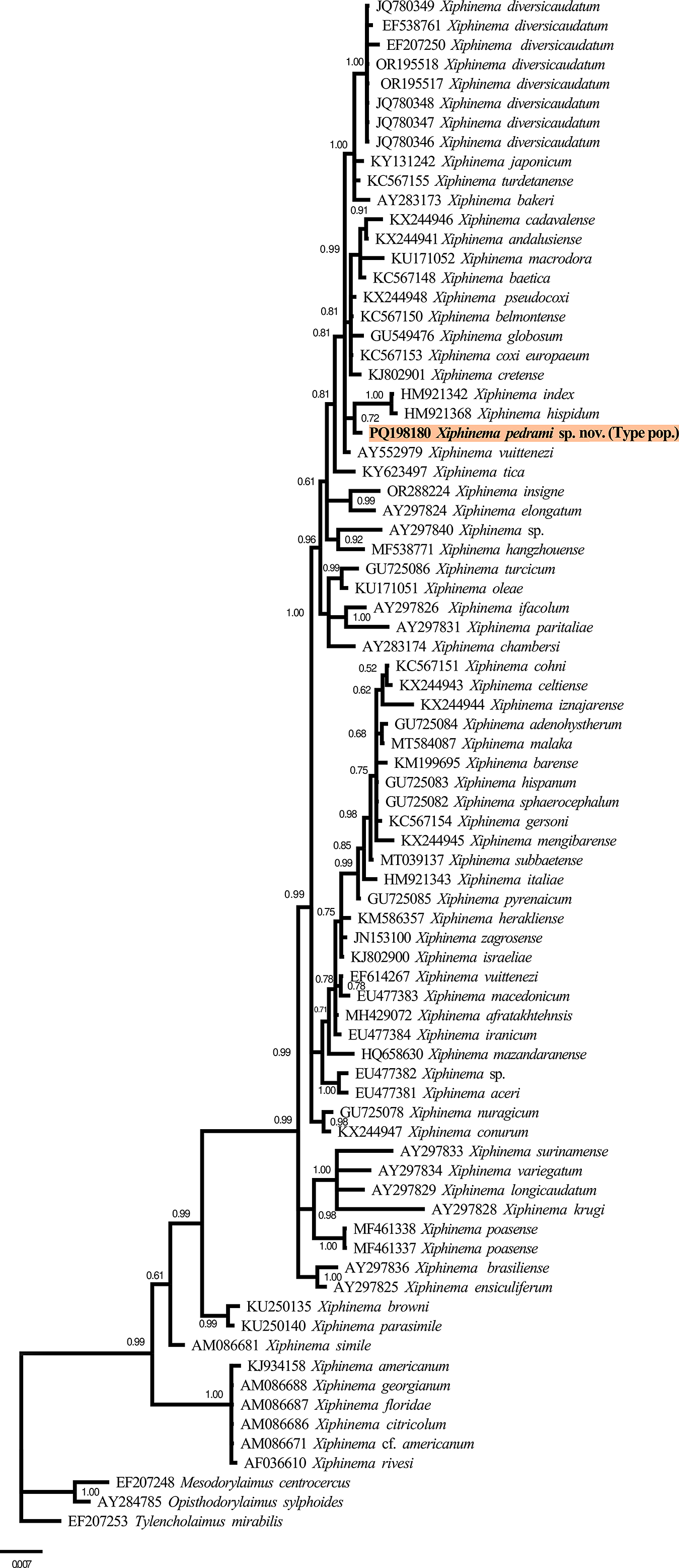
One new SSU sequence from X. pedrami sp. nov. was obtained in the present study (PQ198180). The SSU sequence X. pedrami sp. nov. showed a high nucleotide similarity with the SSU sequences from X. non-americanum group species, being 99% similar to several accessions from this group deposited in GenBank, such as X. andalusiense (KX244941), X. diversicaudatum (JQ780346–JQ780349, OR195517, OR195518), and X. pseudocoxi (KX244948).
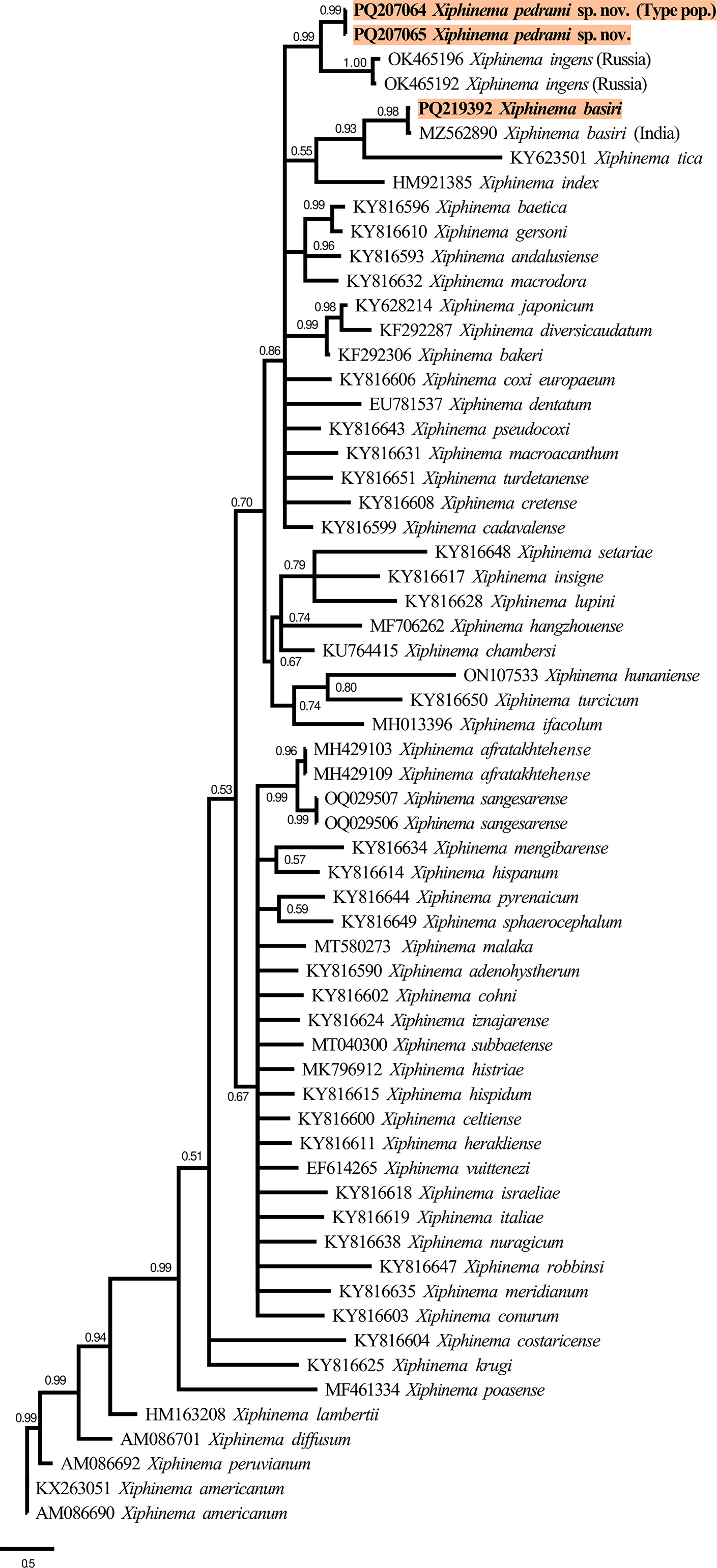
Two new COI sequences from X. pedrami sp. nov. were obtained in the present study (PQ207064, PQ207065). No intraspecific variation for COI sequences of X. pedrami sp. nov. was observed. The COI sequences of X. pedrami sp. nov. showed a relatively low nucleotide similarity with Xiphinema non-americanum group spp. deposited in GenBank, X. ingens (OK465192, OK465196) being the closest species, with 83.6–84.2% similarity, 96% coverage, differing by 68 nucleotides. For X. basiri, one new COI sequence was obtained in the present study (PQ219392). The COI sequence of X. basiri showed 100% similarity with sequence form India (MZ562890).
Phylogenetic relationships of X. pedrami sp. nov. and X. basiri, among Xiphinema non-americanum group species, inferred from Bayesian analyses of D2-D3/LSU, ITS1, SSU, and COI gene sequences are given in Figures 7, 8, 9, and 10, respectively. The 50% majority rule consensus BI tree resulting from D2-D3/LSU sequences showed two major clades, but the relationships between the clades’ inside groups were not fully resolved (Figure 7). In this tree, X. pedrami sp. nov. was placed in a low-supported clade (PP = 0.61) with two other species from the morphospecies group 5 – for example, X. ingens (KJ956388, KJ956389, from Iran), X. artemisiae (MG603036, OR700025), and two unidentified Xiphinema species (MK957242, MG603039). These species are related to X. castilloi (KF446655), Xiphinema sp. (MK957240, MK957243), X. ingens (OK481383, from Russia), and X. macroacanthum (HF546081), which formed another low-supported clade (PP = 0.57). However, using this marker, X. pedrami sp. nov. was well separated from other species of the genus. The Iranian population of X. basiri formed a well-supported clade (PP = 0.99) with other populations of this species from Cuba (AY601629, AY601630) and India (MZ568465).
Phylogenetic relationships of Xiphinema pedrami sp. nov. and X. basiri Siddiqi, Reference Siddiqi1959 within the Xiphinema non-americanum group complex. Bayesian 50% majority rule consensus tree as inferred from D2-D3 expansion segments of the 28S rDNA sequence alignment under the GTR+I+G model. Posterior probabilities more than 0.50 are given for appropriate clades. The newly obtained sequences are in boldface. Scale bar shows the number of expected changes per site.

Phylogenetic relationships of Xiphinema pedrami sp. nov. within the Xiphinema non-americanum group complex. Bayesian 50% majority rule consensus tree as inferred from ITS1 rDNA sequence alignment under the GTR+G model. Posterior probabilities more than 0.50 are given for appropriate clades. The newly obtained sequences are in boldface. Scale bar shows the number of expected changes per site.

Phylogenetic relationships of Xiphinema pedrami sp. nov. within the Xiphinema non-americanum group complex. Bayesian 50% majority rule consensus tree as inferred from 18S rDNA sequence alignment under the GTR+I+G model. Posterior probabilities more than 0.50 are given for appropriate clades. The newly obtained sequences are in boldface. Scale bar shows the number of expected changes per site.

Phylogenetic relationships of Xiphinema pedrami sp. nov. and X. basiri Siddiqi, Reference Siddiqi1959 within the Xiphinema non-americanum group complex. Bayesian 50% majority rule consensus tree as inferred from COI mtDNA sequence alignment under the GTR+I+G model. Posterior probabilities more than 0.50 are given for appropriate clades. The newly obtained sequences are in boldface. Scale bar shows the number of expected changes per site.

For the ITS1 region sequences, the 50% majority rule consensus BI tree is shown in Figure 8. In this tree, X. pedrami sp. nov. formed a well-supported clade (PP = 0.99) with X. artemisiae (MG603040, MG603041, OR700026), X. ingens (KJ956387, KM893399), and three unidentified Xiphinema species (MG603044–MG603046), undoubtedly separated from all other Xiphinema non-americanum group species.
For the SSU sequences, the 50% majority rule consensus BI tree is shown in Figure 9. In this tree, several not-well-resolved clades were achieved, and X. pedrami sp. nov. formed a low-supported clade (PP = 0.72) with X. index (HM921342) and X. hispidum (HM921368).
Finally, the 50% majority rule consensus BI tree based on COI sequences showed several clades that were not well resolved (Figure 10). In this tree, Xiphinema pedrami sp. nov. was phylogenetically related to X. ingens (OK465192, OK465196) in a well-supported clade (PP = 0.99) and clearly separated from other species. The Iranian population of X. basiri formed a well-supported subclade (PP = 0.98) with another population of this species from India (MZ562890), confirming its identity.
Discussion
Several cryptic species within the Xiphinema non-americanum group have been identified, where overlapping morphological traits make species differentiation particularly challenging. For instance, species in the Xiphinema hispanum complex, as well as X. nuragicum Lamberti et al., Reference Lamberti, Castillo, Gomez-Barcina and Agostinelli1992 and X. pyrenaicum Dalmasso, Reference Dalmasso1969, are genetically distinct but morphologically similar (Archidona-Yuste et al. Reference Archidona-Yuste, Cai, Cantalapiedra-Navarrete, Carreira, Rey, Viñegla, Liébanas, Palomares-Rius and Castillo2020a; Gutiérrez-Gutiérrez et al. Reference Gutiérrez-Gutiérrez, Bravo, Santos, Vieira and Mota2016; Tzortzakakis et al. Reference Tzortzakakis, Archidona-Yuste, Cantalapiedra-Navarrete, Nasiou, Lazanaki, Kabourakis, Palomares-Rius and Castillo2014). These cryptic species may be misidentified if only morphological features are used, highlighting the critical role of molecular tools in accurate species delimitation.
Genomic rDNA markers have been extensively applied in the identification and phylogenetic studies of longidorid nematodes. Among these, the D2-D3 expansion segments of the 28S rDNA and the ITS1 region have proven to be reliable markers for species identification. In contrast, the 18S rDNA, due to its slow evolutionary rate, often lacks the resolution required for species-level distinctions (Gutiérrez-Gutiérrez et al. Reference Gutiérrez-Gutiérrez, Palomares-Rius, Cantalapiedra-Navarrete, Landa, Esmenjaud and Castillo2010; Oliveira et al. Reference Oliveira, Hübschen, Brown, Ferraz, Wright and Neilson2004; Palomares-Rius et al. Reference Palomares-Rius, Cantalapiedra-Navarrete, Archidona-Yuste, Subbotin and Castillo2017). Several studies have confirmed the effectiveness of D2-D3 and ITS1 sequences in delineating cryptic species (Gutiérrez-Gutiérrez et al. Reference Gutiérrez-Gutiérrez, Cantalapiedra-Navarrete, Remesal, Palomares-Rius, Navas-Cortés and Castillo2013; Pedram et al. Reference Pedram, Pourjam, Robbins, Ye, Atighi and Decraemer2012; Roshan-Bakhsh et al. Reference Roshan-Bakhsh, Pourjam, Pedram, Robbins and Decraemer2014; Tzortzakakis et al. Reference Tzortzakakis, Archidona-Yuste, Cantalapiedra-Navarrete, Nasiou, Lazanaki, Kabourakis, Palomares-Rius and Castillo2014). Sequencing these regions has proven particularly useful in distinguishing cryptic species within the Xiphinema non-americanum group (Santos et al. 2020). Understanding genetic diversity within this group is essential for evaluating their ecological impact, pathogenic potential, and capacity for virus transmission in crops.
Although these cryptic species may share similar morphological traits, their genetic differences can lead to variations in their host preferences and ecological behavior, which is critical for managing the species in agricultural systems.
In the current study, two populations from Bushehr Province were discovered, exhibiting very similar morphology (e.g., lip region, tail shape, body length and width, and other morphometrics). Despite minor morphometric differences, their identical D2-D3/LSU sequences and closely matching ITS1 sequences support their conspecificity. Based on these data, the two populations are described here as a new species, Xiphinema pedrami sp. nov.
The cryptic nature of species within the Xiphinema non-americanum group raises the question of whether Xiphinema pedrami sp. nov. may represent a cryptic lineage of a previously described species. Among the known taxa, X. hyrcaniense exhibits certain morphological similarities, particularly in tail shape and lip region morphology. However, the molecular data (D2-D3/LSU and ITS1 sequences) clearly distinguish X. pedrami sp. nov. from X. hyrcaniense, supporting its status as a distinct species rather than a cryptic variant. Therefore, while both species may appear morphologically close, molecular evidence rules out conspecificity.
In the present study, phylogenetic analyses based on D2-D3/LSU, ITS1, SSU, and COI markers were conducted. The topologies obtained were consistent with those reported in earlier studies by Mirzaie Fouladvand et al. (Reference Mirzaie Fouladvand, Pourjam, Castillo and Pedram2019), Jahanshahi Afshar et al. (Reference Jahanshahi Afshar, Shahryari, Gharibzadeh, Pourjam and Pedram2019), and Poureskandarian et al. (Reference Poureskandarian, Pourjam and Pedram2023). In D2-D3/LSU, ITS1, and COI phylogenetic trees, X. ingens and X. artemisiae appear to be the closest relatives of the newly described species.
As seen in other recent phylogenetic studies on this genus (e.g., Ghaemi et al. Reference Ghaemi, Pourjam, Pedram, Robbins, Ye and Decraemer2012; Peraza-Padilla et al. Reference Peraza-Padilla, Cantalapiedra-Navarrete, Zamora-Araya, Palomares-Rius, Castillo and Archidona-Yuste2018; Roshan-Bakhsh et al. Reference Roshan-Bakhsh, Pourjam, Pedram, Robbins and Decraemer2014), the observed clustering patterns do not conform to the intrageneric classification framework proposed by Loof and Luc (Reference Loof and Luc1990) for the Xiphinema non-americanum group, suggesting the need for taxonomic revision based on molecular evidence.
The present paper shows the diversity and density of longidorids in southern Iran could still be higher, as recently documented by Monemi et al. (Reference Monemi, Behdani, Mahdikhani-Moghadam, Amirzadi, Atighi, Ye, Jahanshahi Afshar and Pedram2024), and may need to adopt controlling measures.
Acknowledgements
The authors appreciate Dr. Mohsen Asgari and Dr. Majid Pedram for their help with molecular analyses and reading the manuscript, and anonymous reviewers and the editors of this paper for their efforts in reviewing the manuscript and helping improve this study.
Financial support
There are not projects associated with this research.
Competing interests
The authors declare no conflict of interest.
Ethical standard
The authors assert that all procedures contributing to this work comply with the ethical standards of the relevant national and institutional guides on the care and use of laboratory animals.