Introduction
Leishmaniasis is a group of parasitic infections caused by flagellate protozoans of genus Leishmania that are transmitted through the bite of infected female phlebotomine sand fly (Elmahallawy et al., Reference Elmahallawy, Martínez, Rodriguez-Granger, Hoyos-Mallecot, Agil, Mari and Fernández2014). More than 20 species of Leishmania are capable of causing the disease (Georgiadou et al., Reference Georgiadou, Makaritsis and Dalekos2015; WHO, 2020) with clinical manifestations including Cutaneous Leishmaniasis (CL), Mucocutaneous Leishmaniasis, Diffuse Cutaneous Leishmaniasis and Visceral Leishmaniasis (also known as kala-azar) (Desjeux, Reference Desjeux2004; WHO, 2010; Ghatee et al., Reference Ghatee, Taylor and Karamian2020). Leishmaniasis has been reported in 98 countries affecting about 350 million people worldwide, making it one of the seven most common as well important neglected tropical diseases (Torres-Guerrero et al., Reference Torres-Guerrero, Quintanilla-Cedillo, Ruiz-Esmenjaud and Arenas2017; de Souza et al., Reference de Souza, Marins, Mathias, Monteiro, Yukuyama, Scarim, Löbenberg and Bou-Chacra2018). CL being the most common and widely distributed variety claims between 0.7 and 1.2 million cases reported worldwide annually (Alvar et al., Reference Alvar, Vélez, Bern, Herrero, Desjeux, Cano, Jannin and den Boer2012). About 95% of the incidents are reported from three epidemiological regions i.e. South America, the Mediterranean Basin and spread throughout the Middle East to Central Asia (Torres-Guerrero et al., Reference Torres-Guerrero, Quintanilla-Cedillo, Ruiz-Esmenjaud and Arenas2017). More than 85% of new CL cases appeared in Afghanistan, Algeria, Bolivia, Brazil, Colombia, Iran, Iraq, Pakistan, the Syrian Arab Republic and Tunisia during 2018 (Zijlstra, Reference Zijlstra2016; WHO, 2020).
CL is highly endemic in Pakistan and spread extensively due to massive out-migration from endemic to non-endemic areas and vice versa (Kassi et al., Reference Kassi, Kassi, Afghan, Rehman and Kasi2008). CL affects people of low social-economic status and is associated with undernutrition, population displacement, lack of shelter and a weak immune system (WHO, 2010). The burden of disease in Pakistan has been reported to be surpassed by 400 000 cases reported in 2016 that makes about 10% of CL cases globally (Blum et al., Reference Blum, Desjeux, Schwartz, Beck and Hatz2004) with both anthroponotic (ACL) and zoonotic (ZCL) forms of CL (Alvar et al., Reference Alvar, Vélez, Bern, Herrero, Desjeux, Cano, Jannin and den Boer2012). ACL occasionally occurring due to Leishmania tropica, is widespread and most grave public health problem in Pakistan (Hussain et al., Reference Hussain, Munir, Khan, Khan, Ayaz, Jamal, Ahmed, Aziz, Watany and Kasbari2018). The urban areas of Punjab and Sindh provinces are of high CL endemicity (Iftikhar et al., Reference Iftikhar, Bari and Ejaz2003; Kakarsulemankhel, Reference Kakarsulemankhel2004; Katakura, Reference Katakura2009), the Afghan refugee camps in Northwest Frontier Province, now called Khyber Pakhtunkhwa (Simon Brooker et al., Reference Simon Brooker, Adil, Agha, Reithinger, Rowland, Ali and Kolaczinski2004) and the surrounding tribal belt of Federally Administrated Tribal Areas (FATA) (Khan et al., Reference Khan, ul Bari, Hashim, Khan, Muneer, Shah, Wahid, Yardley, O'Neil and Sutherland2016; Qureshi et al., Reference Qureshi, Ali, Rashid and Ali2016; Irum et al., Reference Irum, Aftab, Khan, Naz, Simsek, Habib, Afzal, Nadeem, Qasim and Ahmed2021). CL is also widely distributed in sub-urban localities of Baluchistan province (Bhutto et al., Reference Bhutto, Soomro, Baloch, Matsumoto, Uezato, Hashiguchi and Katakura2009; Ejaz et al., Reference Ejaz, Raza, Din and Bux2008; Hussain et al., Reference Hussain, Munir, Khan, Khan, Ayaz, Jamal, Ahmed, Aziz, Watany and Kasbari2018), Gilgit Baltistan (Ayub et al., Reference Ayub, Gramiccia, Khalid, Mujtaba and Bhutta2003), Azad Jammu and Kashmir (Mughal, Reference Mughal2014; Shaheen et al., Reference Shaheen, Qureshi, Qureshi, Fatima, Afzal and Alhewairini2020). The ZCL type, caused by Leishmania major, has its reservoir populations in feral animals predominantly gerbils such as Rhombomys opimus in rural and sub-urban localities of Punjab, Baluchistan and Sindh provinces (Bhutto et al., Reference Bhutto, Soomro, Nonaka and Hashiguchi2003; Afghan et al., Reference Afghan, Kassi, Kasi, Ayub, Kakar and Marri2011).
Leishmaniasis has diverse clinical manifestations and may appear similar to a large variety of other conditions (Afghan et al., Reference Afghan, Kassi, Kasi, Ayub, Kakar and Marri2011; Antinori et al., Reference Antinori, Schifanella and Corbellino2012). CL is identified by the appearance of one or more well-defined ulcerous lesions (Bacellar et al., Reference Bacellar, Lessa, Schriefer, Machado, de Jesus, Dutra, Gollob and Carvalho2002; Carvalho et al., Reference Carvalho, Filho, Falcao, Rocha Lima and Gontijo2008), usually found on uncovered areas of the body i.e. forearms, legs and face where sand-fly bites occur most often (Salman et al., Reference Salman, Rubeiz and Kibbi1999; Chaudhary et al., Reference Chaudhary, Bilimoria and Katare2008). After 4–12 weeks of a bite, a small erythematous papule appears at the bite site which gives off a seropurulent discharge, which later on dries up and then a ‘volcanic’ nodulo-ulcer appear which is classical for CL (Iddawela et al., Reference Iddawela, Vithana, Atapattu and Wijekoon2018). These lesions can develop into other morphological forms such as lupoid, keloidal, psoriasiform, erysipeloid, verrucous, zosteriform, tumorous, eczematoid and acneform (Bari and Raza, Reference Bari and Raza2010; Shamsuddin et al., Reference Shamsuddin, Mengal, Gazozai, Mandokhail, Kasi, Muhammad and Ahmad2017; Iddawela et al., Reference Iddawela, Vithana, Atapattu and Wijekoon2018). Routinely used diagnostic methods are direct clinical observations and micro-scopical examination of lesion aspiration samples. Alternative diagnostic options include histopathology and polymerase chain reaction (PCR) but due to the low frequency of parasites, PCR may give false-negative results. The practice of using PCR as a diagnostic tool is also uncommon worldwide and not easily applied to clinical health settings (Weirather et al., Reference Weirather, Jeronimo, Gautam, Sundar, Kang, Kurtz, Haque, Schriefer, Talhari, Carvalho and Donelson2011; Tsukayama et al., Reference Tsukayama, Núñez, De Los Santos, Soberón, Lucas, Matlashewski, Llanos-Cuentas, Ore, Baldeviano, Edgel and Lescano2013; de Paiva-Cavalcanti et al., Reference de Paiva-Cavalcanti, de Morais, Pessoa-e-Silva, Trajano-Silva, da Cunha Gonçalves-de-Albuquerque, Tavares, Brelaz-de-Castro, e Silva and Pereira2015). The sensitivity of enzyme-linked immunosorbent assay is 50% in the case of co-infection due to less circulating antibodies in blood (Sinha et al., Reference Sinha, Pandey and Bhattacharya2005; de Souza et al., Reference de Souza, Veras, Welby-Borges, Silva, Leite, Ferraro, Meyer-Fernandes, Barral, Costa and de Freitas2011; Abeijon et al., Reference Abeijon, Kashino, Silva, Costa, Fujiwara, Costa and Campos-Neto2012). The treatment relies on chemotherapy and the first line of treatment for all types of leishmaniasis are the medications containing pentavalent antimonials such as sodium stibogluconate and meglumine antimoniate. But unfortunately, the treatment failure has been reported for this class of drug because the parasite has shown resistance to this class of drug and its authenticity and clinical value has been challenged (Croft et al., Reference Croft, Sundar and Fairlamb2006; Llanos-Cuentas et al., Reference Llanos-Cuentas, Tulliano, Araujo-Castillo, Miranda-Verastegui, Santamaria-Castrellon, Ramirez, Lazo, De Doncker, Boelaert, Robays and Dujardin2008).
CL is one of the neglected diseases in Pakistan and exact figures on the magnitude of the disease are lacking both at national and regional levels and only a few health centres are available for diagnosis of CL. The limitation/absence in the diagnosis and treatment increase the need for an epidemiological survey of the disease in the country (Afghan et al., Reference Afghan, Kassi, Kasi, Ayub, Kakar and Marri2011). Despite the disease endemicity, the information regarding the epidemiology of CL in Pakistan is incomplete (Khan et al., Reference Khan, ul Bari, Hashim, Khan, Muneer, Shah, Wahid, Yardley, O'Neil and Sutherland2016). Baluchistan is one of the provinces located in the southwest of Pakistan and its geographical distribution has made it a camping ground for sand-fly due to disturbances of the habitats and deforestation (Firdous et al., Reference Firdous, Yasinzai and Ranja2009). Because of the major teaching and tertiary care hospitals located in Quetta, most of CL patients have been reported in Quetta (Kassi and Kasi, Reference Kassi and Kasi2005). This may be due to mass migration from adjoining areas of Afghanistan and the absence of field epidemiological surveys (Shakila et al., Reference Shakila, Bilqees, Salim and Moinuddin2006).
The present study was designed with an aim to identify epidemiology and to assess clinical aspects of CL in Baluchistan Province of Pakistan.
Materials and methods
Study area
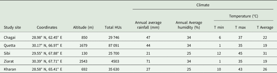
The study was carried out in different districts of Baluchistan covering the surface area of 347 190 km2 and constituting 44% of Pakistan's total landmass located between 30.12N and 67.01E. It has borders to Afghanistan from the north and north-west and to Iran from the south-west and has a population of around 10 million inhabitants predominantly (76%) rural. Physically, the topography is diverse and divided into four distinct zones: upper highlands, lower highlands, plains and deserts altitude ranging from 600 m from the valley floor to 3700 m above the mean sea levels. The climate varied from semi-arid summers to semi-arid winter with temperature as high as 50°C and as low as 1°C where the annual average temperature is about 19°C and average annual rainfall for Quetta is 44 millimetres (Kakarsulemankhel, Reference Kakarsulemankhel2004) (Table 1).
Description of the study sites

masl, meters above sea level; Total HUs, Total housing units; T min, minimum temperature; T max, maximum temperature.
Epidemiological investigation
Due to the lack of ground-level research and treatment limitations, the epidemiological status of CL in the study area was not known. Based on university hospital records between August 2018 and December 2019, 4072 cases of CL were reported from the Baluchistan.
Ethics statement
The study was approved by the IRB & Ethics Committee of National University of Medical Sciences (NUMS), Rawalpindi, Pakistan under reference number 06/R&D/NUMS.
Sample collection and microscopy
A total of 4072 clinically suspected cases of CL visited the Department of Microbiology, Bolan Medical College, Quetta from different areas of Baluchistan. The selected patients were photographed and specific medical explanations for these lesions were obtained from the dermatologist. The samples were collected from the patients after both written and verbal informed consent was obtained. The lesion sample from CL patients was obtained using needle aspiration method. The overlying dry scab was rubbed off with an alcohol gauze pad and approximately 0.1–0.2 mL sterile saline solution injected into the lesion and aspiration fluid was obtained. The slides of smears from each patient were fixed with absolute methyl alcohol and stained with Giemsa stain for parasitological investigation. The slides were examined under the light microscope at 100× magnification for the presence of Leishmania spp. amastigotes (Kassi et al., Reference Kassi, Tareen, Qazi and Kasi2004).
Statistical analysis
The CL data were documented into Microsoft Excel and analysed using SPSS (SPSS Inc., Chicago, Illinois, USA) version 26.0. Prevalence of CL in different age groups and gender was calculated. The Chi-square test (χ 2) and frequencies of demographic characteristic were used for categorical data. Monthly distribution of CL cases for the years of 2018 and 2019 was calculated in form of percentage. Moreover, the prevalence of CL by age groups with 95% confidence limits were analysed. The statistical significance was set for all statistical tests at P value <0.05 (two-sided).
Results
In the present study, a total of 4072 individuals comprising both males and females were studied from the period of August 2018 to December 2019 and analysed with respect to age, gender and body parts affected by CL.
Clinical features of CL patients
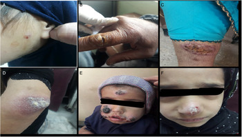
Of the 4072 CL cases, 50.6% (2062/4072) were positive by direct microscopy. The effect of CL in a different age group is depicted in Fig. 1a–f. The lesions were more prevalent among young individuals as compared to older adults. Exposed parts of the body (hand, cheeks and nose) were the main affected areas. Different clinical forms were observed based on clinical morphology of the CL patients' lesion. These lesions diverge from mild to moderate papulonodular form which is <1 cm in diameter to more severe and complex forms. One of the forms was dry type papulonodular lesions with erythematous smooth and superficial papule on the face and size varied from 0.5 to 1 cm (Fig. 1a). Other form presented the clinically infiltrated ulceration which was ulcerated lesion and present the dry type (Fig. 1b). Among CL patients, lupus erythematosus was most common with unusual presentations ranging from scaly and crusted plaque to enlarged erythematous and infiltrated plaque on the elbow wrapped with white and dry scales mimicking psoriasis (Fig. 1c, d and e). A psoriasiform lesion was also observed on the nose of a CL patient which was characterized by an infiltrated plaque (Fig. 1f).
(a–f) Atypical forms of Cutaneous Leishmaniasis (CL) lesions. (a) Papulonodular lesions, dry type; (b) Ulcerated lesion, dry type; (c) Discoid lupus erythematosus, dry type; (d) Psoriasiform lesion, dry type; (e) Mycetomatous, dry type; (f) Erysipeloid, dry type.

Age and gender-wise distribution of CL
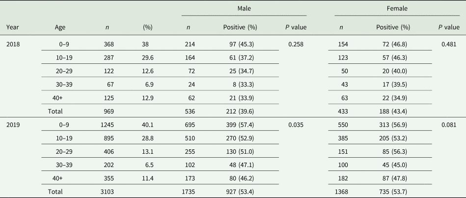
In the present study, the total number of people infected with CL from August to December 2018 was 969. Of the total, 55.3% (536/969) of them were males while 44.7% (433/969) were females. The highest percentage (38%, 368/969) of CL patients was observed aged between 0 and9 years followed by age group 10–19 years which included 29.6% (287/969) of the cases. However, the lowest proportion (6.9%, 67/969) was observed at 30–39 years old (Table 2).
Distribution and prevalence of CL in different age groups and gender for the years 2018 and 2019
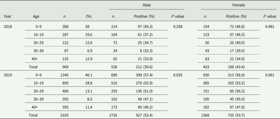
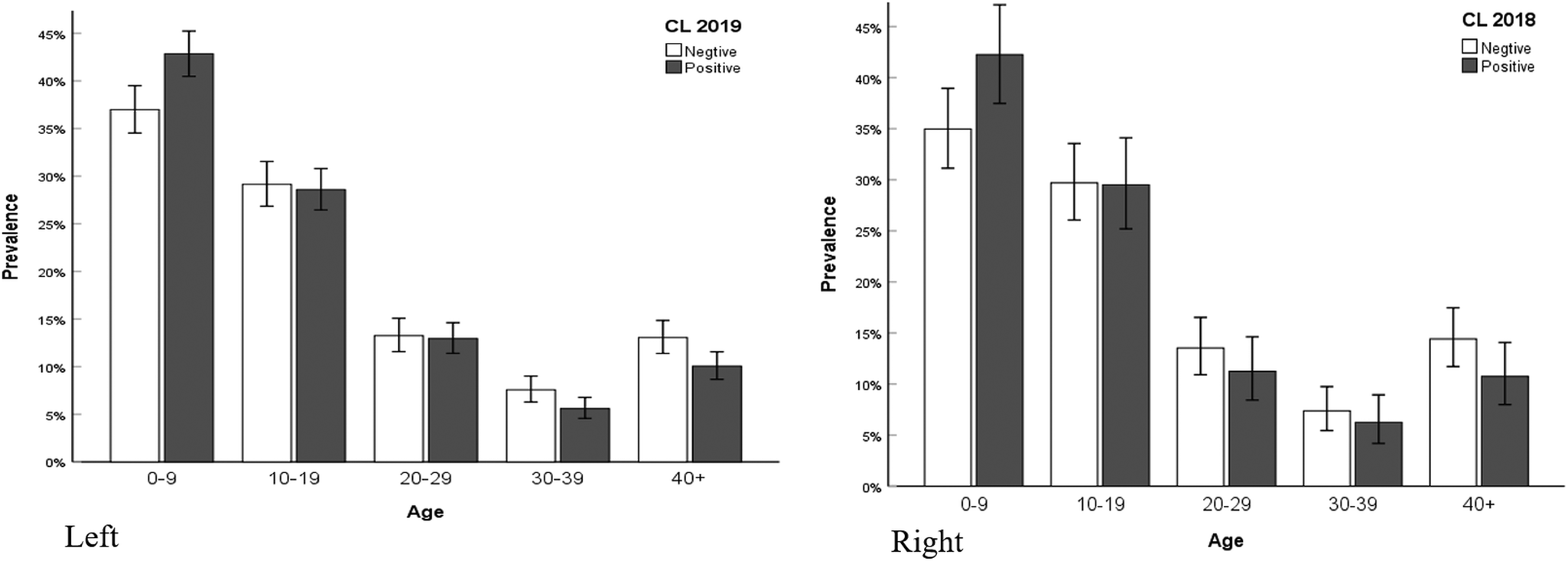
The total number of people infected with CL during the year 2019 was 3103 with the overall CL infection rate during the year 2019 was higher in males (55.9%, 1735/3103) than females (44.1%, 1368/3103). The highest percentage (40.1%, 1245/3103) was observed at 0–9 years old. Only 28.8% (895/3103) of CL patients were observed in 10–19 years old participants followed by 13.1% (406/3103) in 20–29 years old, 6.5% (202/3103) in 30–39 and 11.4% (355/3103) in 40+ years old (Table 2). Based on different age groups, the frequency of males to females CL patients was different. Males (22.4%), (16.44%) and (8.22%) were affected more than females (17.72%), (12.41%) and (4.87%) at the age of 0–9, 10–19 and 20–29 years, respectively. On the other hand, the disease was higher in females (3.22%) and (5.87%) than males (0.29%) and (0.58%) at the age of 30–39 and 40+ years as represented in Fig. 2.
Prevalence of CL according to gender and age Left: Prevalence of CL by age groups with 95% confidence limits for the year 2019. Right: Prevalence of CL by age groups with 95% confidence limits for the year 2018.

Year wise prevalence of CL in different age groups and gender
The findings of the present study revealed that of the total population tested from the period of August–December 2018, 55.3% (536/969) were males and 44.7% (433/969) were females. In total, 41.3% (400/969) of participants were tested positive for lesion aspiration test. Out of which 39.6% (212/536) of males and 43.4% (188/433) of females were positive for lesion aspiration test. The highest percentage (45.3% and 46.8%) in positive lesion aspiration test was found among the participants aged between 0 and 9 years in both males and females, respectively (Table 2). It was observed that the lesion aspiration test was not significantly different by age or gender of participants studied.
In the year 2019, 55.3% (1735/3103) of participants were males and 44.7% (1368/3103) were females. A total of 53% of participants were tested positive for lesion aspiration test and among these 53.4% (927/1735) were males and 53.7% (735/1368) were females. The highest frequency (57%) of lesion aspiration test was tested positive between age 0 and 9 years regardless of age and gender and lowest frequency (47% and 45%) was observed of the participants aged between 30 and 39 years for both males and females, respectively (Table 2).
Site-specific distribution of lesion
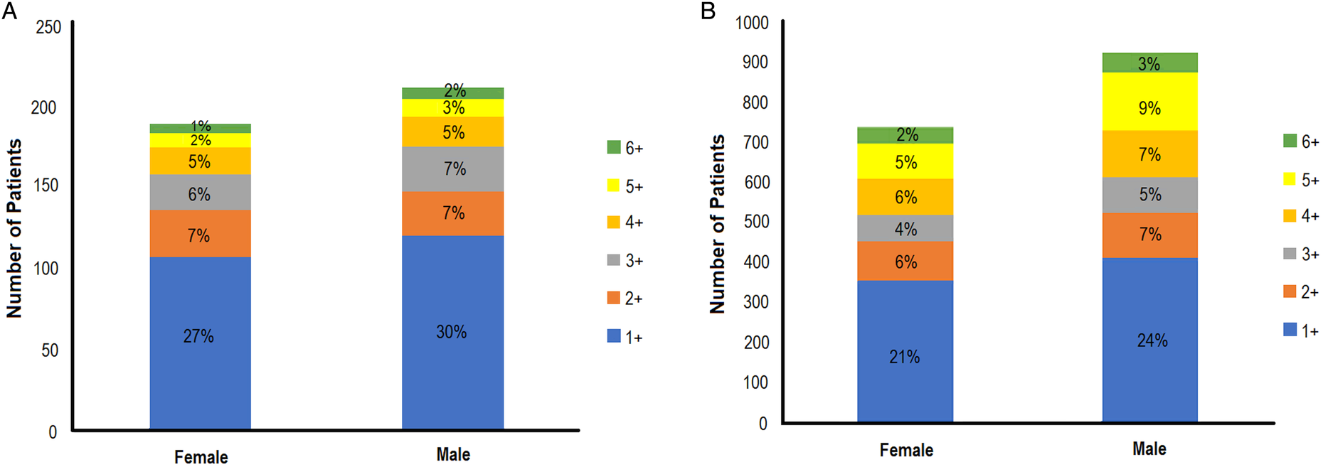
The majority 24.4% (757/3103) of lesions were found on the hands followed by face in which cheeks, ears and nose were most affected. The number of lesions per person ranged from 1 to 6 (Fig. 3). Approximately 50% of the participants had single lesion while 14% of the participants had two and nearly 3% of the participants have six lesions during the study period of 2018 and 2019. Most of the lesions were erythematosus with unusual presentations ranging from scaly and crusted plaque to enlarged erythematous and infiltrated plaque.
Number and percentageof male and female patients with active lesions in 2018 and 2019.

Month-wise distribution of CL
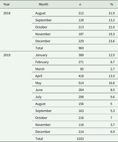
The findings of the study revealed that highest number of CL cases (23.6%, 229/969) was found in December followed by October (22%, 213/969) and August (21.9%, 212/969) during the year 2018. During the year 2019, the highest number of CL cases were observed in May (16.6%, 514/3103) followed by April (13.5%, 418/3103), January (12.5%, 388/3103) and then July (9.6%, 298/3103), February (8.7%, 271/3103) and June (8.5%, 264/3103). The lowest number of CL cases were observed in March (2.7%, 85/3103) and November (3.7%, 116/3103) (Table 3).
Monthly distribution of CL cases for the years of 2018 and 2019

Discussion
CL is one of the prevalent vector-borne diseases in Pakistan which affects people with lower income and is linked to poor housing conditions, migration, inadequate nutrition, vector control failures and immuno-compromised conditions like HIV/AIDS (Alvar et al., Reference Alvar, Yactayo and Bern2006). CL has been reported from all over Pakistan specially from native surrounding areas of FATA and KPK (Khan et al., Reference Khan, ul Bari, Hashim, Khan, Muneer, Shah, Wahid, Yardley, O'Neil and Sutherland2016; Qureshi et al., Reference Qureshi, Ali, Rashid and Ali2016; Khan et al., Reference Khan, Afzal and Ahmed2019), urban-rural areas of Sindh and Punjab (Iftikhar et al., Reference Iftikhar, Bari and Ejaz2003), Azad Jammu and Kashmir (Mughal, Reference Mughal2014; Shaheen et al., Reference Shaheen, Qureshi, Qureshi, Fatima, Afzal and Alhewairini2020) and sub-urban localities of Baluchistan province (Bhutto et al., Reference Bhutto, Soomro, Nonaka and Hashiguchi2003; Ejaz et al., Reference Ejaz, Raza, Din and Bux2008; Rahman and Rehman, Reference Rahman and Rehman2017). It is presumed that uninfected regions with the borders adjacent to endemic areas are at risk and colonization of a large number of populations in areas where sand-fly is endemic is one of the reasons of CL outbreaks (Simon Brooker et al., Reference Simon Brooker, Adil, Agha, Reithinger, Rowland, Ali and Kolaczinski2004).
The current study has been conducted in Baluchistan province which is a high-altitude region and borders to Afghanistan and Iran where CL is endemic (Ghatee et al., Reference Ghatee, Taylor and Karamian2020). The studied area has the highest CL prevalence rate where dominant specie is L. tropica and in addition about 1500 cases of CL were reported in Afghan refugee camps in Pakistan in 2002 (Kakarsulemankhel, Reference Kakarsulemankhel2004). In another study Rahman and Rehman (Reference Rahman and Rehman2017) also reported a significantly high level of CL among Afghans compared to the local Pakistani population (32.5% vs 20.3%). However, the prevalence of CL due to L. major has also been reported from the Southern part of Baluchistan (Bhutto et al., Reference Bhutto, Soomro, Baloch, Matsumoto, Uezato, Hashiguchi and Katakura2009). Thus, the present study was designed for the aim of identifying the epidemiology and assessing the clinical aspects of CL in Baluchistan.
In the current study, CL infection is more prevalent in males (55%) as compared to females with similar findings reported by Shaheen et al. (Reference Shaheen, Qureshi, Qureshi, Fatima, Afzal and Alhewairini2020) in Azad Jammu and Kashmir, by Galgamuwa et al. (Reference Galgamuwa, Sumanasena, Yatawara, Wickramasinghe and Iddawela2017) in Sri Lanka, by Aara et al. (Reference Aara, Khandelwal, Bumb, Mehta, Ghiya, Jakhar, Dodd, Salotra and Satoskar2013) in India by Alavinia et al. (Reference Alavinia, Arzamani, Reihani and Jafari2009) and in Iran. It may be due to the reason that activities of the males are mostly out-door and have maximum chances of being bitten by sand fly while females are confined to indoor activities (Gadisa et al., Reference Gadisa, Tsegaw, Abera, Elnaiem, den Boer, Aseffa and Jorge2015). The current study indicated that 38% of reported CL cases were in children aged 0–9 years followed by adults 10–19 years of age with a prevalence rate of 29% and is in concordance with other studies (Kakarsulemankhel, Reference Kakarsulemankhel2004; Aara et al., Reference Aara, Khandelwal, Bumb, Mehta, Ghiya, Jakhar, Dodd, Salotra and Satoskar2013; Qureshi et al., Reference Qureshi, Ali, Rashid and Ali2016; Yohannes et al., Reference Yohannes, Abebe and Boelee2019; Shaheen et al., Reference Shaheen, Qureshi, Qureshi, Fatima, Afzal and Alhewairini2020). This might be due to a weak immune system and food deprivation of children (Zijlstra, Reference Zijlstra2016). In contrast to this, Aara et al. (Reference Aara, Khandelwal, Bumb, Mehta, Ghiya, Jakhar, Dodd, Salotra and Satoskar2013) reported a higher prevalence of CL cases in 21–30 years age group whereas in our study lower cases of CL were found in 30–39 years old and 40+ as also reported by Nawaz et al. (Reference Nawaz, Khan, Khan and Rauf2010). As lifelong immunity develops following infection with CL, it is understandable why infection is more common in young individuals.
In the present study, most CL cases appeared to have localized symptoms and most lesions were present on the exposed parts of the body particularly hands, nose, ears and cheeks. Similar results were observed in studies carried out in Ethiopia and Turkey where most of the lesions also appeared on the face (Bari, Reference Bari2008; Uzun et al., Reference Uzun, Gürel, Durdu, Akyol, Fettahlıoğlu Karaman, Aksoy, Aytekin, Borlu, İnan Doğan, Doğramacı and Kapıcıoğlu2018; Yohannes et al., Reference Yohannes, Abebe and Boelee2019). For the locations of the lesions, it is of the opinion that it is difficult to cover up the face and is exposed for biting of sand flies at night-time which is a crucial time for the parasite spread (Aara et al., Reference Aara, Khandelwal, Bumb, Mehta, Ghiya, Jakhar, Dodd, Salotra and Satoskar2013). Correspondingly to the findings of Özbilgin et al. (Reference Özbilgin, Töz, Harman, Topal, Uzun, Okudan, Güngör, Erat, Ertabaklar, Ertuğ and Gündüz2019), 77% lesion appeared on uncovered parts of the body. In the present findings, the common unusual presentations were lupus erythematosus followed by the papulonodular lesion, ulcerated lesion and less common psoriasiform lesion. In CL, atypical clinical presentations were progressively observed in Pakistan and it has been speculated that limitations of these atypical cases in certain geographical regions could be due to new strain of Leishmania parasite (Bari, Reference Bari2008). In our study, approximately 50% of the CL cases had a single lesion, 14% had two lesions and remaining had three to six lesions. Our results were similar to the findings of Shaheen et al. (Reference Shaheen, Qureshi, Qureshi, Fatima, Afzal and Alhewairini2020) who also reported the presence of the single lesion in 56% of the CL cases. Other studies conducted by Talari et al. (Reference Talari, Talaei, Shajari, Vakili and Taghaviardakani2006) and Qureshi et al. (Reference Qureshi, Ali, Rashid and Ali2016) also showed the highest number of single lesions in CL cases. CL cases with two or more lesions on the body might be due to exposure to the sandflies for a long time.
Although CL cases were reported throughout the entire year, the highest number of cases were detected in May, followed by April, January and then July, February and June. The lowest number of cases was during March and November. Similar findings have been reported by Shaheen et al. (Reference Shaheen, Qureshi, Qureshi, Fatima, Afzal and Alhewairini2020) that warm months are the peak season of CL in Pakistan. This could be due to the reason that sand flies are more active during the warm weather and consumes more blood for the development of their eggs. The current study has shown rising drift in the number of CL cases in Baluchistan and is a disease of public health importance. It is suggestive that different atypical clinical forms of CL depend on host immune response, number and site of parasites inoculated and host nutritional status (Bari, Reference Bari2008). The study highlights the importance of early detection of erythematic skin lesion and proper management and treatment of CL to prevent a future outbreak of the disease especially in areas where leishmaniasis is endemic. The study also suggests that the use of insecticides, bed nets and new therapies for parasite can also help in the control of leishmaniasis.
Concluding remarks
In conclusion, the current findings suggest that more epidemiological studies are needed in Baluchistan and health education campaigns for population awareness regarding the CL. It is recommended to evaluate the risk factors and to establish control and management strategies to prevent disease at an individual and community level in Baluchistan. It is also recommended to conduct further reservoir studies to understand the vertical transmission of disease among various hosts.
Data
Raw data cannot be made publicly available due to ethical restrictions imposed by the Institutional Ethical Committee on human rights related to research. However, data can be provided on request.
Authors' contributions
HA designed and supervised the study. AK and RS collected the data, drafted and wrote the manuscript along with SN. AH, SG and MTZ contributed to data and sample collection. MQ did the statistical analysis. SS revised the manuscript along with MSA. All authors read and approved the final manuscript.
Financial support
This research was partially supported by Pakistan Science Foundation under Project No. PSF-TUBITAK/Med /P-NUMS(09).
Conflict of interest
The authors declare that they have no conflict of interest.
Ethical standards
All procedures performed in studies involving human participants were in accordance with the ethical standards. The study was approved by the IRB & Ethics Committee under project ‘A new and combined approach for the diagnosis of cutaneous leishmaniasis, a neglected vector-borne disease’ and letter reference number 06/R&D/NUMS.