Pulmonary venous return anomalies exist on a wide anatomical spectrum that results in the abnormal drainage of one, two, three, or all pulmonary veins to the right atrium or the systemic venous system. The hemodynamics and clinic vary according to the number of abnormally draining pulmonary veins. Reference Geva, Van Praagh, Allen, Driscoll, Shaddy and Feltes1
Septum primum malposition, which is an extremely rare congenital cause of abnormal pulmonary venous return, was first reported by Edwards Reference Edward2 in 1953 and confirmed by Moller et al Reference Moller, Nakib, Anderson and Edwards3 in 1967. The deficient development or absence of the septum secundum was found to cause the malposition of the septum primum towards the left, resulting in the abnormal drainage of one or more normally developed pulmonary veins to the morphologically right atrium. Reference Van Praagh, Carrera, Sanders, Mayer and Van Praagh4 In extreme degrees of malposition, left heart hypoplasia might develop because of the lack of left heart filling. Reference Türkvatan, Güzeltaş, Tola and Ergül5 Although this anomaly usually occurs in conjunction with the polysplenia subset of heterotaxy syndrome or hypoplastic left heart syndrome, isolated cases have been reported. Reference Van Praagh, Carrera, Sanders, Mayer and Van Praagh4,Reference Cuttone, Hadeed and Lacour-Gayet6
Diagnoses can be established by transthoracic echocardiography. The deviation of the septum primum is best demonstrated in the subxiphoid coronal, apical four-chamber, and parasternal long-axis views. Reference Tomar, Radhakrishnan and Shrivastava7 Different imaging modalities, such as cardiac CT and MRI, might be needed for the clear delineation of pulmonary venous anatomy in patients with unreasonable isolated right ventricle dilation or in heterotaxic patients with complex congenital cardiac anomalies. Reference Türkvatan, Güzeltaş, Tola and Ergül5
The treatment of septum primum malposition is surgical. Atrial septal displacement by septoplasty without the use of foreign material or by autologous pericardial patch reconstruction is the surgical methods applied to maintain the connection of the pulmonary veins and left atrium, which is the basic treatment. Reference Hiramatsu, Takanashi and Imai8
Very few articles about septum primum malposition defects were reported in the literature. Reference Jhaveri, Erenberg and Yaman9 In this study, the clinical features of septum primum malposition patients, the imaging modalities used, their effects on the surgical approach, and the results of the surgeries were evaluated at our cardiac surgery centre.
Materials and methods
A consecutive series of patients with septum primum malposition were reviewed between 1 January, 2015 and 1 January, 2019. This retrospective study was approved by the local institutional ethics committee and was conducted in accordance with the principles of the Declaration of Helsinki.
The preoperative demographic data (i.e., gender, weight, and additional genetic syndromes), previous history, preoperative and postoperative echocardiography reports, multidetector CT angiography imaging data, surgical data, additional cardiac defects, and clinical follow-up reports of the study group were evaluated retrospectively.
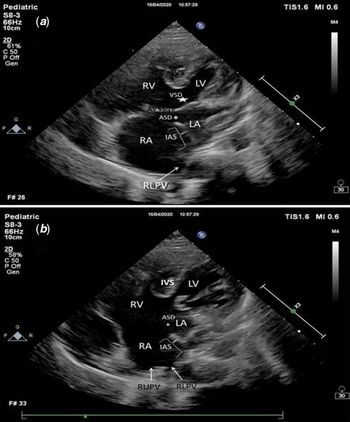
Two-dimensional (2D) echocardiography and a Doppler study were performed (Phillip Medical System, Netherlands, İE33-XMatrix using broadband transducers (S5-1, S8-3) appropriate to the patient size). A thorough echocardiographic examination was performed in each patient by using subxiphoid, apical, parasternal long- and short-axis, and suprasternal views. The abdominal situs, atrial situs, interatrial septum, ventricular looping, morphology of atrioventricular valves, ventricular septum, and great vessels were assessed. The morphologic right atrium was defined by the site of the inferior caval vein connection and the morphology of atrial appendages if possible. These prerequisites for the identification of morphologic right atrium were based on the fact that the right horn of sinus venosus contributes to the orifice of the superior caval vein and inferior caval vein. Although the malposition of the septum primum can allow half of or all the pulmonary veins to drain into the morphologic right atrium, it does not interfere with the identification of morphologic right atrium (Fig 1a and b).
( a and b ) The echocardiographic views of patient 15. The malposition of the primum septum and partial abnormal return of the right pulmonary veins to the right atrium was demonstrated at the subcostal views. ASD = atrial septal defect; IAS = interatrial sepum; IVS = interventricular septum; LA = left atrium; LV = left ventricle; RA = right atrium; RLPV = right lower pulmonary vein; RUPV = right upper pulmonary vein; RV = right ventricle.

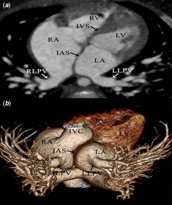
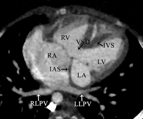
As a part of our clinical protocol, multidetector CT angiography was performed on all patients with pulmonary venous drainage anomaly or suspicion of this anomaly. Multidetector CT angiography examinations were performed using a second-generation dual-source 256-multidetector computed tomographic Somatom® Definition Flash scanner (Siemens Healthcare GmbH; Forchheim, Germany) with a sectional collimation of 2 × 128 × 0.6, a gantry rotation time of 280 ms, and a temporal resolution of 75 ms. A non-electrocardiography-gated protocol with a pitch factor of 3 was used, and every scan was obtained with z-axis modulation technique (CARE Dose; Siemens Healthcare, Erlangen, Germany) in all patients Reference Türkvatan, Tola, Kutlutürk, Güzeltaş and Ergül10 (Figs 2a, b and 3).
( a and b ) The 2D and 3D CT views a patient with SPM. The abnormal drainage of right lower pulmonary vein to right atrium due to malposition of atrial septum where left lower pulmonary vein draining normally to left atrium was demonstrated. ASD = atrial septal defect; IAS = interatrial sepum; IVC = inferior caval vein, IVS = interventricular septum; LA = left atrium; LV = left ventricle; RA = right atrium; RLPV = right lower pulmonary vein; RUPV = right upper pulmonary vein; RV = right ventricle.

The CT view of a patient with abnormal drainage of both right and left lower pulmonary veins to right atrium due to septum primum malposition was demonstrated. ASD = atrial septal defect; IAS = interatrial sepum; IVS = interventricular septum; LA = left atrium; LV = left ventricle; RA = right atrium; RLPV = right lower pulmonary vein; RUPV = right upper pulmonary vein; RV = Right ventricle.

Surgical technique
Under general anaesthesia, the conventional median sternotomy approach was used and the cardiopulmonary bypass was established by aortic and bicaval cannulation. Under moderate hypothermia, the aorta was cross-clamped and antegrade blood cardioplegia (n = 9) or del Nido cardioplegia solution (n = 5) was applied to the aortic root.
Additional cardiac pathologies were corrected first. Ventricular septal defect closure in two patients, total correction of atrioventricular septal defect in one patient, and the total correction of Fallot tetralogy in one patient were performed as presented in the literature. The surgical correction of SPM was then employed.
Anatomically, the absence of the superior limbic band was inspected in all the operated cases, whereas the atrioventricular junction and the inferior component of the interatrial septum (crux cordis) were normal. The septum primum was carefully excised.
An anatomical atrial septation was obtained in 10 cases using an autologous pericardial patch sewn without mitral valve damage or pulmonary venous orifices distortion or stenosis. In four cases, atrial septal displacement was performed by septoplasty without the use of foreign material. The right atrium was closed in standard fashion, and most of the patients were easily weaned from the cardiopulmonary bypass without inotropic support.
Patients with complex cardiac pathology and those who had additional interventions were admitted to the ICU under milrinone infusion (0.5 µg/kg/minute) alone or in combination with a low-dose adrenaline infusion (0.05 µg/kg/minute).
Statistical analysis
The Statistical Package for the Social Sciences for Windows (SPSS) version 15 (SPSS, Chicago, Illinois, United States of America) was used for the statistical analysis. Continuous variables were expressed as the median (minimum–maximum) or mean ± standard deviation, and the categorical variables were expressed in percentages.
Results
There were 15 patients with septum primum malposition. Eight patients were females, and seven were males. The median age was 12 months (2 months–10 years). Nine patients were situs solitus, and six patients were left isomeric. There were bilateral superior caval vein in three patients, right-sided superior caval vein in 11 patients, and left-sided superior caval vein in one patient. There was additional secundum atrial septal defect in 80% of the cases (n = 12). Interatrial communication was found between the septum primum and posterior atrial wall, which was nonrestrictive in all cases.
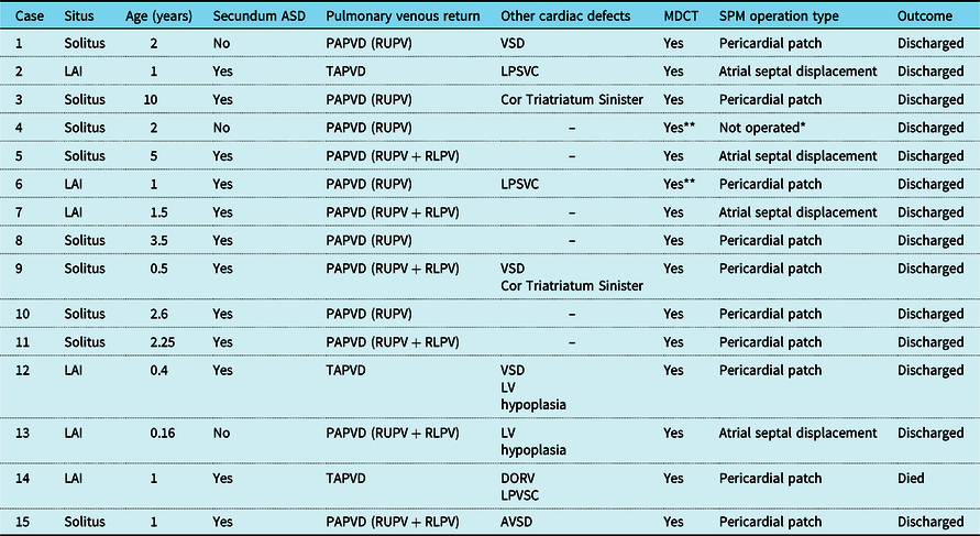
There was total abnormal pulmonary venous drainage in 3 (20%) of the 15 patients. All the total abnormal pulmonary venous drainage patients were also left isomeric. There were partial pulmonary venous drainage in remaining 12 (80%) patients. There was abnormal drainage of the right upper pulmonary veins in six patients. In the remaining six of these patients, all the right pulmonary veins were drained into the right atrium. The situs was solitus in nine patients and isomeric in three of them. Among the three patients with isomerism, all right pulmonary veins were drained anomalously to the right atrium in two patients, whereas in the remaining patient, only the right upper veins were anomalously drained (Table 1).
Demographic features, imaging, and surgical results of the patients

AVSD = atrioventricular septal defect; LAI = left atrial isomerism; PAPVD = partial pulmonary venous drainage; TAPVD = total abnormal pulmonary venous drainage; MDCT = multidetector CT; RLPV = right lower pulmonary vein; RUPV = right upper pulmonary vein; DORV = double outlet right ventricle; LPSVC = left persistent superior caval vein; LV = left ventricle; VSD = ventricular septal defect.
* On follow-up due to drainage anomaly of only one pulmonary vein.
** Differences were present between echocardiographic and MDCT angiographic findings.
There was additional cardiac pathology in 47% of the patients (n = 7). Ventricular septal defect was present in three patients, left ventricular hypoplasia in two patients, cor triatriatum sinister in two patients, atrioventricular septal defect in one patient, and double outlet right ventricle of the tetralogy type in one patient. There was left isomerism in one patient with ventricular septal defect and in one patient with double outlet right ventricle.
Multidetector CT angiography was performed additionally in all the patients. There were conflicting findings from the transthoracic echocardiography and CT angiography in two patients. The route of abnormal drainage of the right lower pulmonary vein in one patient and the presence of a left persistent superior caval vein in another patient could not be delineated clearly by transthoracic echocardiography.
Fourteen patients underwent surgery. Only a patient with abnormal right upper pulmonary vein drainage who had Qp/Qs < 1.5 was not operated. The median cardiopulmonary bypass time was 90 minutes (range, 50–158 minutes). The surgeons reported postoperatively that in three of the operated patients, the surgical strategy was changed for re-septation after the preoperative delineation of septum primum malposition.
The sternum was closed postoperatively in all the patients except the patient with left isomerism + double outlet right ventricle. Two patients with left isomerism + ventricular septal defect had inotrope support of milrinone, one patient with atrioventricular septal defect had milrinone, and one patient with left isomerism + double outlet right ventricle had milrinone + adrenaline. Postoperatively, another 10 patients were admitted to the ICU without the administration of any inotropes.
The mean duration of mechanical ventilation was 4 ± 2.1 hours, the duration of ICU stay was 3 ± 1 days, and the postoperative hospitalisation was 5 ± 1 days.
One patient with left isomerism + double outlet right ventricle needed extracorporeal life support in the fourth postoperative hour because of the persistence of low cardiac output. Extracorporeal life support was ceased at the third postoperative day because of irreversible neurologic sequela and patient-accepted exitus.
Discussion
In this study, patients with septum primum malposition, an aetiology of abnormal pulmonary venous drainage, were evaluated in our cardiac surgery centre. In addition to the varying clinical and anatomical properties of the septum primum malposition patients, especially in complex cases, multidetector CT angiography imaging was observed to provide an additional benefit, although echocardiography is usually adequate for the diagnosis. It was also noted that the septum primum malposition diagnosis might have facilitated or modified the surgical strategy. Among the limited amount of studies with the limited amount of study patients in the literature, our study is one of the first to be taken into account when these features described above and the patient numbers are considered.
Van Pragh et al Reference Van Praagh, Carrera, Sanders, Mayer and Van Praagh4 proposed the following theory to explain this malformation: the malposition of the septum primum might be the result of the poor development or the absence of the superior limbic band of the septum secundum. Septum primum, which normally originates in the sinus venous tissue adjacent to the inferior caval vein – right atrium junction, is attached to the left atrium side of the superior limbic band by the end of its normal growth, and it forms the valve of the foramen ovale during intrauterine life. The attachment of the septum primum to the septum secundum is crucial for the alignment of the common pulmonary vein with the left atrium cavity. When the superior limbic band of the septum secundum fails to develop, the septum primum is carried leftwards in cases of atrial situs solitus or rightwards in cases of atrial situs inversus by the circulating fetal blood, which flows from the right atrium towards the left atrium because the cephalad border of the septum primum remains unattached. This process is finalised by the drainage of the pulmonary veins into the anatomic right atrium despite their normal connection to the posterior wall of the left atrium. Reference Van Praagh, Carrera, Sanders, Mayer and Van Praagh4–Reference Gajjar and Desai11 Recently, new hypotheses have been proposed on the development of atrial septum based on the experimental data. Reference Goddeeris, Rho and Petiet12,Reference Versacci, Pugnaloni and Digilio13 Goddeeris et al Reference Goddeeris, Rho and Petiet12 used a genetic marker and novel magnetic resonance microscopy technique to demonstrate the origins of the dorsal mesenchymal protrusion in the dorsal mesocardium, and its valuable contribution to atrioventricular septation. They showed that sonic hedgehog signalling is required within the dorsal mesocardium for its contribution to the atria. The failure of this step results in an atrioventricular septal defect.
The situs is variable in patients with septum primum malposition. In 1995, Van Pragh et al Reference Van Praagh, Carrera, Sanders, Mayer and Van Praagh4 reported the heterotaxy syndrome to be 92% in the largest series of 36 patients (21 postmortem, 15 alive) with septum primum malposition. In their series of nine patients, Tomar et al Reference Tomar, Radhakrishnan and Shrivastava7 reported situs solitus in four patients (45%) and heterotaxy in five patients (55%). In contrast to previous studies, in our study, situs solitus (60%) was more frequent in patients with septum primum malposition. This finding might be because of technological advancements and the widespread use of imaging systems.
Depending on the degree of septum primum displacement towards the left atrium, half of or all the pulmonary veins may drain into the right atrial cavity despite their normal connection to the left atrium. Reference Türkvatan, Güzeltaş, Tola and Ergül5 Van Pragh et al found partial pulmonary venous drainage in 44% of the patient sample and total abnormal pulmonary venous drainage in 56% of the patient sample, which was caused by the malalignment of the septum primum. Reference Van Praagh, Carrera, Sanders, Mayer and Van Praagh4 Tomar et al reported total abnormal pulmonary venous drainage in 78% and partial pulmonary venous drainage in 22% of their patient series. Reference Tomar, Radhakrishnan and Shrivastava7 Conversely, partial pulmonary venous drainage and total abnormal pulmonary venous drainage frequencies were 80 and 20% (50% in Left isomerism patients), respectively, in our study. Herein, more frequent multidetector CT angiography use might be the reason for higher frequency of partial pulmonary venous drainage diagnosis.
Septum primum malposition might be considered in the morphogenetic spectrum of laterality defects as well as other isolated cardiac malformations. Reference Versacci, Pugnaloni and Digilio13 Different pathologies were reported to accompany septum primum malposition in addition to abnormal pulmonary venous drainage. Reference Prasad, Snyder and Ashwath14 In 2013, Park et al Reference Prasad, Snyder and Ashwath14 described the leftward displacement of the septum primum in patients that presented with hypoplastic left heart syndrome. Tomar et al Reference Tomar, Radhakrishnan and Shrivastava7 reported additional cardiac pathologies, such as double outlet right ventricle and ventricular septal defect, in their septum primum malposition patients with situs solitus or heterotaxy syndrome.
There is no definitive consensus in literature on the definition of the defect at interatrial septum in the presence of septum primum malposition. Reference Tomar, Radhakrishnan and Shrivastava7,Reference Hiramatsu, Takanashi and Imai8 The generally accepted main view is, there is no true secundum atrial septal defect in this entity. This interatrial communication is defined as “Inter-atrial communication associated with absence of septum secundum and malposition of septum primum” and is located between malpositioned primum septum and posterior wall of the atrium. Reference Tomar, Radhakrishnan and Shrivastava7,Reference Hiramatsu, Takanashi and Imai8,Reference Gajjar and Desai11 There were different reported frequencies for atrial septal defect association in literature. While Van Pragh reported occasional atrial septal defect associations, Hiramatsu declared secundum atrial septal defect association in all of his patients. Reference Van Praagh, Carrera, Sanders, Mayer and Van Praagh4,Reference Hiramatsu, Takanashi and Imai8 Besides there were case reports about the rare association of inferior sinus venosus defect and septum primum malposition. Reference Prasad, Snyder and Ashwath14 In addition, it was declared that a partial or complete atrioventricular septal defect with an ostium primum atrial septal defect might develop after the deviation of septum primum to inferior location. Reference Cohen, Weinberg, Coon, Gaynor and Rychik15
In our study, different cardiac pathologies, such as double outlet right ventricle and ventricular septal defect, were determined in addition to septum primum malposition in patients with situs solitus and heterotaxy syndrome. Moreover, the left ventricles were significant although not severely hypoplastic in two patients. The associated atrial septal defect frequency was found as 80%, and inferior sinus venosus type defect was not detected in any of our patients.
Transthoracic echocardiography is infact a sufficient technique for the diagnosis of septum primum malposition. The subxiphoid coronal, apical four-chamber, and parasternal long-axis views mostly adequate to demonstrate the abnormal position of the septum primum. Tomar et al Reference Tomar, Radhakrishnan and Shrivastava7 reported the sufficient demonstration of the specific anatomy with transthoracic echocardiography in all their patients. The cardinal features of this anomaly were reported as follows: the pulmonary veins were connected normally to the morphologic left atrium, to the left of the right superior caval vein, or between two superior caval veins in the case of bilateral superior caval veins; significantly underdeveloped or absent septum secundum together with the abnormal deviation of the septum primum towards morphologic left atrium; anomalous drainage of half or all pulmonary veins into the morphologic right atrium. Reference Tomar, Radhakrishnan and Shrivastava7 But sometimes, transthoracic echocardiography alone might not be an adequate evaluation method because of the poor acoustic window and the poor depiction of extracardiac vascular structures. Reference Türkvatan, Güzeltaş, Tola and Ergül5 Recently, multidetector CT angiography has been increasingly used to assess patients with suspected or known CHD, especially those in whom associated vascular anomalies must be ruled out. Reference Pandey, Sharma and Jagia17 Multidetector CT scanners with a high volume of coverage (i.e., 128 or more slices per gantry rotation) permit faster and more accurate assessment of the cardiac and vascular anatomy with lower radiation exposure compared with first-generation CT scanners. Reference Türkvatan, Tola, Kutlutürk, Güzeltaş and Ergül10,Reference Pandey, Sharma and Jagia17 Türkvatan et al Reference Türkvatan, Güzeltaş, Tola and Ergül5 reported the clear delineation of pulmonary venous return anomalies and total abnormal pulmonary venous drainage anatomy by multidetector CT angiography.
As a standard part of our clinical protocol, multidetector CT angiography was performed in all the patients with pulmonary venous drainage anomaly or suspicion of this anomaly. Pulmonary venous anatomy and other extracardiac vascular structures that were not significant results of echocardiography were clearly described.
The repair of septum primum malposition has generally been reported to be uncomplicated with good results. Reference Hiramatsu, Takanashi and Imai8,Reference Park, Fedderly and Frommelt16 These results are partly due to surgical correction without an intervention in the pulmonary vein orifices or pulmonary veins in contrast to other partial or total pulmonary venous abnormalities. The typical surgical technique consists of the complete resection of the septum primum and the replacement of the excised tissue by a pericardial patch. Complete resection was recommended to avoid pulmonary venous obstruction as a late complication. Furthermore, the atrial septal displacement technique without the substitution by a pericardial patch has been proposed as beneficial because it avoids patch retraction and allows natural septal growth. Reference Cuttone, Hadeed and Lacour-Gayet6,Reference Hiramatsu, Takanashi and Imai8,Reference Park, Fedderly and Frommelt16 In our study, the atrial septal displacement and pericardial patch techniques were both performed for surgical correction.
Limitation
The results of this study have limited generalisability because of the retrospective single-centre nature of the data available for analysis.
Conclusion
Septum primum malposition, which is a cause of abnormal pulmonary venous drainage, should be differentiated from other pulmonary venous abnormalities as accurate diagnosis would facilitate the ideal surgery in the complex cardiac pathologies requiring detailed preoperative preparation, great intraoperative effort, and postoperative care. Although echocardiography would be efficient and adequate in patients with septum primum malposition in experienced hands familiar with the pathology, multidetector CT angiography can be needed for patients in whom the evaluation of pulmonary venous structures is inadequate or in patients with complex CHDs.
Acknowledgement
None.
Financial Support
This research received no specific grant from any funding agency, commercial or not-for-profit sector.
Conflicts of Interest
None.
Ethical Standards
The Ethics and Research Committee from Istanbul Mehmet Akif Ersoy Thoracic and Cardiovascular Surgery Center (number 2019/64) approved this study. The study was performed in accordance with the 1975 Declaration of Helsinki.