Introduction
Physical dormancy (PY) occurs in seeds of 18 angiosperm families (Baskin and Baskin, Reference Baskin and Baskin2014). It results from the presence of one or two layers of palisade cells in the seed coat that have cell walls impregnated with hydrophobic substances, which prevents absorption of water (Baskin et al., Reference Baskin, Baskin and Li2000; Baskin and Baskin, Reference Baskin and Baskin2014). Water impermeability is acquired at the last stage of seed development, during the dehydration process that occurs shortly before dispersal (Werker, Reference Werker1997; Baskin and Baskin, Reference Baskin and Baskin2014). Moreover, the acquisition of water impermeability can also occur after dispersion (Jaganathan et al., Reference Jaganathan, Li, Biddick, Han, Song, Yang, Han and Liu2019; Magalhães and Oliveira, Reference Magalhães and Oliveira2020). Both the occurrence of PY and the proportion of impermeable seeds produced are controlled by genetic (Baskin et al., Reference Baskin, Baskin and Li2000; Baskin and Baskin, Reference Baskin and Baskin2014) and environmental (Baskin et al., Reference Baskin, Baskin and Li2000; Baskin and Baskin, Reference Baskin and Baskin2014; Jaganathan, Reference Jaganathan2016; Jaganathan et al., Reference Jaganathan, Li, Biddick, Han, Song, Yang, Han and Liu2019) factors.
PY is very common for seeds of Leguminosae (Werker, Reference Werker1997; Baskin and Baskin, Reference Baskin and Baskin2014), which is why representatives of this family are widely used as models in studies that address this type of dormancy. Since seeds of this family do not develop a tegmen, water impermeability is conferred by the typical and complex anatomy of the testa, which is composed of two to three distinct regions: endotesta, mesotesta and exotesta (Corner, Reference Corner1951, Reference Corner1976; Gunn, Reference Gunn, Polhill and Raven1981). The endotesta is the innermost one-layered region and can be differentiated or not, and when distinct, it can be formed by hourglass cells (Corner, Reference Corner1951, Reference Corner1976; Gunn, Reference Gunn, Polhill and Raven1981). The mesotesta is the intermediate region and comprises a layer of hourglass cells and some layers of parenchymal cells with vascular tissues commonly being restricted to the rapheal bundle (Corner, Reference Corner1951, Reference Corner1976; Gunn, Reference Gunn, Polhill and Raven1981). The exotesta, the outermost region of the testa, is formed by one layer of palisade cells. The linea lucida is present in this region, which results from variation in the thickening of the palisade cell wall. The testa is covered, externally, by an amorphous stratum that varies in thickness among species and is commonly referred to as a cuticle (Corner, Reference Corner1976; Arechavaleta-Medina and Snyder, Reference Arechavaleta-Medina and Snyder1981; Ma et al., Reference Ma, Cholewa, Mohamed, Peterson and Gijzen2004; Shao et al., Reference Shao, Meyer, Ma, Peterson and Bernards2007; Smýkal et al., Reference Smýkal, Vernoud, Blair, Soukup and Thompson2014; Molizane et al., Reference Molizane, Julio, Carmello-Guerreiro and Barbedo2018). In the seeds of Leguminosae, this stratum is generally pectic in nature (Werker, Reference Werker1997). According to the author (see p. 307), the mucilaginous stratum (she called ‘mucilaginous layer’) ‘is composed of pectic insoluble materials which shrink on desiccation and undergo chemical changes that make them [the seeds] hard and hydrophobic’. Therefore, it should be called a mucilaginous stratum (sensu Boesewinkel and Bouman, Reference Boesewinkel, Bouman and Johri1984), with the term cuticle not being applicable.
In addition to the seed coat structure, another factor that influences PY is seed water content. There is a greater proportion of seeds with PY for a given species when the seed water content is lower than when it is higher (Baskin and Baskin, Reference Baskin and Baskin2014). Variation in PY due to seed water content at the dispersal time (Hyde, Reference Hyde1954; Rolston, Reference Rolston1978; Egley, Reference Egley1979; Murphy et al., Reference Murphy, Gossett and Toler1986; Chinnasamy and Bal, Reference Chinnasamy and Bal2003; Baskin and Baskin, Reference Baskin and Baskin2014; Jaganathan, Reference Jaganathan2016; Molizane et al., Reference Molizane, Julio, Carmello-Guerreiro and Barbedo2018), and after dispersion (Jaganathan et al., Reference Jaganathan, Li, Biddick, Han, Song, Yang, Han and Liu2019), has been reported for many species of Papilionoideae. Papilionoideae species usually have seeds with a hard testa (LPWG, 2017). In this group, the acquisition of PY is related to the ability of seeds to lose water due to hygroscopic movement that occurs through the hilum (Hyde, Reference Hyde1954; Rolston, Reference Rolston1978). The hilum of Papilionoideae seeds has a peculiar structure, including a duplication of the palisade layer (counter palisade), which has a thick cell wall like the exotestal palisade (Corner, Reference Corner1951; Hyde, Reference Hyde1954; Gunn, Reference Gunn, Polhill and Raven1981). This double palisade is interrupted by the hilar fissure formed above the tracheid bar, both longitudinal to the hilum. The tracheid bar located in the sub-hilar region has an obovate transverse section and is composed of short tracheids. When seeds are exposed to low relative humidity (RH), the cell walls of the palisade and counter palisade lose water and open the hilar fissure, through which water vapour escapes from inside of the seed via the tracheid bar (Hyde, Reference Hyde1954). In the presence of high RH, the cell walls of the counter palisade rehydrate and close the hilar fissure (Hyde, Reference Hyde1954). This mechanism favours a decrease in seed water content in an environment with low water availability, which in turn increases the proportion of water-impermeable seeds (Hyde, Reference Hyde1954; Rolston, Reference Rolston1978; Murphy et al., Reference Murphy, Gossett and Toler1986; Baskin and Baskin, Reference Baskin and Baskin2014; Jaganathan, Reference Jaganathan2016; Molizane et al., Reference Molizane, Julio, Carmello-Guerreiro and Barbedo2018; Jaganathan et al., Reference Jaganathan, Li, Biddick, Han, Song, Yang, Han and Liu2019). For this reason, the Papilionoideae clade is a suitable model for studies on this theme. This subfamily has approximately 14,000 species and is the largest subfamily of Leguminosae (LPWG, 2017). Papilionoideae species occur in tropical, subtropical and temperate zones of all the world (Polhill, Reference Polhill, Polhill and Raven1981; Lewis et al., Reference Lewis, Schirire, Mackinder and Lock2005).
Although the relationship between water content and PY is well known, little is known about the mechanisms by which variation in seed moisture changes the structure of the testa and, consequently, impermeability. Molizane et al. (Reference Molizane, Julio, Carmello-Guerreiro and Barbedo2018) demonstrated that, for Erythrina speciosa (Papilionoideae), there is a greater proportion of impermeable seeds when the moisture content is lower. However, they did not verify any anatomical changes to explain this finding. Similarly, Magalhães and Oliveira (Reference Magalhães and Oliveira2020) also did not find any changes in the anatomy of the testa of E. speciosa that explained the differences between permeable and impermeable seeds. However, when comparing fresh seeds (with a higher percentage of permeable seeds) with stored seeds (impermeable), the authors identified small openings in the mucilaginous layer. These small openings, located in the lateral region of the seeds, occurred only in the permeable seeds, and they were on the water gap. However, the work did not address aspects related to seed water content, so it is impossible to associate this change in seed lateral surface with water loss during storage.
In order to clarify the basis of the relationship between moisture a PY in Leguminosae, the present study investigated how variation in RH during the post-dispersion period affects the proportion of E. speciosa seeds with PY from two different populations. Thus, this study tested the hypothesis that the humidity condition to which E. speciosa seeds are exposed after dispersion induces variation in water content of the seeds, which leads to changes in testa structure and influences the proportion of dormant seeds.
Materials and methods
Seed collection and characterization of the environment of the studied populations
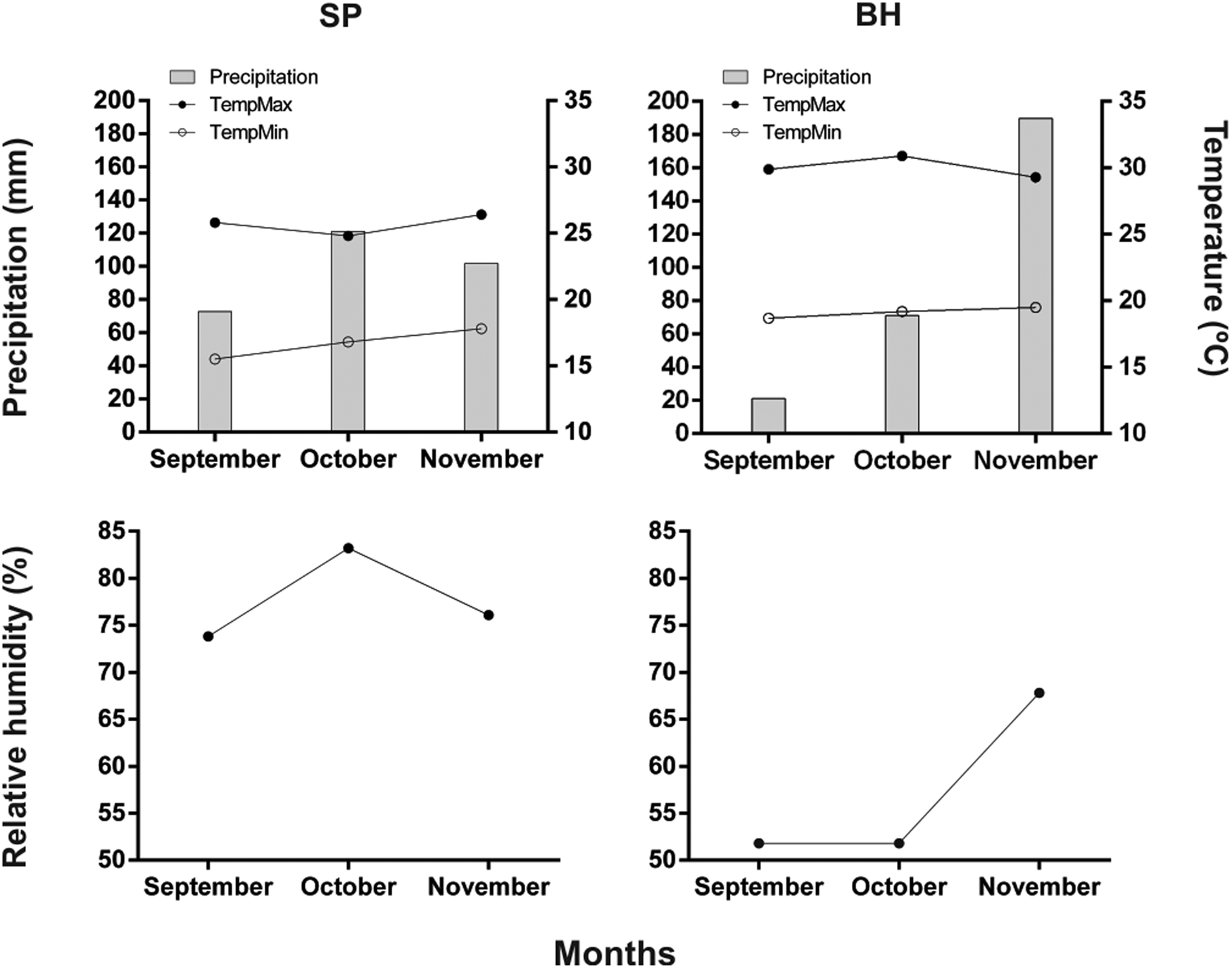
Mature fruits of E. speciosa were collected from two different sites in Brazil: São Paulo (SP) population (23°33′50.3″S, 46°45′38.0″W), whose seeds were collected in November 2018, and Belo Horizonte (BH) population (19°55′12.0″S, 43°56′05.9″W), whose seeds were collected in November 2019. Temperature, precipitation and mean RH data for both locations were obtained from INMET (2020) for the period corresponding to the development, maturation and dispersal time of E. speciosa seeds (September to November, according to Lorenzi (Reference Lorenzi1992)) and are shown in Fig. 1. Data on relative air humidity in the last 10 years in the two studied populations can be seen in Supplementary Fig. S1.
Mean precipitation, mean, maximum and minimum temperatures, and relative humidity in São Paulo (SP) and in Belo Horizonte (BH).

Twenty individuals were selected in each population. Only fruits for which the onset of dehiscence was observed were collected to ensure that seeds were mature and fruits were in the dispersion. The seeds were manually removed from the fruits, packed in paper bags and transported to the PlantSeR Lab of Departamento de Botânica do Instituto de Ciências Biológicas of Universidade Federal de Minas Gerais, Belo Horizonte, MG, Brazil. They were immediately evaluated (initial evaluation) or submitted to storage conditions (intermediate and final evaluations).
Assessment and storage of seeds
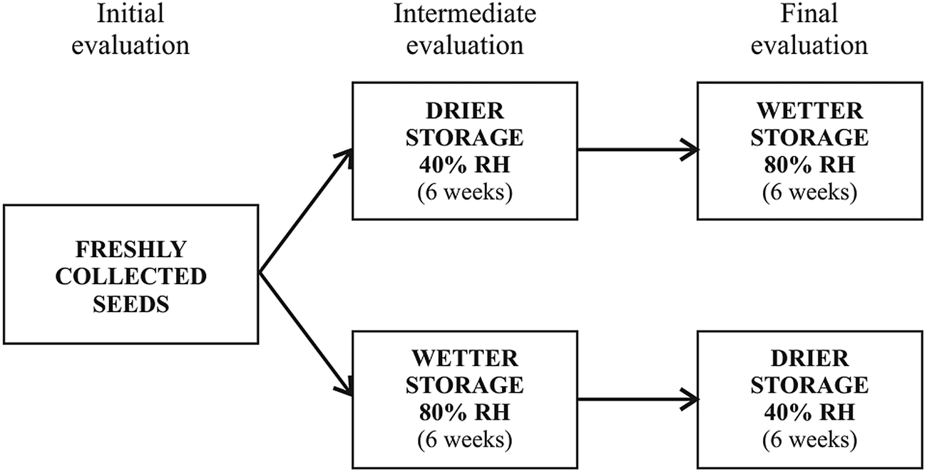
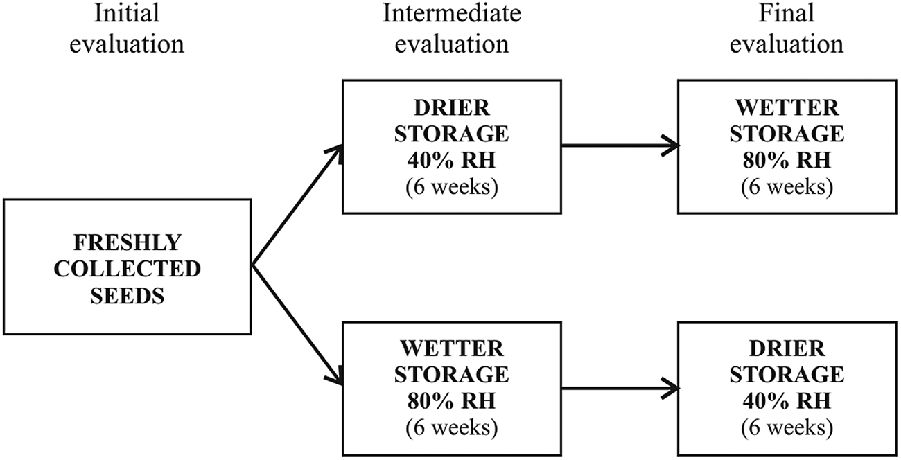
The seeds collected from each population were divided into three lots. The first lot of freshly harvested seeds was submitted to initial evaluation (Fig. 2). To test if the moisture condition influences PY acquisition after dispersion, seed lots were subjected to the alternation of two contrasting humidity conditions. One of these lots was initially stored in an environment with 40% RH (drier environment, DE) for 6 weeks, after which an intermediate evaluation was performed using half the seeds. The other half of this lot of seeds was transferred to an environment with 80% RH (wetter environment, WE), where they remained for another 6 weeks, at which point they were submitted to a final evaluation (see Fig. 2). The second lot of seeds was stored for 6 weeks in WE, and half of them submitted to the intermediate evaluation. The other half of this lot of seeds was transferred to DE, remaining in this condition for 6 weeks when the final evaluation was performed (see Fig. 2).
Summary of the experimental design showing the storage periods and the evaluations performed.
DE and WE were produced in desiccators without a vacuometer. The RH of the DE was maintained at approximately 40% and controlled by the use of silica gel. The RH of the WE was controlled by filter paper soaked in distilled water and was maintained at about 80%. The RH inside the desiccators was checked periodically using a Minipa MT-241 thermo-hygrometer.
The analyses described below were performed for the three evaluations – initial, intermediate and final – using about 240 seeds from the SP population and about 350 seeds from the BH population.
Water content
To analyse seed water content (initial, intermediate and final evaluations), three replicates of 10 seeds from the SP population and five replicates of 10 seeds from the BH population were initially weighed and stored in an oven at 105°C for 24 h. The samples were then kept in a desiccator until they cooled and then were weighed again. Seed water content was calculated according to Brasil (2009).
A portion of the seeds of the BH population was stored, under the conditions described above, after having the hilum of seeds blocked with instant liquid cyanoacrylate glue. Water content was calculated after the seeds passed through the storage cycles (DE followed by WE and WE followed by DE).
Imbibition and germination percentages
Seed imbibition was determined using intact seeds (three replicates of 10 seeds from the SP population and five replicates of 10 seeds from the BH population). The seeds were previously weighed (time 0) and placed to soak in Petri dishes with distilled water at 30°C (12-h photoperiod) and evaluated every 24 h until curve stabilization, which occurred after 240 h (10 days). At the end of this period, the percentage of imbibed seeds of each replicate was calculated according to the formula: Imbibition % = P final – P initial × 100−1, where P final = mass at last weighing (240 h of imbibition) and P initial = mass at first weighing (dry seeds). The number of germinated seeds in each replicate was computed daily during the storage period, with germination percentages being calculated at the end. The seeds that were not imbibed were scarified and exposed to moisture to determine viability.
Surface micromorphology
For micromorphological surface analysis, five replicates of seeds from each population were placed in an oven at 60°C for 24 h to complete dehydration. The seeds were then fixed on metal supports using epoxy adhesive to adjust the positioning of the samples and graphite glue to ensure the conductivity of the material. The positioning of the samples was done to visualize the lateral region of the seeds, which was identified as a water gap by Magalhães and Oliveira (Reference Magalhães and Oliveira2020). The samples were then metalized with 10 nm gold in a Balzers SCD 050 device. Analyses were performed and digital images acquired using a Quanta FEI 200 (Eindhoven, the Netherlands) or a JEOL JSM - 6360LV (Tokyo, Japan) scanning electron microscope (SEM) at Centro de Microscopia of Universidade Federal de Minas Gerais, Belo Horizonte, MG, Brazil.
Anatomical analysis
The anatomical analysis was performed only with seeds of the SP population using five seeds in each evaluation. Before infiltration and embedding, the seeds were sectioned transversely with the aid of a Dremel Rotary tool. The fragments were then dipped in about 1 ml of 100% glycerin and heated on a magnetic stirrer at 100°C for 15 min under agitation, after which they were cooled for 30 min at room temperature (Magalhães and Oliveira, Reference Magalhães and Oliveira2020, with modification). This heating–cooling process was repeated four times. Then, samples were washed three times in 95% ethanol, a solution in which they were stored for 18 h for entirely removing glycerin from the material (Magalhães and Oliveira, Reference Magalhães and Oliveira2020, with modification). After softening the testa without hydration, the material was infiltrated and embedded in Leica (2-hydroxyethyl)-methacrylate (Heidelberg, Germany). The embedding process was carried out for 30 days in a freezer to ensure polymerization inside the samples (Paiva et al., Reference Paiva, Pinho, Oliveira, Chiarini-Garcia and Melo2011, with modification).
The blocks were sectioned to about 8–10 μm in thickness with a Zeiss Hyrax M40 (Walldorf, Germany) rotary microtome. Staining was performed with 0.05% toluidine blue in pH 4.7 acetate buffer (O'Brien et al., Reference O'Brien, Feder and McCully1964, with modification) and mounted with Entellan. Structural analyses were performed and documented using an Olympus U-TV0.5XC-3 photomicroscope (Waltham, Massachusetts, US).
Statistical analysis
Water content, germination and imbibition data were tested for normality (Shapiro–Wilk) and homogeneity (Bartlett). Non-homoscedastic data were ordered and distributed in ranks and statistically evaluated using ANOVA, with detected differences being compared by Tukey's HSD test (P < 0.05). Statistical analyses were performed using JMP 13.2 software (SAS Inst. Inc, Cary, North Carolina, US), and the results were expressed as the mean of four replicates.
Results
Water content
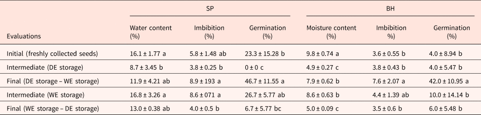
The initial evaluation revealed that the seeds recently harvested from the SP population had a water content of 16.1% (Table 1). The water content of seeds stored in DE for 6 weeks decreased significantly (P < 0.005), reaching 8.7% (Table 1). After transferring from the DE to the WE (final evaluation), the water content increased to 11.9% (Table 1). The seeds of the SP population submitted to 6 weeks in WE had a water content of 16.8% (intermediate evaluation) and 13.0% after being kept for 6 weeks in DE (final evaluation), with no significant difference between these treatments and the freshly harvested seeds (P = 0.0156; Table 1).
Moisture content (%), imbibition percentage and germination percentage (means ± SD) of Erythrina speciosa seeds performed for populations of São Paulo (SP) and Belo Horizonte (BH), at the three evaluation times (initial, intermediate and final)

Values are means ± SD of three (SP) or five (BH) replicates. Different letters within the columns indicate significant differences (Tukey test, P < 0.05).
DE, drier environment (40% Relative Humidity); WE, wetter environment (80% Relative Humidity).
The water content of freshly seeds harvested from the BH population was 9.8%, which differed significantly from the water content found after all storage treatments (P < 0.0001; Table 1). The water content of the seeds decreased significantly (P < 0.005) after DE storage (4.9%, intermediate evaluation), which then increased during the subsequent WE storage (7.9%, final evaluation), without, however, reaching the same water content of the initial evaluation (Table 1). After storage in the WE, the water content was 8.6% (intermediate evaluation), reaching 5.0% after being kept in the DE (final evaluation), which were significantly different values (P<0.005; Table 1). The water content of seeds with their hilum blocked (BH population) after both storage treatments (DE followed by WE and WE followed by DE) showed a small variation relating to freshly harvested seeds (range 9–9.6%).
Imbibition and germination percentages
The imbibition percentage for seeds from the SP population did not differ among freshly harvested (initial evaluation), DE storage (intermediate evaluation) and WE storage, followed by DE storage (final evaluation). Seeds whose treatments ended with WE storage had similar imbibition percentages to newly harvested seeds but significantly higher than the treatments that ended with DE storage (P < 0.005; Table 1).
Seeds from the BH population had a similar imbibition response to that of seeds from the SP population. The imbibition percentage of freshly harvested seeds did not differ from that of seeds exposed to the DE immediately before the evaluation. It was significantly different from that for the WE treatment for 6 weeks (intermediate evaluation). However, DE treatment followed by WE treatment did not differ from the other treatments.
Germinability for seeds freshly harvested from the SP population was 23.3%. The seeds stored in the DE did not germinate, but there was a significant increase in germination (46.7%) after exposure to the WE (final evaluation, Table 1). Germination reached 26.7% after 6 weeks of WE storage, then decreased to 6.7% after subsequent DE storage (final evaluation, Table 1).
Seeds recently harvested from the BH population had a germinability of 4.0%. DE storage did not alter seed germinability (intermediate evaluation). However, after seeds passed through WE storage, germination increased to 42.0% (final evaluation, Table 1). Germination for seeds initially stored in the WE was 10.0% (intermediate evaluation) and 6.0% after being exposed to the DE (final evaluation), with no significant difference between these evaluations and newly harvested seeds (Table 1).
Surface micromorphology
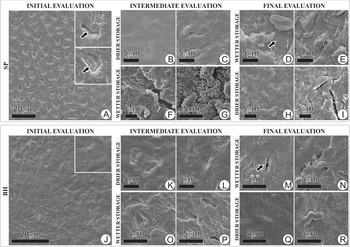
The initial evaluation of freshly seeds harvested from the SP population revealed numerous superficial depressions in the mucilaginous stratum, where cracks were observed (Fig. 3a). The intermediate evaluation revealed that, after storage in the DE, the seeds had a rough surface, but more continuous than the surface of newly harvested seeds with few depressions (Fig. 3b); superficial cracks could be observed in these few depressions (Fig. 3c). The final evaluation revealed that, after changing from the DE to the WE, the surface of the seeds became discontinuous and with cracks (Fig. 3d), in addition to the formation of lumps in the mucilaginous stratum (Fig. 3e).
Surface micromorphology of seeds of Erythrina speciosa. (a–i) Seeds collected from the SP population. (a) Freshly harvested seed. (b−c) Intermediate evaluation of a seed stored in a drier environment. (d and e) Final evaluation of a seed stored in a wetter environment after storage in a drier environment. (f and g) Intermediate evaluation of a seed stored in a wetter environment. (h and i) Final evaluation of a seed stored in a drier environment after storage in a wetter environment. (j–r) Seeds collected from the BH population. (j) freshly harvested seed. (k and l) Intermediate evaluation of a seed stored in a drier environment. (m and n) Final evaluation of a seed stored in a wetter environment after storage in a drier environment. (o and p) Intermediate evaluation of a seed stored in a wetter environment. (q and r) Final evaluation of a seed stored in a drier environment after storage in a wetter environment. Arrows indicate cracks.

After WE storage, the intermediate evaluation revealed the seeds to have a rough surface with cracks (Fig. 3f) and the formation of lumps in the mucilaginous stratum (Fig. 3g). The final evaluation revealed that the surface remained rough (Fig. 3h) after transferring from the WE to the DE. There were thicker cracks in only one of the replicates than those observed in the initial evaluation (Fig. 3i).
Initial and intermediate evaluations of seeds from the BH population revealed that recently harvested seeds and seeds stored in a DE had surfaces with few undulations and a continuous mucilaginous stratum (Fig. 3j–l). After transferring from DE to WE storage (final evaluation), the seeds showed cracks in the mucilaginous stratum (Fig. 3m). In some regions, the mucilaginous stratum was detached from the testa (Fig. 3n). Seeds stored in the WE (intermediate evaluation) showed surface undulations (Fig. 3o, p). After transferring from the WE to the DE (final evaluation), the seed surface became similar to that observed in the initial evaluation in some regions (Fig. 3q) and with more undulations in others (Fig. 3r).
Anatomical analysis
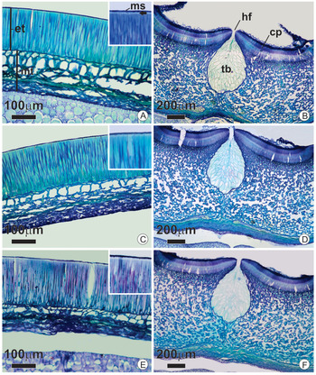
There were no differences in the organization and shape of the cells that make up the testa between freshly harvested seeds (Fig. 4a, b) and seeds stored in any treatments (Fig. 4c–f). The testa is composed of the exotesta, formed by a layer of palisade cells covering approximately half the thickness of the testa; externally, they are covered by the mucilaginous stratum (Fig. 4a, c, e). The linea lucida is observed close to the external periclinal face of the palisade. This layer has cells with thick walls of pectocellulosic nature, containing phenolic compounds in the lumen (Fig. 4a, c, e). Internally, adjacent to the palisade, the mesotesta consists of a well-spaced hourglass-cell layer with pectocellulosic walls and phenolic content; additional layers of parenchymatic cells also occur in the mesotesta. The mesotestal parenchyma has several layers of compressed cells that could not be individualized (Fig. 4a–c).
Transverse sections of seeds of Erythrina speciosa. (a and b) Respectively, lateral and hilum regions of seeds at initial evaluation. (c and d) Respectively, lateral region and hilum of seeds at the intermediate evaluation. (e and f) Respectively, lateral region and hilum of seeds at final evaluation. Arrow, linea lucida; co, cotyledon; cp, counter palisade; et, exotesta; hf, hilar fissure; me, mucilaginous stratum; mt, mesotesta; tb, tracheid bar.

In the hilum region, the palisade is double, and the counter-palisade cells have a thicker wall than the palisade cells (Fig. 4b, d, f). The sub-hilum region cells form a thick band with several layers of parenchymal cells that delimit many intercellular spaces, making it looks spongy (Fig. 4b, d, f). Inserted in this region, and internal to the hilum fissure, is the tracheid bar, a structure longitudinal to the hilum, with an obovate transverse section and composed only of tracheids (Fig. 4b, d, f).
Discussion
For the first time, this study showed that the proportion of E. speciosa seeds with PY after dispersion varies according to the humidity of the environment. The results obtained with seeds from two populations revealed that DE storage, at both intermediate and final evaluations, had the consequence of decreasing seed water content and imbibition and germination percentages. These results corroborate Molizane et al. (Reference Molizane, Julio, Carmello-Guerreiro and Barbedo2018), who found a relationship between lower water content and a higher proportion of impermeable seeds for E. speciosa, a relationship also described for other species of Papilionoideae. Murphy et al. (Reference Murphy, Gossett and Toler1986) showed that Vigna unguiculata seeds with the moisture content of 6.9 and 11.4% had germination percentages of 11 and 88%, respectively. Similarly, Jaganathan et al. (Reference Jaganathan, Li, Biddick, Han, Song, Yang, Han and Liu2019) showed that reduced seed water content resulted from post-dispersion drying, induced PY for Astragalus adsurgens. Thus, the results described in the literature and those obtained by the present study corroborate that an increased proportion of PY seeds is related to decreased seed water content.
The present results showed that DE storage led to an increase in the proportion of seeds with PY (population SP) or kept this proportion low (population BH). Besides, the data demonstrated that water leaves the seeds through the hilum, since seeds with a blocked hilum did not show a significant decrease in water content after DE and WE treatments, corroborating that described by Hyde (Reference Hyde1954). The present study evidenced the rehydration of seeds and decreased the proportion of water-impermeable seeds for both populations after exposure to WE storage, followed by DE storage. Considering that WE storage (intermediate evaluation) did not result in variation in the proportion of dormant seeds, our results indicate that the break in PY occurs only if there is initial dehydration followed by rehydration. However, additional studies are needed to understand how this process occurs. Our results indicate that, for E. speciosa, the hilum directly participates in the rehydration of seeds since seeds with a blocked hilum did not present an increase in water content.
The present study found a difference between the two populations of E. speciosa relating to germination percentage of freshly harvested seeds (SP 23.3%; BH 4%) and, consequently, in the proportion of impermeable seeds. The parental environment can explain this difference, that is, the environment to which seeds were exposed during maturation (Fig. 1). Jaganathan et al. (Reference Jaganathan, Li, Biddick, Han, Song, Yang, Han and Liu2019) indicated that seeds are dispersed with less water content in drier and warmer environments, in which there is a higher proportion of water-impermeable seeds. During seed development (see Supplementary Fig. S1), the E. speciosa seeds from SP population were exposed to higher RH (~80%) than the BH population (~40%), suggesting that the parental environment determined the differences between the newly harvested seeds of these populations. The range of temperature of the two collection sites was also different, lower in the SP population (see Fig. 1).
Although the relationship between decreased seed water content and PY acquisition, or an increase in the proportion of seeds with PY, in Papilionoideae is clear, there are no reports of how seed dehydration interferes with the structure of the testa, making it impermeable to water. Molizane et al. (Reference Molizane, Julio, Carmello-Guerreiro and Barbedo2018) did not observe any changes in the organization of cells that form the testa when comparing dormant and non-dormant seeds of E. speciosa, data corroborated here by the analyses with light microscopy. However, with scanning electron microscopy, the present surface morphology analysis revealed structural variation in the mucilaginous stratum distribution, corroborating Magalhães and Oliveira (Reference Magalhães and Oliveira2020). This portion, being tenuous and amorphous, is difficult to observe under light microscopy, which is probably why it was poorly documented in previous works with E. speciosa. Many studies have suggested that the amorphous layer covering the testa of legume seeds would be a cuticle. However, authors usually did not carry out tests to identify the lipid nature of the cuticle, which, if present, could play a recognized role in seed impermeabilization (Arechavaleta-Medina and Snyder, Reference Arechavaleta-Medina and Snyder1981; Ma et al., Reference Ma, Cholewa, Mohamed, Peterson and Gijzen2004; Smýkal et al., Reference Smýkal, Vernoud, Blair, Soukup and Thompson2014; Molizane et al., Reference Molizane, Julio, Carmello-Guerreiro and Barbedo2018) and, consequently, in the proportion of seeds with PY.
There are reports that the storage of seeds of Leguminosae (Caesalpinioideae and Papilionoideae) in a WE, whether in the laboratory or field, causes PY to break (van Klinken et al., Reference van Klinken, Lukitsch and Cook2008; Renzi et al., Reference Renzi, Chantre and Cantamutto2016). However, a significant gap in understanding how the fluctuation in RH acts on this mechanism still persists. For a species of Papilionoideae, Ma et al. (Reference Ma, Cholewa, Mohamed, Peterson and Gijzen2004) suggested that fluctuation in RH during pre-dispersion desiccation and seed storage is responsible for forming cracks through which water enters during imbibition. The present results corroborate this interpretation since the formation of superficial cracks was observed in seeds of both populations after being transferred from the DE to the WE, which coincided with a decrease in the proportion of seeds with PY. Furthermore, superficial cracks were no longer observed after transferring from the WE to the DE for seeds of both populations. This indicates that variation in RH influences not only the formation of the cracks they affect seed permeability. The cracks make seeds permeable to water, and, when they are closed, consequently culminate in the impermeabilization of the testa.
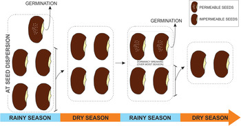
Plants of E. speciosa reproduce once a year, and the seed dispersal occurs during the rainy season (Lorenzi, Reference Lorenzi1992). The observed capacity for dehydration/rehydration in this study, and the consequent variation in the proportion of seeds with PY, due to environmental humidity fluctuation seems to constitute an important ecological strategy; this strategy also can be seen in natural conditions (see Supplementary Fig. S1). This strategy favours the germination and colonization of an environment over at least two humid seasons and two dry seasons (along 2 years), as shown in Fig. 5. This conceptual model shows that some seeds of E. speciosa are dispersed with a water-permeable testa, therefore being able to germinate immediately after dispersal (rainy season). The seeds that do not germinate (PY) will pass through the dry season, and in the subsequent rainy season, some seeds will germinate. An increase of humidity promotes the formation of the cracks in the mucilaginous stratum, which induce the breaking of dormancy and become seeds capable of starting the germination process (Fig. 5). There is no information regarding how many times this process of dehydration acquisition of PY and rehydration-overcoming of PY can occur without compromising seed viability. However, considering that seeds of E. speciosa can remain viable for several years (Mello et al., Reference Mello, Barbedo, Salatino and Figueiredo-Ribeiro2010), this process is likely to occur several times.
Conceptual model of the processes to which seeds of Erythrina speciosa were subjected during two subsequent cycles of alternating humid and dry periods.

Supplementary material
To view supplementary material for this article, please visit: https://doi.org/10.1017/S0960258520000495.
Acknowledgements
The authors thank Alison Pelri Menezes and Marcelo Tome Kubo for help with seeds collection, and Marcelo Pedrosa Gomes for help with statistical analysis, and the Center of Microscopy at the Universidade Federal de Minas Gerais (http://www.microscopia.ufmg.br) for providing the equipment and technical support for experiments involving electron microscopy. This study was financed in part by the Coordenação de Aperfeiçoamento de Pessoal de Nível Superior, Brazil (CAPES, Finance Code 001). We thank the Brazilian Conselho Nacional de Desenvolvimento Científico e Tecnológico (CNPq) for the research productivity grant from D.M.T. Oliveira (process 305686/2018-6) and Q.S. Garcia (process 304387/2016-9).
Conflicts of interest
None declared.