Lysophosphatidic acid (LPA) and sphingosine 1-phosphate (S1P) are lipid derivatives that can act as potent signalling molecules. Strong biochemical evidence has implicated LPA and S1P in the modulation of processes that can contribute to tumourigenesis, such as proliferation and angiogenesis. Signalling by these molecules is altered in cancer, as are the levels of the enzymes that generate them and the receptors through which they act. Furthermore, manipulating the receptor levels and the production and metabolism of lysophospholipids inhibits many of the hallmarks of cancer. Recent anticancer therapies have successfully used monoclonal antibodies that target aberrant receptor signalling. New antibodies under development against LPA and S1P might likewise alter levels of signalling. In addition, drugs targeting LPA and S1P receptors hold promise, given the success of many existing pharmaceuticals that target similar G-protein-coupled receptors (GPCRs). Thus, LPA and S1P have emerged as important potential targets in cancer therapy, and a range of compounds that can modulate their generation and action are being assessed with a view to clinical application.
LPA and S1P, their metabolic enzymes, and their receptors
LPA
LPA is an important bioactive lipid mediator found circulating in the blood. Normal levels of LPA are produced in vital physiological human processes, such as wound healing (Ref. Reference Lee, Goetzl and An1) and ion channel regulation (Ref. Reference Chemin2), by activated platelets, adipocytes, peritoneal mesothelial cells and fibroblasts (Refs Reference Gaits3, Reference Pages4, Reference Ren5). Healthy women generally have a slightly higher serum concentration of LPA than men do (Ref. Reference Baker6), probably because of the role that LPA plays in fertility and fetal development (Refs Reference Ye7, Reference Tokumura8).
LPA has been implicated in the aetiology of human cancer for several reasons. First, a large amount of evidence from in vitro experiments has verified LPA stimulation of cell growth, proliferation, differentiation, motility, survival and invasion at the molecular level (Ref. Reference Mills and Moolenaar9). Second, in vivo studies suggest that reducing the amount of LPA by increasing its extracellular hydrolysis or decreasing LPA-producing enzymes decreases tumour progression (Ref. Reference Tanyi10). Third, LPA levels are markedly elevated in the ascites fluid of ovarian cancer patients, and in the blood of patients with ovarian and other gynaecological cancers (Refs Reference Xu11, Reference Yoon, Kim and Cho12) and multiple myeloma (Ref. Reference Sasagawa13).
In women with ovarian cancer, abnormal LPA production occurs in cells of ascites fluid in the abdominal cavity. Peritoneal mesothelial cells, in addition to producing LPA, might also contribute to ascites formation and the production of vascular endothelial growth factor (VEGF). Up to several litres of fluid can accumulate in the abdominal cavity, within which ovarian cells generate their own lipid growth factors to create an autocrine and paracrine signalling network (Refs Reference Xu14, Reference Xie, Gibbs and Meier15). This fluid has significantly higher levels of LPA than normal: the mean total LPA level has been reported to be 1.8 µmol/l in controls and 9.7 µmol/l in ovarian cancer (Ref. Reference Baker16), with some patients having levels as high as 80 µmol/l (Ref. Reference Westermann17).
Autotaxin
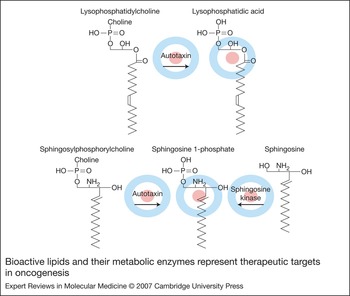
Originally known as exo-enzyme or ecto-nucleotide pyrophosphatase/phosphodiesterase 2 (ENPP2), autotaxin (ATX) was identified as a tumour motility-stimulating protein after isolation from the conditioned medium of A2058 melanoma cells (Ref. Reference Stracke18). A decade after this discovery, ATX was linked to LPA production (Ref. Reference Umezu-Goto19) and its role in catalysing cell motility was uncovered. At the molecular level, ATX cleaves choline from lysophosphatidylcholine and sphingosylphosphorylcholine to produce LPA and S1P, respectively (Fig. 1) (Ref. Reference Clair20). ATX is a major contributor to circulating LPA levels. Indeed, LPA levels in mice lacking ATX (Enpp2-knockout mice) are approximately half that in control animals. The contribution of ATX to S1P production in vivo, if any, remains unclear. Research on the functional outcomes of ATX expression has shown that a combination of ATX overexpression and Ras transformation in NIH3T3 mouse fibroblasts significantly enhances metastatic potential compared with Ras transformation alone, suggesting that ATX contributes to tumour invasiveness and aggressiveness (Ref. Reference Nam21). Furthermore, in an in vivo Matrigel-plug assay, Ras-transformed NIH3T3 cells overexpressing ATX formed more new blood vessels than control cells did, thereby establishing a role for ATX in angiogenesis (Ref. Reference Nam22).
Bioactive lipids and their metabolic enzymes represent therapeutic targets in oncogenesis. This simplified schematic shows chemical structures for lysophosphatidylcholine (LPC), lysophosphatidic acid (LPA), sphingosine 1-phosphate (S1P), sphingosylphosphorylcholine (SPC) and sphingosine, and the enzymes involved in their metabolism: autotaxin (ATX) and sphingosine kinase (SphK). The choline head group is hydrolysed from LPC and SPC by ATX to produce LPA and S1P, respectively. A phosphate group is attached to sphingosine by SphK, producing S1P. The enzymes and directional arrows represent one route (but not all routes) of lipid synthesis. Background targets indicate molecules that are current therapeutic targets by industrial and academic groups; see Table 2 and the text for more details.

Indeed, ATX has a crucial and obligatory function in the establishment of the embryonic vasculature. In Enpp2-knockout mice, early blood vessels failed to form into mature vessels, resulting in lethality at around embryonic day 10 (Ref. Reference Tanaka23). In addition, vascular defects were discovered in the yolk sac, neural tube and allantois of these embryos (Ref. Reference van Meeteren24). Presumably because it facilitates early developmental processes, the serum ATX level, like LPA, is higher in women of reproductive age than in men (Ref. Reference Nakamura25). Owing to its function in angiogenesis and its natural presence in serum, ATX must be held under tight regulation to maintain homeostasis because the human adult system may not require neovascularisation. ATX is inhibited by the presence of at least three molecules – cyclic phosphatidic acid, LPA and S1P – which participate in negative feedback regulation (Refs Reference Tsuda26, Reference van Meeteren27). This arrangement allows the lipids LPA and S1P some regulatory control over their own abundance in biological fluids.
Several cancer types exhibit increased ATX protein expression or activity, and in many carcinomas the highest levels of ATX expression correlate with metastasis (Ref. Reference Zhang28). Breast cancer tissue has higher ATX levels than normal breast tissue (Ref. Reference Yang29) and in non-small-cell lung cancer, 58% of the human cell lines studied demonstrated a higher ATX expression level than that of normal cells (Ref. Reference Yang30). In renal cell carcinoma tissues, suppression subtractive hybridisation on patient-matched tissues identified ENPP2 as one of several variably regulated genes (Ref. Reference Stassar31). Furthermore, the activity of ATX was significantly increased in undifferentiated anaplastic thyroid carcinoma cells compared with follicular thyroid cells and goitre tissue controls (Ref. Reference Kehlen32). In glioblastoma multiforme – the most malignant type of glioma – ATX is highly expressed, and central nervous system cell lines collectively have higher levels of ATX expression than other tested tumour types (Ref. Reference Kishi33). Also of interest is the finding through gene expression analysis that Epstein–Barr virus infection in Hodgkin lymphoma cells triggers the induction of ATX production (Ref. Reference Baumforth34). Together, these studies suggest that changes in ATX expression occur in various types of cancer and that ATX may have a role in one of many genetic or mutational events or a cascade of events that facilitate tumour progression.
S1P
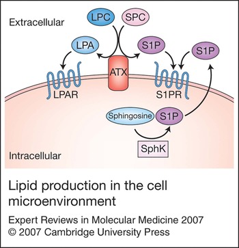
S1P is a bioactive lysophospholipid that elicits a number of in vitro cellular responses such as migration, growth, invasion and survival while also mediating wound healing, immune responses, neurogenesis and angiogenesis in vivo (Refs Reference Mizugishi35, Reference Lee36, Reference Spiegel and Milstien37). Indeed, the functions of LPA and S1P are remarkably similar – they activate related intracellular signalling molecules and share a potential for more direct receptor crosstalk. S1P is formed in the cytosol of many cells, but it can also be produced in the extracellular environment (Fig. 2) (Ref. Reference Ancellin38). In this way, S1P can activate its receptors extracellularly and can also signal intracellularly independently of the receptors and regulate Ca2+ levels (Ref. Reference Spiegel and Milstien37). Normal S1P secretion can occur from epithelial cells (Ref. Reference Johnson39), cerebellar granule cells (Ref. Reference Anelli40), cerebellar astrocytes (Ref. Reference Anelli40), thrombocytes (Ref. Reference Hanel, Andreani and Graler41) and platelets (Ref. Reference English42). Although circulating erythrocytes do not produce S1P, they are capable of storing and releasing this molecule and thus contribute to regulating the amount of S1P in the circulation and protecting it from degradation (Ref. Reference Hanel, Andreani and Graler41).
Lipid production in the cell microenvironment. The production and localisation of the receptors, lipids and enzymes discussed in this review, including autotaxin (ATX), lysophosphatidylcholine (LPC), lysophosphotidic acid (LPA), an LPA receptor (LPAR), sphingosine 1-phosphate (S1P), sphingosylphosphorylcholine (SPC), sphingosine, sphingosine kinase (SphK) and an S1P receptor (S1PR). These are the essential components of lipid production. Most represent major targets in cancer therapy.

Like LPA, S1P is associated with ovarian cancer because it was detected in ascites fluid (Ref. Reference Hong, Baudhuin and Xu43) and is a component of the follicular fluid that stimulates ovarian angiogenesis (Ref. Reference von Otte44). Similarly to ATX, S1P contributes to angiogenesis (Ref. Reference Lee36). The physiological process of angiogenesis can become pathophysiological when it allows a tumour to establish an independent blood supply with which to nourish the carcinogenic cells. A study of mice with knocked out S1P receptors confirmed the necessity of S1P signalling in the embryonic development of the vascular system (Ref. Reference Kono45). In addition, dramatic suppression of tumour growth can be achieved by inhibiting S1P-mediated signalling, because this impedes vascular stabilisation (Refs Reference Visentin46, Reference Chae47). The relative roles of intracellular and extracellular S1P as well as the relative roles of S1P on receptor signalling and lipid metabolism in the pathophysiology of cancer remain to be determined.
Sphingosine kinase
A major component in the biosynthesis of S1P is sphingosine kinase (SphK). Both isoenzymes – SphK1 and SphK2 – phosphorylate the precursor molecule sphingosine, producing S1P, and are the most essential elements in sphingolipid metabolism because they maintain the balance between apoptotic ceramide and anti-apoptotic S1P. This mechanism is referred to as the ‘sphingolipid rheostat’. More is known about the molecular function of SphK1 than SphK2 in cancer, but these isoenzymes may have opposing functions in sphingolipid metabolism (Ref. Reference Maceyka48). Sphk1 inhibits apoptosis and initiates cell growth, whereas Sphk2 promotes apoptosis and inhibits cell growth. These divergent outcomes mediated by the sphingosine kinases are probably due to the differences in cellular localisation and localised S1P production (Ref. Reference Maceyka48).
Compelling evidence suggests that SPHK1 is an oncogene. In one study, overexpression of SPHK1 in nontransformed NIH3T3 cells dramatically enhanced cell growth even in serum-free medium, prolonged cell survival, eliminated contact inhibition when cells in culture had reached saturation density, enhanced tumour development in NOD/SCID (nonobese diabetic/severe combined immunodeficiency disease) mice within 3–4 weeks of injection, and was involved in Ras transformation (Ref. Reference Xia49). The expression of SPHK1 mRNA is elevated in cancers of the breast, uterus, ovary, lung, colon, small intestine and rectum (Ref. Reference Johnson50). The expression of SPHK1 in glioblastoma multiforme correlates with a shorter patient survival time, from a median time of 357 days for patients with low levels of SphK1 to 102 days for patients with high levels of SphK1 (Ref. Reference Van Brocklyn51). In addition, a recent study suggested that SPHK1 expression is required for intestinal tumour cell proliferation and recommends that SphK1 inhibitors be investigated for potential use in controlling intestinal cancer (Ref. Reference Kohno52).
Of particular relevance to cancer treatment, SphK1 is suspected of playing a role in the resistance of tumourigenic cells to chemotherapy. The knockdown of SphK1 activity with small-interfering RNA (siRNA) enhances the sensitivity of melanoma cells to apoptosis induced by Fas and ceramide, suggesting that SphK is involved in the chemotherapy resistance of malignant melanoma (Ref. Reference Bektas53). Other evidence indicates that enforced expression of SPHK1 in PC-3 and LNCaP prostate cancer cells reduces the efficacy of chemotherapy because it affects the delicate sphingolipid rheostat (Ref. Reference Pchejetski54). In this example, high levels of S1P prevented ceramide from initiating apoptosis under pro-apoptotic conditions. Another study suggested that PC-3 cells are resistant to camptothecin because of the higher SphK1 expression and activity levels and the elevated expression of S1P1 and S1P3 receptors (Ref. Reference Akao55). Because SphK can be exported from the cell and catalyse S1P formation extracellularly (Ref. Reference Ancellin38), SphK is poised to control the rate of S1P receptor stimulation, which affects many processes that enhance tumour progression, including cell survival, proliferation, angiogenesis, vasculogenesis and resistance to chemotherapy.
LPA and S1P receptors
The bioactive lipids LPA and S1P mediate many of their effects by activating transmembrane and intracellular receptors. For both lipids, there are at least five receptors that are stimulated in response to binding each agonist: LPA1, LPA2, LPA3, GPR23/LPA4, GPR92/LPA5, PPARγ (peroxisome proliferator-activated receptor γ)/LPA, S1P1, S1P2, S1P3, S1P4 and S1P5. Most of these are cell-surface GPCRs connected to intracellular signalling pathways through G proteins that become activated and initiate second-messenger signalling cascades. In addition to activating G proteins, GPCRs can recruit and activate other molecules through intracellular domains such as PDZ-binding sites. Furthermore, cell-surface GPCRs can dimerise and heterodimerise and thus alter the outcomes of receptor ligation. Through this mechanism, extracellular lipids are able to signal intracellular events that lead to functional outcomes. LPA and S1P receptor expression can be elevated in cancer cell lines and tumour samples. For example, a high expression level of S1P receptors may be involved in tumour progression in thyroid cancer (Ref. Reference Balthasar56), gastric cancer (Ref. Reference Yamashita57) and angiogenesis (Ref. Reference Chae47). In addition, an increased number of LPA receptors are produced when cells become malignant in ovarian cancer (Ref. Reference Nakamoto58), colon cancer (Refs Reference Yun59, Reference Shida60), breast cancer (Ref. Reference Kitayama61), intestinal cancer (Ref. Reference Yamashita62) and glioma (Ref. Reference Kishi33).
Several studies have indicated that manipulating LPA or S1P receptor expression alters the hallmarks of cancer in vitro and in vivo. These changes include altered tumour growth, and increased cell motility, invasion, metastasis and neovascularisation. Receptors can link preferentially to specific cellular functions, and some receptors can function as negative regulators. The eventual functional outcomes probably represent the balance between receptor ligation, additional extracellular cues and the intracellular machinery.
Clinical implications and applications: future targeted therapeutic agents
Drugs targeting receptors
GPCRs are accessible to extracellular drugs and have key effects on cellular processes; they are thus considered exceptionally useful targets for drug intervention. Indeed, more than half of the currently approved pharmaceuticals on the market target GPCRs (Ref. Reference Schlyer and Horuk63) and nearly a third of the top 20 best-selling prescription drugs in 2003 targeted GPCRs (e.g. Zyprexa®, Advair diskus®, Plavix®, Risperdal® and OxyContin®) (Ref. Reference Schlyer and Horuk63). High-throughput screening and computational modelling have made rational drug design for these receptors even more manageable. Medications such as Claritin® and Allegra®, which are used to treat allergies, or Zantac® and Pepcid®, which are used to treat ulcers, all target histamine GPCRs.
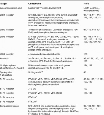
A number of compounds have been developed worldwide to activate and antagonise the 11 known receptors for LPA and S1P ligands. The inhibitors are summarised in Table 1, and some are discussed below. Most of these compounds have not undergone testing in clinical trials and have not been extensively evaluated in preclinical tumour models. So far, many of these antagonists have been used as in vitro tools to examine other biological questions. It is important to bear in mind that use of antagonists lacking specificity to one receptor may have unexpected consequences.
Selective antagonists of lysophosphatidic acid (LPA) and sphingosine 1-phosphate (S1P) signalling pathways

aReceptor agonist also functions as an antagonist. Abbreviations: FDP, farnesyl diphosphate; FMP, farnesyl phosphate kinase; FR, farnesol; SDP, serinediamide phosphate; SKI, sphingosine kinase inhibitor.
Drugs targeting S1P receptors
The S1P-receptor-agonist-analogue compound FTY720 potently inhibits angiogenesis and tumour vascularisation in vivo and decreases tumour growth overall (Ref. Reference LaMontagne64). In vivo, SphK phosphorylates FTY720, thereby generating a compound that structurally mimics S1P and binds with high affinity to the S1P1, S1P3, S1P4 and S1P5 receptors, which results in internalisation and downregulation of these receptors (Refs Reference Brinkmann65, Reference Brinkmann, Cyster and Hla66). FTY720 has not yet entered into clinical trials to determine its relevance in human cancer. However, it has been evaluated in clinical trials to determine its efficacy in preventing graft rejections after renal transplantation. One model for the net effect of FTY720 treatment after transplantation is the sequestration of lymphocytes in the lymphatic system (Ref. Reference Luo67), thus preventing their recirculation into the bloodstream where they could reach newly transplanted organs and cause rejection.
The first Phase I clinical trial demonstrated that FTY720 significantly reduced the number of blood lymphocytes without affecting renal function or increasing overall toxicity, except for the occurrence of bradycardia (Ref. Reference Kahan68). The subsequent Phase II clinical trial compared FTY720 with mycophenolate mofetil (an immunosuppressive agent) in combination with cyclosporine and corticosteroids (Ref. Reference Tedesco-Silva69). The results indicated that FTY720 was as effective as mycophenolate mofetil in preventing acute rejection after renal transplantation. Adverse effects observed with other immunosuppressant therapies (e.g. abdominal pain, serious infections, cytomegalovirus infection and neutropaenia) were not noted with FTY720, but it did increase lymphopaenia and reduced the heart rate (Ref. Reference Tedesco-Silva69). Another Phase II trial compared FTY720 with other immunosuppressive drugs and similarly concluded that FTY720 was safe and effective for de novo renal transplant patients (Ref. Reference Mulgaonkar70).
Larger, international Phase III clinical trials began assessing FTY720 in combination with immunosuppressive drugs. However, these trials were discontinued because of safety concerns such as higher incidences of worsening graft function, reduced heart rate, macular oedema and pulmonary adverse reactions (Ref. Reference Tedesco Silva71). Although FTY720 may no longer be considered for use against graft rejection after renal transplantation, it is currently under investigation in Phase II clinical trials for patients with relapsing multiple sclerosis (Ref. Reference Budde72). The clinical concerns for FTY720 – or any receptor-targeting agent – are its lack of specificity and its adverse effects (e.g. the inhibition of normal, homeostatic receptor functions such as in the regulation of heart rate).
The therapeutic potential of targeting S1P receptors in cancer requires further evaluation. The utility of any drug is determined by its therapeutic index: the ratio of drug efficacy to toxicity. Since many forms of primary cancer do not currently have viable therapeutic approaches, the balance between benefit and risk may allow the acceptance of significant toxicity and subsequently a narrower therapeutic index. Indeed, many of the current surgical, chemotherapeutic and radiotherapeutic approaches to cancer treatment are highly toxic, and the risk of life-threatening outcomes is real. Nevertheless, it is not clear whether a pan-receptor inhibitor such as FTY720 will have a sufficiently high therapeutic index to justify its use. Although the effects of FTY720 are probably mediated through the S1P1 receptor (Ref. Reference Schmid73), FTY720 may not demonstrate sufficient receptor specificity to allow selective receptor inhibition.
More-specific receptor inhibitors could offer a better trade-off between efficacy and toxicity. The S1P2 receptor has been reported to inhibit many cellular functions associated with tumourigenesis, including motility (although it may enhance invasion and angiogenesis in glioma cells) (Ref. Reference Young and Van Brocklyn74). Inhibiting other S1P receptors – particularly S1P1 – while sparing S1P2 may increase the efficacy of a cancer treatment. An antagonist against the S1P1 and S1P3 receptors, VPC-23019, inhibits ovarian (Ref. Reference Park75) and thyroid (Ref. Reference Balthasar56) cancer cell migration. The S1P1 receptor is induced in tumour vessels, and its inhibition by RNA interference significantly reduces tumour growth (Ref. Reference Chae47). By contrast, enforced expression of the S1P2 receptor suppresses the migration of glioblastoma cells (Ref. Reference Lepley76) and negatively regulates morphogenesis and angiogenesis in mouse vascular endothelial cells (Ref. Reference Inoki77). In vivo testing should be conducted to assess the efficacy and toxicity of using a combination of agonists and antagonists against these S1P receptors.
Drugs targeting LPA receptors
More compounds have been developed as agonists and antagonists of LPA receptors than of S1P receptors (Table 1). In vitro and in vivo work studying breast cancer has implicated the LPA1 receptor in the enhancement of metastatic progression. siRNA targeting LPA1 receptors or a pharmacological blockade of LPA1 receptor activity with the LPA1/LPA3 antagonist Ki16425 inhibits metastasis to bone in vivo (Ref. Reference Boucharaba78). In support of this idea, work using cDNA array analysis on breast cancer isotypes that suppress genes involved in invasion and motility has determined that the LPA1 receptor plays a major role in the cellular potential for invasion and motility (Ref. Reference Stadler79). These studies suggest that LPA1-receptor-specific inhibitors may be useful in preventing breast cancer metastasis. In addition, in vitro data have suggested that the LPA1 receptor may mediate metastatic behaviour in pancreatic cancer cells (Ref. Reference Yamada80).
The LPA2 receptor may have a niche in some cancers such that specific inhibitors could be of great benefit. The LPA2 receptor is preferentially expressed in more than half of bowel cancers, and its expression is correlated with a higher rate of invasion and metastasis (Ref. Reference Yamashita62). Colon cancer cells are unique in that they are the only major cancer type that preferentially expresses abundant LPA2 receptor levels and little or none of the other LPA receptors. In these cells, the ratio of LPA2 to LPA1 receptors increases 18-fold during malignant transformation (Ref. Reference Shida60). In primary cultures of thyroid cancers, LPA2 receptor expression was increased to three times normal thyroid or goitre levels (Ref. Reference Schulte81). LPA-induced stimulation of the chemokine growth-regulated oncogene α contributes to the angiogenesis and tumourigenesis of ovarian cancer cells, but is reduced after transfection with LPA2-receptor-targeting siRNA (Ref. Reference Lee82).
More agonists and antagonists have been developed for the LPA3 receptor than for the other LPA receptors because the LPA3 receptor has a unique and advantageous basic residue near the phosphate headgroup (Ref. Reference Fujiwara83). The greatest impediment to further drug development is the redundant signalling of all LPA receptors and the lack of definitive evidence to corroborate the LPA3 receptor with any specific function required for tumour progression. To date, the only wholly unique function of the LPA3 receptor is its role in fertility. LPA3 receptor expression in mouse uteri during early pregnancy is regulated by oestrogen and progesterone (Ref. Reference Hama84), and embryo implantation and proper spacing in litters of mice are regulated by the LPA3 receptor through its effects on generation of prostaglandins by cyclooxygenase 2 (Ref. Reference Ye7). Further research is needed to determine whether the LPA3 receptor, and also the newly identified LPA4 and LPA5 receptors, have independent actions that influence tumour progression.
Crosstalk
A potential complication in implementing LPA and S1P receptor antagonists as anticancer therapy is the crosstalk that occurs among GPCRs and between GPCRs and other growth-factor-stimulated receptors. For example, both LPA and S1P participate in transactivation with the epidermal growth factor (EGF) receptor (Refs Reference Daub85, Reference Kim86) and ErbB2/Her2 (Ref. Reference Shida87). S1P mediates the crosstalk between the S1P3 receptor and platelet-derived growth factor (PDGF) receptor in mouse embryonic fibroblasts. It also induces membrane ruffling through the VEGF receptor in human vein endothelial cells (Refs Reference Baudhuin88, Reference Endo89), and may regulate tumour progression because the S1P3 receptor is required for oestrogen-induced EGF receptor transactivation in MCF-7 cells (Ref. Reference Sukocheva90). Similarly, studies have suggested that LPA induces functional outcomes via VEGF receptor 2 in epithelial ovarian cancer cells (Ref. Reference So91) and PDGF receptor β in human bronchial epithelial cells (Ref. Reference Wang92). Interestingly, the LPA1–3 receptors form strong interactions with each other as homodimers and also as differential heterodimers of LPA1–3 and S1P1–3 receptors. S1P1–3 receptors can also form homodimers (Ref. Reference Zaslavsky93).
In many cancers, aberrant signalling of EGF receptors contributes to tumour progression, and overactive LPA production may contribute as well. PDGF receptor and VEGF receptor signalling effects include cell proliferation, cell migration, and angiogenesis. Such interwoven signalling cascades can complicate therapeutic intervention, but the outcome could be improved if these signalling events were stifled concurrently. There are many growth factor and tyrosine-kinase receptor inhibitors on the market in development and in clinical trials.
Drugs targeting lipids
Some recent breakthroughs in cancer therapy are the development of monoclonal antibodies targeting tyrosine-kinase receptors and growth factors involved in aberrant signalling pathways. These breakthroughs have enhanced the arsenal of agents available to treat patients with cancer and other diseases and have made individualised medicine a reality. Of these agents, notable mentions include Erbitux®, which targets the EGF receptor; Avastin®, which targets circulating VEGF; Rituxan®, which targets CD20; and Herceptin®, which targets the ErbB2/Her2 receptor. In addition, Revlimid® – a thalidomide analogue, which targets tumour necrosis factor α – is highly effective in the treatment of multiple myeloma. Because of the success in combining these targeted agents with other therapies and their high degree of specificity, monoclonal antibodies will continue to be developed, introduced into clinical trials and eventually approved by the US Food and Drug Administration (FDA) for clinical use.
Lpath Therapeutics (San Diego, CA, USA) has successfully created SphingomabTM – a monoclonal antibody that binds to S1P. This antibody sequesters the lipid and prevents it from activating its receptors. SphingomabTM can significantly reduce tumour progression by inhibiting neovascularisation, decrease the ability of S1P to protect cells from apoptosis and eliminate tumours in some models (Ref. Reference Visentin46). This monoclonal antibody is expected to receive approval from the FDA for the commencement of a Phase I clinical trial by the end of 2007. Lpath is also the first company to generate a monoclonal antibody that is functionally active against LPA: LpathomabTM. Although SphingomabTM and LpathomabTM are highly specific to S1P and LPA, these antibodies will target all the extracellular effects of S1P and LPA, including action on receptors implicated in increasing or decreasing cancer growth. Although the antibodies will not enter cells, they may alter the intracellular functions of S1P and LPA by preventing cellular uptake or perhaps by acting as an extracellular sink. More work is needed to determine whether these antibodies will be useful against human disease.
Drugs targeting enzymes
Drugs that target enzymes represent a larger market share than those targeting GPCRs and lipids because more enzyme targets are available. The pharmaceutical industry has had many successes with drugs targeting enzymes, including Lipitor®, Zocor®, Nexium® and Prevacid®. One of the first major breakthroughs against a cancer-causing enzyme was Gleevec®, a small-molecule kinase inhibitor that is approved for treating chronic myelogenous leukaemia and gastrointestinal stromal tumours. Gleevec® works by inhibiting the Abelson tyrosine kinase and Kit receptors that cause those two types of cancer, respectively, and is in clinical trials to assess its effectiveness against additional types of cancer. Other enzymes that anomalously appear and signal in various cancers contribute to the inappropriate activation of molecular pathways and are highly attractive targets for re-establishing regulation. The lipid pathways discussed in this review are rich in enzymes and kinases that may be suitable targets for therapy.
Drugs targeting ATX
ATX has a documented role in the invasiveness, aggressiveness and progression of tumours. Originally isolated from slime mould, cyclic phosphatidic acid (cPA) is a bioactive lipid analogue of LPA and a natural inhibitor of ATX (Ref. Reference Kobayashi94). LPA increases cell cycle progression, cell proliferation and tumour cell invasion, and cPA inhibits these biological responses in vitro (Ref. Reference Murakami-Murofushi95). Because cPA bound to human serum albumin is generated by ATX in the blood (Refs Reference Tsuda26, Reference Kobayashi94), cPA is a natural inhibitor that could be manipulated to enhance its preventative effects on ATX. cPA has been investigated in both xenograft and carcinogen-induced tumour models. In both cases, cPA has had limited activity against the primary tumour but potent activity in preventing metastases. The utility of a molecule that prevents metastases in cancer patients is not clear because most of the challenge with metastasis is its pre-existence when the patient presents. However, as local tumour control improves, approaches that prevent secondary or delayed metastasis may play a significant role in cancer therapy. ATX inhibitors could help define this role.
Chemically synthesised inhibitors of ATX are created by fine-tuning the structure of cPA by adding a methylene (carba) group to increase its natural inhibitory properties and to create compounds more potent than parental inhibitors. These cPA analogues indeed appear to be more potent inhibitors of ATX activity, LPA production, in vitro cell invasion and in vivo cell metastasis (Ref. Reference Baker96). In addition, the carba-cPA compounds are highly specific and extremely potent inhibitors of ATX. By contrast, Darmstoff analogues (a family of gut smooth-muscle-stimulating acetal phosphatidic acids) are weak ATX inhibitors and weak LPA receptor agonists (Ref. Reference Gududuru97), and l-histidine compounds may lack both efficacy and specificity for ATX (Ref. Reference Clair98). Research is under way to determine the effectiveness of carba-cPA compounds in vivo and to establish whether they are suitable candidates to test in clinical trials.
Drugs targeting SphK
SphK inhibitors are under development because of the direct evidence that SphK is an oncogene product associated with tumour progression and can control the levels of S1P activating a cell. Library screening of synthetic compounds using human recombinant SphK has identified several structurally diverse SphK inhibitors, including SKI-I, SKI-II and SKI-V (Ref. Reference French99). These compounds inhibit Akt phosphorylation, S1P formation and average tumour volume in vivo without exhibiting detectable toxicity (Ref. Reference French100). Even though they have demonstrated intriguing preclinical activity, these SphK inhibitors are not currently undergoing clinical trials.
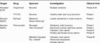
Although they inhibit SphK1, safingol (l-threo-dihydrosphingosine) and dimethylsphingosine are not specific to SphK1 because they also inhibit protein kinase C (Ref. Reference Igarashi101). In a pilot Phase I study, safingol demonstrated a pharmacologically active, safe dose without toxicity (Ref. Reference Schwartz102). Safingol is currently under investigation in another Phase I clinical trial (Table 2) to determine whether, in combination with cisplatin, it can enhance the treatment of patients with locally advanced or metastatic solid tumours (Clinical trial ID: NCT00084812).
Clinical trials of compounds targeting the sphingosine 1-phosphate pathwaya

aFor more information, see http://www.clinicaltrials.gov/
bCombinatorial drug therapy.
Clinical trials are ongoing with phenoxodiol – an experimental isoflavone derivative that initiates apoptosis, inhibits SphK and has specificity for tumour cells (Ref. Reference Schwartz102). This drug is being examined in the treatment of ovarian, prostate, fallopian tube, advanced and solid cancers (Refs Reference Gamble103, Reference de Souza104, Reference Choueiri105). Results from Phase I trials in patients with advanced cancer have indicated that most patients demonstrate progressive disease and that phenoxodiol is safe and has minimal toxicity (nausea, lymphocytopaenia and increased concentrations of alkaline phosphatase and transaminases) (Ref. Reference de Souza104). Another Phase I clinical trail of phenoxodiol in patients with solid tumours reported that several patients achieved stabilised disease (Ref. Reference Choueiri105). Studies are currently recruiting patients to test this agent in a Phase III clinical trial of recurrent or persistent advanced fallopian tube cancer (Clinical trial ID: NCT00382811) and a Phase II clinical trial examining the combination of phenoxodiol with docetaxel in patients with recurrent or persistent advanced ovarian epithelial, fallopian tube, or primary peritoneal cavity cancer (Clinical trial ID: NCT00303888).
Research in progress and outstanding research questions
Despite the wealth of indirect evidence associating S1P and LPA signalling with tumourigenesis, no definitive evidence has directly implicated LPA or S1P in the initiation of cancer. Genomic mutations, rearrangements, amplifications, deletions and methylation of either of these genes have not been discovered in any cancer. Thus, the aberrations in LPA and S1P observed in cancer are probably secondary events. In addition, owing to their genomic instability, cancer cells may be able to bypass the effects of LPA or S1P inhibition. However, S1P or LPA inhibitors may target the stroma rather than the tumour directly, and because the stromal genome is inherently more stable than the genome of tumour cells, inhibiting stromal functions may be of benefit.
Many of the outstanding questions will be answered as preclinical animal models and human trials are initiated. Because it has been extremely difficult to estimate the therapeutic index from animal studies, human trials will be required. Early clinical trials suggest that targeting some aspects of the LPA and S1P pathways can be accomplished with acceptable toxicity. The initiation and completion of single-agent and combinatorial trials are eagerly awaited. Several avenues that alter the function of the LPA and S1P pathways are under investigation; these trials have demonstrated different levels of efficacy, toxicity and disease-specific activity. One of the basic precepts of targeted therapy is matching the underlying genetic defects in cancer patients to the appropriate therapeutic agents. However, a lack of understanding of the driving genomic events makes this tactic difficult. Although assessing circulating or tumour levels of ligands, receptors or enzymes may identify patients most likely to respond to a therapeutic regimen, no preclinical data have supported this approach. Thus, tissue and serum samples obtained during clinical trials will need to be assessed to determine whether there are genomic, transcriptomic, proteomic, lipidomic or metabolomic profiles that will be able to identify patients most likely to respond to a particular therapeutic regimen.
LPA and S1P are highly implicated in the regulation of vascularisation and may be expressed in tumours in which Avastin®, Nexavar® and Sutent® (polyspecific kinase inhibitors targeting VEGF and VEGF receptors) have demonstrated activity. Although these drugs have remarkable activity in a number of tumour lineages, the vast majority of patients eventually develop drug resistance with these therapies. Targeting LPA or S1P may prove effective in these patients, and their targeting may eventually be part of a combination therapy to increase treatment efficacy or to suppress treatment resistance. Because S1P plays an important role in inducing vascularisation by multiple neovascularising factors, inhibiting S1P production or action may prevent resistance due to endothelial or tumour plasticity.
Perhaps the most interesting and highly anticipated route for LPA and S1P signalling pathway inhibitors lies in biospecific monoclonal antibody development. Although SphingomabTM effectively inhibits tumour growth, endothelial cell migration, S1P-induced cell proliferation, cytokine release and angiogenesis (Ref. Reference Visentin46), it will be intriguing to see whether LpathomabTM can inhibit tumour progression as well. It will be exciting to determine whether SphingomabTM alone or with other therapeutic agents will demonstrate activity in humans. Another important question to resolve is whether combinatorial monoclonal antibody treatment against multiple circulating growth factors can improve patient outcomes. Thus far, broad patent protection has been granted for SphingomabTM, and a clinical Phase I trial of this ‘humanised’ antibody is expected to be initiated by the end of 2007. The results will determine its metabolism, side effects, dosage and efficacy in targeting circulating S1P and whether it holds promise in anticancer therapy.
LPA and S1P in the blood mediate a number of emerging mechanisms that regulate the concentrations of these lipids. For example, ATX participates in regulatory feedback loops (Ref. Reference van Meeteren27), and erythrocytes are capable of storing S1P (Ref. Reference Hanel, Andreani and Graler106). An unanswered question surrounding the abundance of circulating lipids is what triggers the dysregulation of the molecular systems that contribute to the aetiology of cancer. Is there one event or a series of primary events that causes overactive lipid signalling? Will one or all of these events need to be targeted?
Possible secondary sources of lipid dysregulation are the metabolic enzymes ATX and SphK. This possibility would help explain why although LPA, S1P and cPA can inhibit ATX, this enzyme is still overactive and overexpressed in some cancers. Future work should continue to address the use of ATX enzyme inhibitors, such as carba-cPA analogues, against tumour metastasis (Ref. Reference Baker96) and SphK inhibitors against intestinal and other cancer progression (Ref. Reference Kohno52). If natural or small-molecule inhibitors for these proteins are able to block the functional outcomes related to cancer in humans, then a dominant role for metabolising enzymes would be validated.
Finally, it is not clear why certain types – or indeed, which types – of cancer are driven by LPA and S1P anomalies. Common abnormalities include the increased expression of the LPA1 receptor and ATX in cancers of the central nervous system, the likelihood of LPA3 receptor expression in ovarian cancer, and a dominance of exclusive LPA2 receptor expression in colon cancer. The mechanism for developing deviating features might be common to specific cell types. Receptor antagonists may be useful against identifiable tumour types showing these trends in gene expression. The safety and specificity of LPA and S1P receptor antagonists need to be established to address this question. Some cancer patients taking drugs that target growth factor receptors – such as Herceptin® – eventually relapse. In these cases, it may be worth testing whether targeting S1P and LPA receptors could be a last resort for treating patients who cannot continue on Herceptin® yet still show evidence of a growth factor response. Future research should tackle these questions, as they could bring us closer to a solution for treating cancers with abnormal LPA- and S1P-mediated signalling.
Acknowledgements and funding
This work was supported by a training fellowship to M.M. from the Keck Center Pharmacoinformatics Training Program of the Gulf Coast Consortia, NIH Grant No. 1 T90 070109-01. We thank Elizabeth Hess for critically editing this work and the peer reviewers for examining the article.