Introduction
Tympanosclerosis is an inflammatory process that is histopathologically characterised by increased connective tissue hyalinisation, calcium accumulation, decreased vascularisation, fibroblast infiltration, collagen deposition, and cartilage and/or bone-like structure formation.Reference Sorensen and True1 Any inflammatory event in the middle ear or tympanic membrane may contribute to the development of tympanosclerosis. However, it is difficult to identify what type of inflammatory event induces tympanosclerosis.Reference Ferlito2 Any events affecting inflammation, such as acute or chronic otitis media, or the placement of a ventilation tube, may play a role in the aetiology.Reference da Costa, Paparella, Schachern, Yoon and Kimberley3, Reference Tos, Bonding and Poulsen4
The relevant literature includes several experimental studies on interleukin-6, nitric acid, free oxygen radicals and other substances considered to have an antioxidant feature; these studies investigated whether inflammatory agents play a role in tympanosclerosis.Reference Forseni Flodin and Hultcrantz5–Reference Kaptan, Emir, Gocmen, Uzunkulaoglu, Karakas and Senes9 Recent studies suggest that matrix metalloproteinases have an effect on the development of tympanosclerosis.Reference Aslan, Basoglu, Senturk, Ozbay, Katilmis and Ozturkcan10
Clarithromycin is a macrolide antibiotic with antioxidant, anti-inflammatory and immunomodulatory effects, as well as an antimicrobial effect.Reference Hardy, Rios, Chavez-Bueno, Jafri, Hatfield and Rogers11, Reference Nakajima, Hishikari, Ogawa, Watanabe, Suzuki and Nagashima12 In this study, we hypothesised that clarithromycin which was added to the treatment at an early period would suppress the inflammatory process that results in tympanosclerosis. Accordingly, we investigated the effect of systematic clarithromycin therapy on the development of tympanosclerosis in an in vitro tympanosclerosis model.
Materials and methods
This study was performed with the approval of the local ethics committee for animal experiments.
Experimental design
The study included 28 adult Wistar albino rats, ranging from 250 g to 300 g in weight. The rats had normal eardrums and external auditory canals bilaterally, according to the otomicroscopic examination.
The rats were randomly allocated to one of three groups: a clarithromycin group, a non-clarithromycin group and a negative control group. In the clarithromycin group (12 rats), a tympanosclerosis model was experimentally created and clarithromycin therapy was delivered orally. In the non-clarithromycin group (12 rats), a tympanosclerosis model was experimentally created, but no therapy was given. In the negative control group (four rats), a tympanosclerosis model was not experimentally created, and no surgical procedure or medical treatment was administered.
The rats in the clarithromycin and non-clarithromycin groups received 50 mg/kg of ketamine hydrochloride and 5 mg/kg of xylazine hydrochloride intraperitoneally for anaesthesia. These rats were placed in a suitable position so that a 1 mm diameter myringotomy could be performed on the upper-superior portion of the right and left eardrums. These procedures were conducted with a myringotomy knife, using a sterile set and a surgical microscope. A single dose of 0.03 ml 109 CFU/ml type 3 Streptococcus pneumoniae strain was then injected into the middle ear through the myringotomy opening in order to create acute otitis media.
Otomicroscopic examinations were conducted at day 3 and at the end of week 1. Further myringotomies were performed on weeks one, two, three, four and five, on the same site as the previous myringotomy, in order to induce tympanosclerosis, but no strains of bacteria were injected.
In this way, acute otitis media was developed in both of the ears of the rats in the clarithromycin and non-clarithromycin groups at week one, and an experimental tympanosclerosis model was created by performing repeat myringotomies each week. The negative control group underwent no procedures.
At the end of week one, a single dose of 100 mg/kg/day clarithromycin was administered by oral gavage in the clarithromycin group; this treatment was continued for five weeks. The non-clarithromycin group and the negative control group received no medical treatment. The rats in all of the groups were sacrificed at the end of week six.
Otomicroscopic evaluation
All eardrums of rats in the clarithromycin and non-clarithromycin groups were examined with an otomicroscope on day 3 (after inoculation) and at the end of week 1, to evaluate the development of acute otitis media.
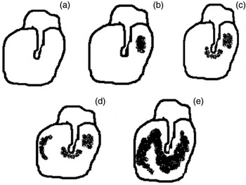
At the end of week six, the eardrums of the rats in all groups were examined, before the rats were sacrificed. The sclerotic foci were ranked on a five-point scale, ranging from no myringosclerosis to very severe myringosclerosis, using a modification of the classifications of Ozcan et al.Reference Ozcan, Gorur, Cinel, Talas, Unal and Cinel13 (Figure 1).
Ranking of sclerotic foci in the eardrums of the rats: (a) normal eardrum (classified as no myringosclerosis); (b) limited myringosclerosis only on the back-upper quadrant (mild myringosclerosis); (c) horseshoe-shaped myringosclerosis on the back-upper quadrant and at the tip of the manubrium mallei (moderate myringosclerosis); (d) horseshoe-shaped myringosclerosis on the back-upper quadrant and at the tip of the manubrium mallei, and arrow-shaped myringosclerosis on the anterior annulus (severe myringosclerosis); and (e) diffuse myringosclerosis on all quadrants (very severe myringosclerosis).

Histopathological evaluation
The temporal bones of the sacrificed rats were bilaterally dissected and the materials were fixed in a 10 per cent buffered neutral formalin solution. They were decalcified for two weeks using a formic acid-sodium method. Suitable sections were extracted from tissue samples passing through the ear and eardrum. The extracted sections were immersed in paraffin blocks. The paraffin blocks were sectioned to a thickness of 4–6 µm, and stained using the Lillie–Mayer haematoxylin and eosin method. Two specialist pathologists, who were not informed about the study groupings, evaluated the sections using a light microscope.
The sections were histopathologically classified according to the degree of middle-ear inflammation and eardrum thickness. Inflammation was ranked as follows: (1) no inflammation; (2) mild inflammation (inflammatory cells were present in the perivascular area); (3) moderate inflammation (inflammation was present in the perivascular and subepithelial area); and (4) severe inflammation (subepithelial diffuse infiltration). The measurements for eardrum thickness (in micrometres) were taken from 10 different portions of stained sections, and the means were recorded.
Statistical analysis
The analysis of eardrum thickness was conducted using the Statistical Analysis System (2009) software general linear procedure (the ‘PROC GLM’ function) (SAS, Cary, North Carolina, USA). Myringosclerosis and inflammation scores were evaluated using the Statistical Analysis System ‘identity link’ function in the generalised linear model (‘GENMOD’) equivalence analysis. Convergence was assumed when the difference between sequential analyses was 10−6. All analyses yielded a convergence.
Duncan's multiple comparisons test was used to evaluate the difference between the three groups in terms of mean eardrum thickness. The differences between the scores for the ears with myringosclerosis and the scores for the ears with inflammation were evaluated in terms of contrast structure. Compliance failure was investigated using deviance values and scaled deviances (Statistical Analysis System software).
Results
Otomicroscopy findings
Vascularity was significantly increased in the eardrums of all rats in the clarithromycin and non-clarithromycin groups, particularly around the malleus, on day 3 of streptococcus inoculation. The eardrums appeared camber and hyperaemic at the end of week one, and all ears developed acute otitis media.
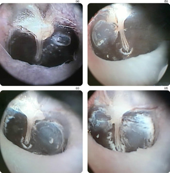
Myringosclerosis development was evaluated at the end of week six; the findings are presented in Table I. According to the otomicroscopic examination, all eardrums in the clarithromycin and non-clarithromycin groups had myringosclerosis, whereas no myringosclerosis was present in any of the eardrums in the negative control group. Otomicroscopic images that best reflect the different categories of myringosclerosis severity are presented in Figure 2.
Otomicroscopic images of the different categories of myringosclerosis severity observed on the eardrum of rats, showing: (a) mild, (b) moderate, (c) severe and (d) very severe myringosclerosis.

Severity of myringosclerosis in the eardrum*

Chi square (2 degrees of freedom) = 23.62; p=0.001.
* At the end of week six.
The number of severe and very severe myringosclerosis cases in the non-clarithromycin group was statistically significantly higher than that in the clarithromycin group (p < 0.001).
Histopathology findings
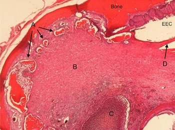
The number of ears with no inflammation or mild inflammation in the clarithromycin group was statistically significantly higher than that in the non-clarithromycin group (p < 0.001). One ear in the negative control group had mild inflammation; no inflammation was present in the other ears in this group. The findings are presented in Table II and histopathological findings of inflammation are shown in the microscopic image in Figure 3.
Photomicrograph of thickened, hyalinised middle-ear mucosa, with findings of severe inflammation, in a rat from the non-clarithromycin group; this image shows: newly formed bone tissue (A), dense hyalinisation and fibrosis areas in the subepithelial connective tissue (B), heavy pus formation in the middle-ear space (C), and a thickened eardrum (D). EEC = external ear canal (H&E; ×40)

Severity of inflammation in the middle ear*

Chi square (2 degrees of freedom) = 69.90; p=0.001.
* At the end of week six.
The mean eardrum thickness in the non-clarithromycin group was statistically significantly higher than that in the clarithromycin group. In addition, mean eardrum thickness was statistically significantly lower in the negative control group when compared with the clarithromycin and non-clarithromycin groups (p < 0.05). The distribution of mean eardrum thicknesses by group is presented in Table III.
Mean eardrum thickness

*Difference between the group means; p<0.05. †No observation was made of one ear for one animal. SD = standard deviation; min = minimum; max = maximum
Discussion
The incidence of myringosclerosis due to prolonged otitis media with effusion in children with a ventilation tube inserted has been reported to be between 23 and 53 per cent.Reference Maw and Bawden14–Reference Pereira, Pereira and Costa18
There are currently two arguments regarding the treatment of acute otitis media. One is to keep the infection under control by initiating antibiotic therapy as soon as it is diagnosed. The second is to recommend antibiotic therapy when there is deterioration in clinical factors during follow up or if risk factors are present. This latter conservative approach to antibiotic therapy in the treatment of acute otitis media, which is the approach suggested in developed countries,Reference Pappas and Owen Hendley19–Reference Tähtinen, Laine, Huovinen, Jalava, Ruuskanen and Ruohola22 is the result of rapidly developing antibiotic resistance. However, clarithromycin may be preferred, not only to keep the infection under control, but also to suppress inflammation in consideration of the likelihood of developing tympanosclerosis.
Nakajima et al. reported that clarithromycin prevented myocardial ischaemia-reperfusion damage by regulating matrix metalloproteinases.Reference Nakajima, Hishikari, Ogawa, Watanabe, Suzuki and Nagashima12 Ozdemir et al. reported that clarithromycin reduced the levels of free oxygen radicals, thereby inhibiting intestinal hypoxia and reoxygenation damage.Reference Ozdemir, Ergin, Yenisey, Turk and Simsek23 Free oxygen radicals are known to be involved in the aetiopathogenesis of tympanosclerosis.Reference Mattsson, Magnuson and Hellstrom6, Reference Mattsson, Marklund and Hellstrom7 It has recently been suggested that matrix metalloproteinases could have effects on the aetiopathogenesis of tympanosclerosis.Reference Aslan, Basoglu, Senturk, Ozbay, Katilmis and Ozturkcan10 In a study by Ozcan et al. on 25 guinea pigs with experimentally induced tympanosclerosis, at the end of week 6 the authors observed more sclerotic foci in the middle-ear mucosa and eardrums of the guinea pigs that had not been administered topical doxycycline. They pointed out that the inhibiting effect of matrix metalloproteinases could be a significant factor for this effect of doxycycline.Reference Ozcan, Selcuk, Ozcan, Akdogan, Giray and Dere24
In this study, we used clarithromycin to prevent tympanosclerosis. This antibiotic is widely used in clinical settings. It has the anti-inflammatory, antioxidant and immunomodulatory features of agents used in previous studies to prevent tympanosclerosis. The findings demonstrated that systematically administered clarithromycin significantly diminished the severity of middle-ear mucosa inflammation and eardrum myringosclerosis. Furthermore, the eardrum thickness was statistically greater in the non-clarithromycin group, which did not receive clarithromycin. There was diffuse hyalinisation with few inflammatory cells in the subepithelial area of thickened eardrums. We observed that eardrum thickness was increased by this hyalinisation resulting from inflammation.
• Any inflammatory event in the middle ear or tympanic membrane (e.g. acute or chronic otitis media, or ventilation tube placement) may contribute to tympanosclerosis development
• Clarithromycin has antioxidant, anti-inflammatory, immunomodulatory and antimicrobial effects
• Clarithromycin may be the preferred therapy for controlling infection and suppressing inflammation, to prevent tympanosclerosis developing
The literature contains no definite information as to how many days clarithromycin should be taken to achieve the anti-inflammatory effect. Nakajima et al. demonstrated that clarithromycin regulates matrix metalloproteinases and prevents ischaemia-reperfusion damage.Reference Nakajima, Hishikari, Ogawa, Watanabe, Suzuki and Nagashima12 These authors administered systemic clarithromycin therapy at a dose of 100 mg/kg/day; this therapy was initiated one week before the ischaemia-reperfusion damage was produced in the hearts of the rats, and terminated at the end of week three. There are publications indicating that long-term use of clarithromycin is beneficial for patients with nasal polyps and chronic rhinosinusitis because of its anti-inflammatory feature.Reference Peric, Vojvodic, Baletic, Peric and Miljanovic25, Reference Hashiba and Baba26 These studies suggest that long-term use of clarithromycin would be helpful in treating pathologies that involve chronic inflammation. Clarithromycin therapy was administered for five weeks in our study because the pathogenesis of tympanosclerosis included ongoing inflammation.
We derived no definite findings regarding what feature of clarithromycin suppresses the sclerosis. We postulate that this effect is due to clarithromycin's antibiotic, antioxidant and anti-inflammatory properties, and its regulatory effects on matrix metalloproteinases.
There are no studies in the literature that have investigated the mechanism of action, the duration or the dosage of clarithromycin to be used for preventing tympanosclerosis. Further investigation of both the prevention of sclerosis and the aetiology of sclerosis should be the focus of future studies.
In conclusion, this study demonstrated that clarithromycin inhibited the development of tympanosclerosis. Thus, we consider that clarithromycin may be the preferred therapy for preventing ear diseases, tympanosclerosis in particular, because of its potential anti-inflammatory feature in addition to its antibiotic feature.