INTRODUCTION
The tapeworm Echinococcus granulosus presents a major public health concern, since its larval stage causes the life-threatening zoonotic disease called cystic echinococcosis in many parts of the world. Its intermediate hosts, which suffer from parasitic cysts in the liver, lungs and other organ systems, include both wild and domestic animals, and also humans. The definitive hosts of the parasite, which harbour the tiny adult tapeworm, are primarily wild and domestic canids, including grey wolves (Canis lupus) and domestic dogs. Based on the analyses of mitochondrial genes, E. granulosus has been reported to comprise ‘genotypes’ G1-G10 (Bowles et al. Reference Bowles, Blair and McManus1992, Reference Bowles, Blair and McManus1994; Scott et al. Reference Scott, Stefaniak, Pawlowski and McManus1997; Lavikainen et al. Reference Lavikainen, Lehtinen, Meri, Hirvelä-Koski and Meri2003). Recently, it has been suggested that some of these genotypes may deserve the taxonomic status of species: G4 as E. equinus, G5 as E. ortleppi (Thompson and McManus, Reference Thompson and McManus2002), G6-G10 as E. canadensis and G1-G3 as E. granulosus sensu stricto (Nakao et al. Reference Nakao, McManus, Schantz, Craig and Ito2007). Allocating different E. granulosus genotypes to particular species has also been supported through phylogenetic analyses, inferring that they are not monophyletic (Bowles et al. Reference Bowles, Blair and McManus1994; Thompson et al. Reference Thompson, Lymbery and Constantine1995). Indeed, recent phylogenetic studies have placed 2 other species of Echinococcus, namely E. multilocularis and E. shiquicus, between the mitochondrial E. granulosus G1-G4 and G5-G10 (Lavikainen et al. Reference Lavikainen, Lehtinen, Meri, Hirvelä-Koski and Meri2003; Reference Lavikainen, Lehtinen, Laaksonen, Ågren, Oksanen and Meri2006; Xiao et al. Reference Xiao, Qiu, Nakao, Li, Yang, Chen, Schantz, Craig and Ito2005; Thompson et al. Reference Thompson, Boxell, Ralston, Constantine, Hobbs, Shury and Olson2006; Bhattacharya et al. Reference Bhattacharya, Bera, Bera, Maity and Das2007; Nakao et al. Reference Nakao, McManus, Schantz, Craig and Ito2007).
Echinococcus granulosus in cervids is sustained by a predator-prey cycle, involving mostly wolf and large cervids such as moose (Alces alces), elk (Cervus elaphus) and reindeer (Rangifer tarandus) in Eurasia and North America (Rausch, Reference Rausch1967). To date, E. granulosus G8 and G10 have been recorded in cervids. While G10 is found in cervid hosts in Eurasia (Lavikainen et al. Reference Lavikainen, Lehtinen, Meri, Hirvelä-Koski and Meri2003, Reference Lavikainen, Lehtinen, Laaksonen, Ågren, Oksanen and Meri2006; Moks et al. Reference Moks, Jõgisalu, Saarma, Talvik, Järvis and Valdmann2006), both E. granulosus G8 and G10 have been recorded in these hosts in North America (Thompson et al. Reference Thompson, Boxell, Ralston, Constantine, Hobbs, Shury and Olson2006). Several phylogenetic studies have shown that G8 and G10 are close genetically to E. granulosus G6 and G7, with all 4 clustering together with G5 (E. ortleppi) and forming a monophyletic group G5-G10 (Lavikainen et al. Reference Lavikainen, Lehtinen, Meri, Hirvelä-Koski and Meri2003, Reference Lavikainen, Lehtinen, Laaksonen, Ågren, Oksanen and Meri2006; Xiao et al. Reference Xiao, Qiu, Nakao, Li, Yang, Chen, Schantz, Craig and Ito2005; Thompson et al. Reference Thompson, Boxell, Ralston, Constantine, Hobbs, Shury and Olson2006; Bhattacharya et al. Reference Bhattacharya, Bera, Bera, Maity and Das2007). However, while the basal position of G5 in relation to G6-G10 was recognized in these studies, the phylogeny of E. granulosus G6-G10 has remained unresolved.
Two competing hypotheses have emerged from previous analyses. On one hand, based on analyses of mitochondrial cytochrome c oxidase subunit 1 (cox1) and ATPase subunit 6 (atp6) sequence data, Thompson et al. (Reference Thompson, Boxell, Ralston, Constantine, Hobbs, Shury and Olson2006) suggested that E. granulosus G6/7 and G10 are sister taxa, G8 being placed adjacent to G6/7-G10. This hypothesis is supported by recent studies based on the analyses of mitochondrial NADH dehydrogenase subunit (nad1 and nad3), cox1 and atp6 data sets (Lavikainen et al. Reference Lavikainen, Lehtinen, Laaksonen, Ågren, Oksanen and Meri2006) or based on cox1 data alone (Bhattacharya et al. Reference Bhattacharya, Bera, Bera, Maity and Das2007) (Fig. 1A). However, evidence from nad1 (Thompson et al. Reference Thompson, Boxell, Ralston, Constantine, Hobbs, Shury and Olson2006) and nad1, cox1 and atp6 gene sequence data (Xiao et al. Reference Xiao, Qiu, Nakao, Li, Yang, Chen, Schantz, Craig and Ito2005) has suggested that E. granulosus G8 and G10 could be sister taxa and that G6/7 could be regarded as adjacent to G8-G10 (Fig. 1B). However, currently, there is no robust statistical evidence with which to favour one hypothesis over the other.
(A and B) Two competing hypotheses for the phylogeny of Echinococcus granulosus ‘genotypes’ G5-G10. (A) Hypothesis 1 states that G6/7 and G10 are sister taxa while G8 can be regarded as adjacent to G6/7-G10; and (B) hypothesis 2 states that G8 and G10 are sister taxa and G6/7 is placed adjacent to G8-G10. (C) Bayesian tree of Echinococcus granulosus ‘genotypes’ G5-G10 based on the analysis of concatenated mitochondrial gene sequences atp6, nad1 and cox1; 1028 bp in total). E. multilocularis was used as outgroup. Statistical supports for genetic groupings are in bold; two values above are: posterior probability of Bayesian inference (BI) and bootstrap support obtained by maximum-likelihood (ML); values below are: bootstrap supports obtained by maximum-parsimony (MP) and neighbour-joining (NJ) methods. Support values for nodes are shown in light italics. E. granulosus isolates from Estonia are shown by an asterisk (cf. Table 1). (D) Median joining network illustrating relationships of Echinococcus granulosus ‘genotypes’G5-G10, on the analysis of concatenated mitochondrial gene sequences (atp6, nad1 and cox1; 1028 bp in total). Filled circles are median vectors (haplotypes not sampled or extinct). One bar equals one mutation; otherwise, mutations are indicated by number. Note that the number of mutations and the length of bars (or broken bars, which designate numerous mutations) are not to scale.

In Estonia, the first detailed study of E. granulosus was carried out in the 1950s, when this parasite was frequently recorded in farm pigs (Lesinš, Reference Lesinš1955). Since then, E. granulosus has been detected neither in domestic animals (even though they have been subjected to regular veterinary inspections) nor in wild cervids, whose helminth fauna was studied during the 1970s when the number of ungulates in Estonia was very high (Järvis, 1993 ). However, E. granulosus was detected again in 2004 in a single grey wolf. Based on a partial sequence of the nad1 gene, the E. granulosus isolate from that wolf was shown to be genetically identical to the G10 isolate described in Fennoscandia (Moks et al. Reference Moks, Jõgisalu, Saarma, Talvik, Järvis and Valdmann2006).
As genetic variation within E. granulosus can reflect differences in infectivity for distinct host groups or species (Thompson and McManus, Reference Thompson and McManus2002), and since several ‘genotypes’ are potentially infective to humans, it is of great significance to genetically characterize E. granulosus isolates and establish their phylogenetic status. The aims of this study were to investigate the presence of E. granulosus in wild ungulates in Estonia, to genetically characterize these E. granulosus isolates using mitochondrial gene sequences and also to reappraise the phylogeny of E. granulosus G5-G10 employing these sequence data.
MATERIALS AND METHODS
Parasite material
The carcasses of 3524 legally hunted, wild ungulates were collected throughout Estonia during the hunting seasons (September–December) of 2004 and 2005. This number of ungulates represented 2038 moose, 1044 roe deer (Capreolus capreolus) and 442 wild boars (Sus scrofa). Parasitological examinations of the carcasses were carried out by a private veterinary company (Delimeat LLC), with the species identification of helminth parasites being based on morphological criteria (Abuladze, Reference Abuladze1964). One cyst from each animal infected with E. granulosus was stored in 96% ethanol and retained for further analyses. In addition, an isolate of E. granulosus, collected during a helminthological survey of wolves (Moks et al. Reference Moks, Jõgisalu, Saarma, Talvik, Järvis and Valdmann2006), was also included in the study.
Genomic DNA purification, enzymatic amplification and sequencing
Both protoscoleces and cyst walls were used for DNA extraction. Total genomic DNA was isolated using a High Pure PCR Template Preparation Kit (Roche Diagnostics GmbH, Germany) following the manufacturer's instructions. Mitochondrial gene regions in the genes atp6, nad1 and cox1 were individually amplified by the polymerase chain reaction (PCR) from genomic DNAs from all moose isolates of E. granulosus using the oligonucleotide primers described by Lavikainen et al. (Reference Lavikainen, Lehtinen, Laaksonen, Ågren, Oksanen and Meri2006) for atp6, Bowles and McManus (Reference Bowles and McManus1993) for nad1 and Bowles et al. (Reference Bowles, Blair and McManus1994) for cox1. In addition to the nad1 sequence obtained in a previous study (Moks et al. Reference Moks, Jõgisalu, Saarma, Talvik, Järvis and Valdmann2006), the atp6 and cox1 gene regions were sequenced for the E. granulosus isolate from wolf. The PCRs were performed in a volume of 20 μl containing 1×BD Advantage 2 PCR buffer, 1U BD Advantage 2 Polymerase mix (BD Biosciences, USA), 0·2 mm dNTP (Fermentas, Lithuania), 4 pmol of each primer and 20–80 ng of purified DNA. The cycling parameters were: 95°C for 1 min, 40 cycles at 95°C for 30 s, 50°C (nad1 and cox1) or 55°C (atp6) for 30 s and 68°C for 45 s, followed by a final extension step at 68°C for 7 min. The PCR products were resolved in 1·2% agarose gels (Fermentas, Lithuania) and stained with ethidium bromide. Individual PCR products were incubated with 1 unit of shrimp alkaline phosphatase/exonuclease I (USB, USA) for 30 min at 37°C, and then heated to 80°C for 15 min to inactivate the enzymes.
Sequencing of amplicons was performed using DYEnamic ET Terminator Cycle Sequencing Kit (Amersham Biosciences, Sweden), employing 33 cycles (95°C 20 s, 50°C 20 s and 60°C 1 min) in a volume of 10 μl. Both strands of DNA were sequenced with the same primers as used for the primary PCR. Sequences were resolved using an ABI PRISM 377 automated DNA sequencer (Applied Biosystems, USA). Each unique mitochondrial gene sequence was defined as a ‘haplotype’ (according to Itagaki et al. Reference Itagaki, Kikawa, Sakaguchi, Shimo, Terasaki, Shibahara and Fukuda2005; Beveridge et al. Reference Beveridge, Shamsi, Hu, Chilton and Gasser2007).
Phylogenetic analyses
Consensus sequences were assembled with the program Consed employing the sequences of both DNA strands (Gordon et al. Reference Gordon, Abajian and Green1998). Sequences were verified by eye, edited manually in BioEdit (Hall, Reference Hall1999) and aligned using Clustal W (Thompson et al. Reference Thompson, Higgins and Gibson1994). All of the sequences conceptually translated using the program Mega 4 to ensure that they had open reading frames (Tamura et al. Reference Tamura, Dudley, Nei and Kumar2007).
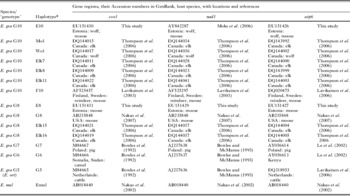
For the analysis of phylogenetic relationships of the E. granulosus G5-G10, the dataset was expanded by including mitochondrial nad1, cox1 and atp6 sequence data for E. granulosus and E. multilocularis (=outgroup) (Table 1). The 3 gene fragments were merged and sequences were re-aligned. In the final analyses, 13 Echinococcus sequences were used (see Table 1). Phylogenetic trees were generated using a concatenated dataset of 3 mitochondrial gene segments employing 4 methods: maximum parsimony (MP), maximum likelihood (ML) and neighbour-joining (NJ) using PAUP*4.0b10 (Swofford, Reference Swofford2003) and Bayesian inference (BI) using MrBayes 3.1 (Ronquist and Huelsenbeck, Reference Ronquist and Huelsenbeck2003). To evaluate possible conflicts between 3 mitochondrial gene regions, the partition homogeneity tests were carried out in PAUP*4.0b10 (Swofford, Reference Swofford2003), employing the random addition heuristic search option using 1000 replicates. MP analysis consisted of branch and bound searches (1000 bootstrap pseudoreplicates), with the options ‘multrees’, and branches being collapsed if they had a length of zero. For the NJ method, the HKY85 parameter model was used with 1000 bootstrap pseudoreplicates. ML analysis was carried out with the model of nucleotide substitution and parameters determined by Modeltest 3.7 to best fit the data set using the corrected Akaike Information Criterion (AICc) (Posada and Crandall, Reference Posada and Crandall1998). For ML analysis, heuristic searches with 100 replicates of random, stepwise addition and tree bisection–reconnection (TBR) branch swapping were used. Nodal support in the ML trees was evaluated using 1000 bootstrap pseudoreplicates. For the BI, the data were partitioned according to mitochondrial gene regions, and the appropriate model of sequence evolution for each partition was predicted using the AIC test, implemented using the program Modeltest 3.7. BI calculations were carried out by combining 3 data partitions and applying the appropriate substitution model. Searches were conducted with 4 simultaneous Markov Chains (3 heated and 1 cold chain) with over 2×10⁶ generations, sampling every 100 generations; burn-in trees were discarded and a 50% majority rule consensus tree was calculated. To ensure that the BI was not trapped in local optima, the analysis was performed 3 times. Phylogenetic trees were visualized with TreeView 1.6.6 (Page, Reference Page1996). A median joining network was calculated with Network 4.201 (Bandelt et al. Reference Bandelt, Forster and Röhl1999) using the default settings.
Species, ‘genotype’ and mitochondrial haplotypes for members of the genus Echinococcus, their hosts, geographical origins,mitochondrial gene sequences with Accession numbers in GenBank and sources
(* Identity as in Fig. 1C and D. E. gra – E. granulosus; E. ort – E. ortleppi; E. mul – E. multilocularis.)

RESULTS
In total, 36 E. granulosus cysts were found in 16 moose, while no cysts were detected in roe deer or wild boar. Thus, 0·8% of moose were infected with this parasite. The number of cysts varied among individual animals, ranging from 1 to 6, with most cysts being found in the lungs.
Amplicons representing the 3 different mitochondrial gene regions (in atp6, nad1 and cox1) were PCR-amplified and sequenced for each of 16 isolates of E. granulosus from moose. All of the sequences had open reading frames after conceptual translation. The lengths of sequences used for further phylogenetic analyses and their corresponding nucleotide positions (in parenthesis), according to reference sequence of E. multilocularis (Accession number AB018440; Nakao et al. Reference Nakao, Yokoyama, Sako, Fukunaga and Ito2002) were for 468 bp (5865–6332) for atp6, 251 bp (7647–7897) for nad1 and 309 bp (9915–10223) for cox1. These Estonian isolates of E. granulosus from moose represented E. granulosus G8 (haplotype E8: 11 samples, 69%) and G10 (haplotype E10: 5 samples, 31%). The single specimen of E. granulosus from the wolf also represented haplotype E10. No intraspecific sequence variation was detected in any of the gene loci for both haplotypes E8 and E10 of E. granulosus from Estonia. The sequences have been submitted to GenBank and are publicly available under the Accession numbers listed in Table 1. The alignment of all 13 concatenated sequences (listed in Table 1) is comprised of 75 parsimony informative sites (available from authors upon request).
On the basis of AICc, the HKY+I model was selected for each of the 3 different gene regions, as well as for the concatenated dataset. Therefore, the concatenated data set of 3 mitochondrial gene regions (1028 bp) was used in the phylogenetic analysis employing each of the 4 methods (MP, ML, NJ and BI); the nucleotide substitution model HKY+I was applied when BI (lset nst=2 rates=propinv) and ML methods were used. All 4 analyses yielded essentially the same tree topology, albeit with slightly different support for some nodes (Fig. 1C). Partition homogeneity tests revealed no conflicts between 3 mitochondrial gene regions.
The combined phylogenetic analysis of 3 mitochondrial genes indicated, with statistical support, that G6/7 form a clade with G10 and that G8 is a sister taxon to E. granulosus G6/7-G10 (Fig. 1C). The median joining network provided additional insight into the phylogenetic relationships of E. granulosus G5-G10, also revealing that G6/7 and G10 are most closely related, whereas E. granulosus G8 and G5 are more distant (Fig. 1D).
DISCUSSION
This and another recent study (Moks et al. Reference Moks, Jõgisalu, Saarma, Talvik, Järvis and Valdmann2006) have demonstrated that the sylvatic cycle of E. granulosus in Estonia involves moose as a primary intermediate host and wolf as a definitive host. However, at this stage it is not possible to exclude other host species. Although not considered in the present study, red deer may also be an intermediate host of E. granulosus in Estonia and additional epidemiological studies are warranted. Also, further research is required to estimate the prevalence of the parasite in the wolf population, since, to date, only a single wolf (of 26 individuals examined) has been found to be infected with E. granulosus (see Moks et al. Reference Moks, Jõgisalu, Saarma, Talvik, Järvis and Valdmann2006). Further investigation should also focus on the role of feral dogs in rural areas, as they are known to feed on internal organs from moose shot by hunters. Therefore, such dogs could serve as a definitive host for the dissemination of the parasite, potentially posing a risk to both domestic animals and humans. Thus, given our incomplete knowledge and understanding of the of definitive host spectrum, it is not yet entirely clear how the life-cycle of E. granulosus is maintained in Estonia.
It is somewhat surprising that, following the study in the 1950s (Lesinš, Reference Lesinš1955), no cysts of E. granulosus were found until 2004 (Moks et al. Reference Moks, Jõgisalu, Saarma, Talvik, Järvis and Valdmann2006), despite regular veterinary monitoring of the muscles and internal organs of domestic and wild animals. This situation is similar to that in Finland, where no cysts were detected for about a decade after 1992 (Hirvelä-Koski et al. Reference Hirvelä-Koski, Haukisalmi, Kilpelä, Nylund and Koski2003). However, as also argued by Hirvelä-Koski et al. (Reference Hirvelä-Koski, Haukisalmi, Kilpelä, Nylund and Koski2003), it is not possible to verify whether the recent detection of E. granulosus is attributable to a real increase of the parasite in wildlife populations or to increased awareness of and focus on this parasite.
Molecular genetic studies of E. granulosus have defined E. granulosus ‘genotypes’ G8 and G10. Both occur in North America (Bowles et al. Reference Bowles, Blair and McManus1994; Thompson et al. Reference Thompson, Boxell, Ralston, Constantine, Hobbs, Shury and Olson2006), whereas, to date, only G10 has been found in Fennoscandia (Lavikainen et al. Reference Lavikainen, Lehtinen, Meri, Hirvelä-Koski and Meri2003, Reference Lavikainen, Lehtinen, Laaksonen, Ågren, Oksanen and Meri2006). In the present investigation, we report the occurrence of E. granulosus G8 for the first time in Eurasia. The prevalence of haplotype E8 (member of E. granulosus G8) was greater than that of E10 (E. granulosus G10) in the moose samples studied herein (11 isolates of E8 versus 5 isolates of E10). These data show that the genetic differentiation within E. granulosus from cervid hosts is not associated with their geographical distribution, as both E. granulosus G8 and G10 have been found in Eurasia (Estonia and Finland) and in North America (Lavikainen et al. Reference Lavikainen, Lehtinen, Meri, Hirvelä-Koski and Meri2003, Reference Lavikainen, Lehtinen, Laaksonen, Ågren, Oksanen and Meri2006; Moks et al. Reference Moks, Jõgisalu, Saarma, Talvik, Järvis and Valdmann2006; Thompson et al. Reference Thompson, Boxell, Ralston, Constantine, Hobbs, Shury and Olson2006 and this study). Thus, the results of the present study support the proposal that E. granulosus G8 and G10 can both be distributed over most of the Holarctic region (Thompson et al. Reference Thompson, Boxell, Ralston, Constantine, Hobbs, Shury and Olson2006).
Recent studies have proposed that the mitochondrial clade G5-G10 of E. granulosus is monophyletic and thus forms a single cluster on a phylogenetic tree (Thompson et al. Reference Thompson, Boxell, Ralston, Constantine, Hobbs, Shury and Olson2006; Lavikainen et al. Reference Lavikainen, Lehtinen, Laaksonen, Ågren, Oksanen and Meri2006; Nakao et al. Reference Nakao, McManus, Schantz, Craig and Ito2007). A very low pairwise divergence between G6 and G7, determined in a recent analysis of entire mitochondrial genome data set (Nakao et al. Reference Nakao, McManus, Schantz, Craig and Ito2007), suggests that E. granulosus G6 and G7 should be regarded as a single genetic group. The existence of ‘genotype’ G9 of E. granulosus in humans (Scott et al. Reference Scott, Stefaniak, Pawlowski and McManus1997) seems controversial. Through an analysis of mitochondrial nad1 sequence data, Kedra et al. (Reference Kedra, Swiderski, Tkach, Dubinsky, Pawlowski, Stefaniak and Pawlowski1999) found no significant differences between isolates from pigs and humans. Considering the very low pairwise divergence in whole mitochondrial sequence dataset between G6 and G7 (Nakao et al. Reference Nakao, McManus, Schantz, Craig and Ito2007) and very similar sequences of G7 and G9, they all are proposed (based on mtDNA data) to represent E. granulosus G6/7/9.
Previous phylogenetic analyses have raised two competing hypotheses concerning the interrelationships of E. granulosus G6/7, G8 and G10 (see Introduction section and Fig. 1A and B). However, neither of these hypotheses has received reasonable statistical support, and it has thus not been possible to favour one hypothesis over the other. In this study, we demonstrated with strong statistical support that E. granulosus G6/7 and G10 are sister taxa (BI and NJ gave 100% support, while MP and ML gave 99% and 93%, respectively) and G8 is adjacent to G6/7 and G10 (ML support was 91%, while the other 3 methods provided 100% support). As all 4 methods yielded phylogenetic trees with congruent topology and with strong statistical support, it is evident that the amount of phylogenetic information yielded by 3 mitochondrial gene regions (length: 1028 bp), provides confidence regarding the phylogenetic relationships. Also, the median joining network supports a close relationship of E. granulosus G6/7 and G10. However, additional sampling and sequence data sets are required for an adequate reconstruction of the phylogeny within G8 and G10, as not all methods provided sufficient statistical support for relevant nodes (Fig. 1C, in light italic).
Several proposals have been made concerning the taxonomy of the ‘mitochondrial clade’ G5-G10. According to the most recent, E. granulosus G5 was regarded as E. ortleppi (see Thompson and McManus, Reference Thompson and McManus2002; Nakao et al. Reference Nakao, McManus, Schantz, Craig and Ito2007) and clade G6-G10 as a single species, named E. canadensis (see Nakao et al. Reference Nakao, McManus, Schantz, Craig and Ito2007). Recently, based on molecular data, Lavikainen et al. (Reference Lavikainen, Lehtinen, Laaksonen, Ågren, Oksanen and Meri2006) agreed that the classification of G6-G10 as a single species is justified, but argued for some caution regarding the species status of G5 (i.e. whether it should represent a separate species or should be combined with G6-G10 into a single species). The results of the present study, showing that the G6/7 is consistently positioned between G8 and G10 on the phylogenetic trees, provide support for the proposal of combining G6/7, G8 and G10 into a single species. However, we take the cautious approach of Lavikainen et al. (Reference Lavikainen, Lehtinen, Laaksonen, Ågren, Oksanen and Meri2006) and consider the status of G5 as presently unresolved.
We thank Jaak Kinnas and John Davison for their generous help. This work was supported by grants from the Estonian Ministry of Education and Sciences (target financing grant TBGZH 1432A), the Estonian Science Foundation (grant 7040), the Estonian Ministry of Agriculture (grant LBGZH 07073) and from the Environmental Investment Centre (project no. 16, 04-04-9/871 and project no. 9, 05-05-9/591).