How do early care experiences shape development, and to what extent can later experiences remediate the effects of early adverse care? These questions have tremendous importance for our understanding of developmental psychopathology (Cicchetti, 1989). The goals of research on early adversity have never been to accumulate lists of all the ways in which early adversity may limit or impair children. Rather, the objectives have been to better understand developmental mechanisms of early adversity and, armed with this information, to intervene to improve children's outcomes. Unfortunately, there is a large distance between embarking on the first objective and achieving the second one. Bridging this distance is a primary goal of translational research (Strauman & Merrill, 2004).
In the collaborations that have formed the core of our Early Experiences, Stress, and Prevention Science Research Network, we have focused on one facet of this translational challenge: applying basic research on the developmental neurobiology of stress to the understanding of how early neglect and abuse shapes vulnerability to emotional and behavioral disorders and then using this information to inform preventive interventions for maltreated children. Our experiences in attempting to move from bench to bedside have led us to recognize the many challenges that face translational researchers. These challenges will need to be overcome if we are to adequately integrate basic neuroscience into developmental psychopathology and effective preventive treatments (Cicchetti & Tucker, 1994). In what follows, we will describe the paths we have taken and some of the issues with which we have grappled. We wish that we could conclude by demonstrating that we have accomplished the task we set ourselves, fully moving research on early experiences and stress neurobiology into effective preventive interventions for children. However, at this stage, what we have to offer is a progress report on where we are and the obstacles we and others have encountered thus far.
We begin this review by briefly summarizing the data on behavioral outcomes of neglected and abused children and the major psychological orientations that have been applied to explicate why these outcomes occur. Next, we very briefly synopsize the vast literature, spanning the last half-century, on early experience and stress neurobiology using rodent models. From this work we have distilled key issues that provide a framework for our translational efforts. We then review what is currently known about the impact of childhood neglect and abuse on stress neurobiology in human adults and children. Based on similarities in primate and human development, we extend this review to nonhuman primate studies. Finally, we discuss how this basic work has informed the evaluation of our preventive interventions with neglected and maltreated children, and considerations that may move the field forward to more fully integrate an understanding of stress neurobiology into preventive intervention strategies.
Early Neglect and Abuse: Increased Risk of Behavioral and Emotional Problems
Decades of research have provided unequivocal evidence that childhood maltreatment increases the risk of psychological and behavioral disorders (e.g., Manly, Kim, Rogosch, & Cicchetti, 2001). Although some children who suffer maltreatment are resilient, becoming competent adults despite the odds (Kinard, 1998), many others suffer from a variety of clinical disorders in adulthood including substance abuse and affective and personality disorders (e.g., Putnam, 2003). Research examining why some individuals are resilient has identified protective factors, but cannot wholly explain resilience among maltreated children (e.g., Cicchetti & Rogosch, 1997; Kinard, 1998). Similarly, efforts to determine maltreatment factors associated with different types of disorders has examined such characteristics as the timing, dose, duration, and type of maltreatment (e.g., Manly et al., 2001). These studies indicate that more severe, multiple, and prolonged maltreatment results in increased symptoms; however, a coherent and consistent model of the differential effects of specific subtypes of maltreatment on specific symptom clusters or mental health disorders does not appear to have emerged. Interestingly, one challenge to identifying associations between the type and timing of maltreatment and disordered outcomes may be that outcomes vary over the course of development. Externalizing problems appear to predominate during childhood, while substance abuse and affective disorders are observed in adulthood (e.g., Tieman, van der Ende, & Verhulst, 2005). The developmental trajectories associated with these varied outcomes are at present not understood, due in part to the complexity and cost of prospective longitudinal designs.
Until recently, theoretical perspectives on maltreatment have been solely psychological, reflecting two dominant lenses: the developmental–organizational perspective, and the social learning theory perspective. From a developmental–organizational perspective, maltreatment disturbs resolution of stage-salient developmental issues. If the experience occurs early in development, attachment relationships are likely to be disturbed (Page, 1999). Studies have revealed that exposures to frightening or overwhelming behaviors from the caregiver are associated with the development of a disorganized/disoriented (Type D) attachment, characterized by freezing and dissociative behavior and heightened risk for numerous poor outcomes in childhood and beyond (Lyons-Ruth, 2003).
Through the lens of social learning theory, learning principles account for behaviors exhibited by neglected and abused children (Reid & Kavanagh, 1985). Behaviors on the part of the child that are less likely to elicit abusive treatment may be adaptive in the context of abuse, but when generalized to other settings may disrupt the child's relationships (e.g., with teachers, peers, and so on). Indeed, poor peer relations associated with a tendency to attribute hostile intent to others has been noted for maltreated children (Price & Glad, 2003). Maladaptive social behavior may lead children into antisocial peer groups, increasing the risk of conduct disorders, and substance abuse (Patterson, DeBaryshe, & Ramsey, 1989). Maltreated children have been consistently shown to be at high risk for externalizing behavior, conduct problems and substance abuse (Egeland, Yates, Appleyard, & van Dulmen, 2002).
To date, preventive interventions for maltreated children also have been based almost exclusively on the two psychological perspectives describe above. Early interventions based on developmental–organizational perspectives have focused on attachment, attempting to increase parental sensitivity, and responsiveness to foster more secure parent–child relationships and alter the child's inner working models (Cicchetti, 2005; Dozier, 2003). Interventions based on social learning theory have attempted to avert child behavior problems by training parents to use clear, consistent, nonhostile guidance and discipline techniques (Fisher, Burraston, & Pears, 2005). The effectiveness of these psychosocial interventions is typically evaluated at the group level; however, there are always differences in effectiveness at the level of the individual. These psychosocial models often fall short of fully explicating why some individuals respond and others do not.
Early Experience and Stress Neurobiology
Recently, researchers have turned to neurobiological models to explain how maltreatment may affect brain development and heighten risk of psychological disorders (see for reviews, Bremner & Vermetten, 2001; De Bellis, 2005; Heim, Owen, Plotsky, & Nemeroff, 1997; Shea, Walsh, Macmillan, & Steiner, 2005; Teicher, Andersen, Polcarri, Anderson, & Navalta, 2002). Neurobiological models may hold keys to mechanisms through which psychosocial interventions operate and to explanations of individual differences in response to treatment. These neurobiological models are largely based on animal studies of the effects of early life adversity on stress neurobiology and brain development. The largest body of such early experience research has been conducted on rodents, notably the rat.
Over the past 50 years, rodent early experience studies have shown that early parental care profoundly influences brain development, regulates gene expression, and shapes the neural systems that in humans are involved in vulnerability to affective disorders in response to later stressful life events (e.g., Levine, 2005a). Recent studies also indicate that interventions in the postinfancy period may help ameliorate some, but not all, of the impacts of early inadequate parental care (Francis, Diorio, Plotsky, & Meaney, 2002). From the beginning, this rodent research was conducted to inform the understanding of human development and psychopathology. Below we review the rodent literature and arrive at a core list of critical bridging themes. Thorough understanding of these themes, however, requires a brief description of the neurobiology of stress.
The Neurobiology of Stress
Stress neurobiology is organized at three levels (see Figure 1): a corticolimbic level that orchestrates responses to anticipated threat, a hypothalamic–brainstem level that coordinates central and peripheral responses in response to corticolimbic input (and also in response to less processed input regarding threats to homeostasis), and a neural to adrenal gland level that effects increases in stress-sensitive hormones (glucocorticoids and epinephrine; Herman & Cullinan, 1997). Corticotropin-releasing factor (CRF) is a neuroactive peptide that operates at the first two levels to coordinate behavioral, emotional, autonomic, and endocrine facets of stress and defensive responding (Heinrichs & Koob, 2004). Two loosely coupled CRF pathways are involved. One, termed the hypothalamic CRF pathway, involves CRF-producing neurons in the paraventricular nuclei of the hypothalamus (PVN), which regulates activity of the hypothalamic–pituitary–adrenocortical (HPA) axis (see Figure 2). CRF secreted from these neurons travels through a small blood connection to the anterior pituitary where it, along with other cosecretagogues (e.g., arginine vasopressin, AVP), stimulates the production and release of adrenocorticotropic hormone (ACTH). ACTH released into general circulation stimulates cells in cortex of the adrenal glands to produce and release glucocorticoids.

The three levels of neurobiological organization of the stress system that are responsive to psychological stressors. The corticolimbic level of organization involves the anterior cingulate (ACC) and orbital frontal cortex (OFC), which relay information to subcortitical structures involved in the stress response. The ACC and OFC are reciprocally interconnected with each other and with the amygdala, which has connections with the hippocampus and BNST. The hypothalamic–brainstem level of organization involve the hippocampus and brainstem structures such as the locus coeruleus, which releases NE to brain areas involved in alerting. The BNST provides pathways into the PVN of the hypothalamus, which produces corticotrophin-releasing hormone (CRH) and arginine vasopressin (AVP), whereas the hippocampus and regions in the medial frontal cortex (e.g., ACC) maintain feedback control on the paraventricular nucleus (PVN). Considering the neural to adrenal level of analysis, nuclei in the lateral hypothalamus activate highly interconnected nuclei in the brainstem, including the parabrachial nuclei, that regulate the sympathetic (NE and epinephrine, EPI) and parasympathetic (acetylcholine, Ach) nervous systems via pathways traveling through the spinal cord to preganglionic nuclei or to target organs (e.g., the adrenal medulla). The production of CRH and AVP by the PVN regulates activity of the HPA axis and the production of glucocorticoids (GCs) as depicted more fully in Figure 2. Adapted from Gunnar and Davis (2003).

The brain structures that are central to the activation and inhibition of the HPA endocrine stress response: (A) the main brain areas that participate in the regulation of the HPA axis and (B) a schematic representation of the HPA endocrine stress response initiated by the release of CRH/AVP from the medial parvocellular region of the paraventricular nucleus (mpPVN) in the hypothalamus. Glucocorticoids inhibit this system acting at the level of the pituitary, hypothalamus, and hippocampus. GABA, gamma aminobutyric acid; CRH, corticotropin-releasing hormone. From “Stress Neurobiology and Developmental Psychopathology,” by M. Gunnar and D. M. Vazquez. In D. Cicchetti and D. Cohen (Eds.), Developmental Psychopathology: Developmental Neuroscience (2nd ed., Vol. 2), 2006, New York: Wiley. Copyright 2006 John Wiley & Sons, Inc. Reprinted with permission.
Glucocorticoids (predominantly cortisol in primates and corticosterone in rodents) are steroid hormones that affect almost every organ and tissue of the body (Sapolsky, Romero, & Munck, 2000). In the brain, glucocorticoids operate through two types of receptors with distinct functions (de Kloet, Vreugdenhil, Oitzl, & Joels, 1998). At basal levels glucocorticoids operate predominantly through Type I or mineralocorticoid receptors to maintain the sensitivity of neurons to their neurotransmitters and maintain the capacity of the brain to respond to conditions that potentially threaten the organism's viability. At elevated or stress levels, glucocorticoids operate through Type II or glucocorticoid receptors (GR) to counteract the impact of other stress processes, sculpt neural systems to retain information about threats to well-being, and return the organism to prestress levels of functioning. GR-mediated neurochemical events, if well timed, acute, and contained, support organism viability; however, these events are potentially damaging if too frequent or too prolonged (Sapolsky et al., 2000). Multiple mechanisms operate in response to elevated glucocorticoids to suppress or downregulate the HPA system including negative feedback mechanisms that regulate acute responses and genomic alterations at various levels of the system that affect the responsiveness following prolonged or frequent activation.
The second CRF pathway involves CRF-producing neurons in the central nucleus of the amygdala (CeA; Van Bockstaele, Colago, & Valentino, 1998). The CeA receives information through multiple pathways, many of which involve cortical systems that support the integration of past and present experiences, thereby allowing the anticipation of threat (see Figure 1). Psychosocial stressors operate through these pathways converging on the CeA to orchestrate behavioral, autonomic, and neuroendocrine reactions in anticipation of threat (Heinrichs & Koob, 2004). Efferent CeA–CRF pathways project indirectly to PVN–CRF neurons through the bed nucleus of the stria terminalis (BNST) to stimulate the HPA axis, and directly to the locus coeruleus to stimulate release of norepinephrine (NE) into the terminal fields of the ascending noradrenergic system, supporting cognitive arousal and focusing, and behavioral and emotional components of fight/fight/freeze responses (Van Bockstaele et al., 1998). Bidirectional connections of the amygdala and regions in the medial prefrontal cortex (mPFC; orbital frontal cortex or OFC and anterior cingulate cortex or ACC) support the modulation of behavior and cognition in relation to expectations of rewards and punishments. These corticolimbic pathways influences the balance between responding based on rapid, habitual, emotionally charged modes of acting and more nuanced, considered, and dispassionate modes (Sullivan & Gratton, 2002). Notably, chronic intravenous infusions of glucocorticoids tend to upregulate CeA–CRF activity, biasing functioning toward rapid, emotion-charged fight/flight/freeze responses, while at the same time downregulating PVN–CRF, resulting in normal to hyporesponsiveness of the HPA axis (Schulkin, McEwen, & Gold, 1994). In addition, particularly during development, CRF and glucocorticoids interact with other neuropeptides (e.g., oxytocin) and neurotransmitter systems (e.g., serotonin and dopamine), resulting in widespread influences of frequent stress level activation of the CeA–CRF and PVN–CRF systems on emotional and cognitive development (Roceri et al., 2004).
Genetic variability appears to modify these effects. As only one example, individuals carrying at least one short allele of the serotonin transporter gene polymorphism exhibit larger amygdala responses to threat stimuli (Hariri et al., 2002). In addition, these individuals show larger cortisol responses to psychosocial stressors (Barr et al., 2004; Sanchez, Noble, et al., 2005). They are also at higher risk for depression following early experiences of abuse and neglect (Caspi et al., 2003; Kaufman et al., 2004).
Key Translational Issues
Models derived from rodent studies of adverse early experience can be employed to provide a biological level of explanation for the above noted association in children between early maltreatment and later heightened risk for emotional and behavioral disorders. At the most general level, the rodent studies show that disturbances in early care exert a pervasive and lasting impact on the two neural pathways described above, and that alterations in these systems has potential to compromise subsequent development (Meaney & Szyf, 2005; Sanchez, Ladd, & Plotsky, 2001). However, if we are to move beyond these broad generalities to inform specific intervention strategies, we need to determine whether and how particular elements of the rodent early experience models apply to human development. This, in turn, requires attention to the critical details of the rodent findings. As we discuss in this section, we have identified four dimensions of the rodent models that appear critical to building the translational bridge:
- developmental timing of adversity,
- the presence (or absence) of a relative stress hyporesponsive period (SHRP) during human development,
- parental or caregiver mediation of any SHRP, and
- the impact of caregiving that mediates the SHRP on the development of corticolimbic stress response organization.
Timing
The effects of experiences on brain development depend on the maturity of the brain when the events are experienced. Timing is critical. Rat pups are born young relative to human infants; that is, birth occurs in the rodent when the brain is much less mature than the brain of the full-term human infant. Indeed, the first week of the rodent's life are often equated with development of the human infant during the last trimester of gestation (Dobbing, 1981). However, comparing brain development across mammalian species is difficult. Although the general pattern of brain development is probably comparable across mammals, specifics likely vary and specifics are required to translate mechanisms of early experience effects.
For example, methylation of the GR gene expressed in the hippocampus appears to be an important mechanism through which early experiences in the rodent influence later stress reactivity and vulnerability (for review, see, Meaney & Szyf, 2005). At birth, GR genes in the rodent hippocampus are heavily methylated. This means that they are not available for transcription. The GR gene in the brain transcribes the proteins needed to produce the GR, which, in turn, mediates many components of the stress response, including negative feedback regulation of the HPA axis in response to psychosocial stressors. Maternal care in the infant rodent determines patterns of GR gene demethylation with higher levels of maternal stimulation (i.e., licking and grooming) being associated with more demethylation. Accordingly, the offspring of high licking and grooming mothers have more operational GR genes in the brain and regulate stress more effectively. These demethylations of the GR gene occur most prominently during the first week of life in the rat pup, consistent with evidence that manipulations that decrease maternal care have more profound effects if they are imposed beginning in the first rather than the second postnatal week. Manipulations in the later peripubertal period that reduce many of these early experiences effects do not affect GR methylation, suggesting that this early experience effect is more or less permanent (Francis et al., 2002). GR methylation can be affected in adulthood in the rat through pharmacological manipulations (Weaver et al., 2005). Importantly, pharmacological manipulations that demethylate the GR gene also result in changes in other aspects of stress neurobiology, suggesting that GR methylation is not only relatively permanent but also fairly critical to the evidence that early experiences in the rodent have lifelong consequences for adult stress vulnerability and resilience.
Translating the results of rodent GR gene studies requires that we know when in human development adult levels of central GR methylation are determined. As far as we know, this information simply is not available for humans or any other primate. Indeed, our level of knowledge of human developmental neurobiology is sufficiently limited that in many cases, even when basic studies provide a target for early experience effects, that knowledge does not help us pin point comparable periods in the development of stress neurobiology in humans. Nonetheless, given the importance of the first postnatal week in rodents and its rough comparability to the last trimester of gestation in humans, it is prudent to broaden our early experience-translational window to include the prenatal period.
Certainly there is abundant evidence that many children who suffer adverse care during postnatal development often are the products of high risk or stressed pregnancies (Thompson et al., 1994). There is increasing evidence that maternal stress and anxiety during pregnancy are associated with lower birth weight infants, in part through increased maternal HPA activity, upregulation of placental CRH production, with resulting decreases in birth weight and length of gestation (Wadhwa, 2005). Lower birth weight is a known risk factor for poor developmental outcomes, many of which overlap with problems observed for neglected and abused children (e.g., Indredavikm, Vik, Heyerdahl, Kulseng, & Brubakk, 2005). There is also increasing evidence that lower birth weight is associated in adults with increased risk of metabolic syndrome (i.e., high blood pressure, high cholesterol, abdominal fat, type II diabetes) and that elevated and poorly regulated glucocorticoids may mediate this risk (Phillips et al., 2000). Thus, as we proceed with our work on postnatal maltreatment, we should keep in mind that although the rodent findings appear promising in explaining the impact of postnatal neglect and abuse in human children, some of the mechanisms might actually translate more directly to the impact of adversity on prenatal human development.
Relative stress hyporesponsive period
In the rodent, the most profound effects produced by systematically altering parental care are observed in the first 2 weeks of the pup's life. This roughly corresponds to a developmental epoch in rodents that has been referred to as the relative stress hyporesponsive period (SHRP). During the SHRP, the rat pup's HPA axis (the adrenal cortex in particular) shows very little reactivity when challenged by a variety of stressors (Sapolsky & Meaney, 1986). The SHRP may have evolved to protect the rapidly developing brain from the impact of elevated glucocorticoids. Indeed, there is considerable evidence that although basal activity of the HPA axis is necessary for normal development of the central nervous system, elevated levels of glucocorticoids and CRH during this period in the rodent result in significant apoptosis (cell death) and alter the development of brain regions that play critical roles in learning, memory, and stress resilience (Sapolsky & Meaney, 1986). In building a translational bridge, therefore, we need to know whether there is a period in human development that is functionally comparable to the SHRP in the rodent. That is, is there a period during human development when it is difficult to produce elevations in cortisol to stressors, and if so, when? Disturbances in care during this time would be hypothesized to have the greatest impact on the development of stress neurobiology.
Evidence is accumulating that in human children there may be a roughly comparable period that emerges in infancy and extends throughout most of childhood (see for review, Gunnar, 2003). At birth, cortisol elevates readily to a wide variety of stressors from noninvasive stressors like a physical examination to invasive stressors such as a heel lance. The HPA system appears to remain highly responsive for several months postbirth, exhibiting elevations to physical examinations and to childhood inoculations. However, over the course of the first year it becomes increasingly difficult to produce elevations in cortisol to acute stressors, including physical examinations, brief separations (e.g., 3–5 min), inoculations, approach by strangers, and other events that are capable of eliciting increases in heart rate and behavioral distress.
Although it seems fairly clear that the human stress hyporesponsive period emerges gradually over the first year, it is not as clear how long it extends. Recent studies suggest that by puberty laboratory stressor tasks produce elevations in cortisol with increasing response levels observed over the pubertal transition (reviewed in Gunnar & Vazquez, 2006). Similar to the rodent, where corticosterone levels rise as the animal emerges out of the SHRP, a rise in basal levels of cortisol has been reported in human children around this point in development (see for review, Gunnar & Vazquez, 2006). An increase in basal activity of the axis and a corresponding increase in HPA reactivity to stressors around the pubertal transition would be consistent with puberty marking the close of a relative stress hyporesponsive period in human development. If the HPA stress hyporesponsive period demarcates the period when stress neurobiology is open to be shaped by experience, then in humans that period may extend throughout childhood.
Caregiving Mediation
In the rodent, the evidence is fairly conclusive that the SHRP is maintained by maternal care. Specifically, maternal licking and grooming and milk into the gut are the stimuli that buffer the rat pup's HPA axis (Rosenfeld, Suchecki, & Levine, 1992). If these stimuli are removed for a number of hours, the buffering mechanism is disturbed and large increases in corticosterone, ACTH, and CRF can be observed. If there is a functionally equivalent SHRP in humans, are there comparable social stimuli that help to maintain the HPA system in its relatively buffered state? In rodents, the role of maternal stimulation in maintaining the SHRP was identified by removing maternal stimulation for periods of time and then, for some pups, replacing elements of maternal care during separation. Brief periods (up to several hours) of separation do not induce elevations in corticosterone or ACTH in rat pups, but after 6- to 12-hr elevations are observed. Artificially providing licking and grooming and milk maintain the SHRP (Suchecki, Rosenfeld, & Levine, 1993). Needless to say, there are challenges in examining parallel phenomena in humans; however, cortisol levels for children when they are in full-day out-of-home child care may provide some insight (for review, see Gunnar & Donzella, 2002). Although child care is not equivalent to the separation paradigms examined in the animal studies, child care often involves a reduction in individualized care. Comparable to the rodent findings, cortisol levels are not elevated in the first few hours of the child care day; however, by late afternoon, levels are higher than are noted at home on nonchild care days. Increases over home levels are largest for toddlers, but are still significant in cross-sectional studies until children are 5 or 6 years old.
Similar to the rodent findings, children with child care providers who engage them with focused attention and responsive stimulation do not exhibit these elevated levels. Further evidence for caregiver mediation of the human functional equivalent of the SHRP comes from studies of attachment in toddlers during a fear-eliciting stress paradigm (Nachmias, Gunnar, Mangelsdorf, Parritz, & Buss, 1996). Toddlers in secure attachment relationships show no elevations in cortisol, whereas toddlers in insecure attachment relationships exhibit significant cortisol elevations to events that produce fearful behavior. If patterns of caregiving are important in maintaining the relative hyporesponsiveness of the stress system early in development, then identifying the ingredients of stress buffering caregiving will be necessary to translate the basic research to preventive interventions. Consistent with Dozier's Attachment and Biobehavioral Catch-up intervention (Dozier, Peloso, Sepulveda, et al., in press), we have examined whether the caregiver's sensitivity and responsiveness affects the development of stress neurobiology. Parents who were low in sensitivity and responsiveness during medical exams when their children were 2, 4, and 6 months of age had children who as toddlers exhibited larger cortisol responses to childhood immunizations (Gunnar, Broderson, Nachmias, Buss, & Rigatuso, 1996). Recently, Hane and Fox (in press) extended these findings to include measures of frontal EEG asymmetry. Right frontal EEG asymmetry is associated with withdrawal emotions (e.g., fear, sadness) and risk for anxiety and depression (Davidson, 2002). Hane and Fox (in press) found that mothers who were low responsive had infants who exhibited more right frontal EEG asymmetry and were also more fearful, whereas the infants of high responsive mothers exhibited a left frontal EEG pattern and were more bold. Studies with rhesus monkeys have demonstrated that greater right frontal EEG patterns are not only associated with greater behavioral fearfulness, but also with higher cortisol reactions to psychosocial challenge (Kalin, Larson, Shelton, & Davidson, 1998). Taken together, these findings suggest that in early childhood, at least, caregiver sensitivity and responsiveness may play the role that maternal licking and grooming in rodents does to maintain a relatively buffered or hyporesponsive neuroendocrine stress system.
Corticolimbic level of stress organization
Although early experience research in rats focused initially on development of the HPA axis (Levine, 2005b), recently there has also been interest in the impact of early experiences on development of the mPFC and stress-mediating corticolimbic circuits. Repeated separations in rats that result in increased vulnerability to stress also affect the development of the mPFC. Rat pups exposed to disturbances in maternal care exhibit lower levels of neurotrophins that support neural plasticity, with decreases most pronounced in the prefrontal cortex (PFC; Roceri et al., 2004). Maternally deprived pups also exhibit deficient attention, particularly problems with set shifting tasks that are dependent on the mPFC (Lovic & Fleming, 2004). In addition, they display alterations in the responsiveness of mesocortical dopamine neurons to stress and psychostimulants (Brake, Zhang, Diorio, Meaney, & Gratton, 2004). As with work on the HPA axis, these effects appear to be associated with maternal behavior, particularly maternal licking and grooming. The co-occurrence of problems in mPFC function and stress responsiveness likely reflect interactions between neuroendocrine stress systems and frontal functioning throughout development. The PFC is also a target of glucocorticoids, and there is evidence that both chronic stress and prolonged glucocorticoid infusions remodel the dendrites of the mPFC (Brown, Henning, & Wellman, 2005). As noted earlier, the mPFC plays a significant role in regulating behavioral, endocrine, and autonomic responses to stressors (Sullivan & Gratton, 2002).
Relatively few studies of maltreated children have employed neurocognitive tests that can identify specific neurological deficits (Pollak, 2005). General cognitive impairments have been associated with neglect, with abused children sometimes performing more competently than neglected children (Pears & Fisher, 2005). Among abused children with posttraumatic stress disorder (PTSD), both general deficits and deficits in executive functions have been noted (De Bellis, 2005). These deficits are similar to ones reported for nonhuman primates and rats reared under conditions of social isolation (see for review, Sanchez et al., 2001). Social, as opposed to stimulus, deprivation may underlie these effects, as only social deprivation in rats has been shown to influence functions associated with the mPFC (Schrijver, Pallier, Brown, & Wurbel, 2004). Notably, few studies of maltreated children or nonhuman primates have observed problems in memory processes associated with hippocampal functioning, problems frequently noted in rodent studies of maternal deprivation and attributed to disturbances in regulation of the HPA axis. Of particular note for abused and neglected children is evidence that early adverse care increases the risk of inattention and overactivity (Kreppner, O'Connor, & Rutter, 2001). These problems are associated with disturbances in frontostriatal circuitry (Casey et al., 1997). Problems on tasks subserved by frontostriatal circuits are noted for children neglected early in life even after, through improvements in care, the children's general cognitive functioning has returned to the normal range (Bruce, Tarullo, & Gunnar, 2005). Imaging studies also tend to support impacts on the development of the PFC in maltreated children. In addition to overall reductions in brain volume, reduced white matter in the PFC and corpus collosum have been noted (see for review, De Bellis, 2005). In at least one study, particularly marked disturbances were reported in the mPFC. Imaging studies of socially deprived rhesus infants have reported similar findings, particularly with regard to reduction in white matter volume in the frontal and parietal cortices and corpus collosum (see for review, Sanchez et al., 2001).
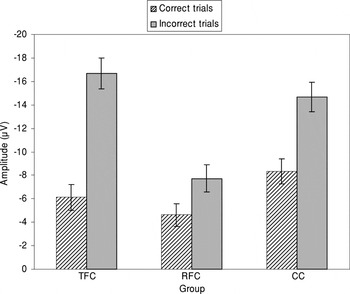
What is not clear is whether disturbances in mPFC functioning are produced by similar mechanisms to those producing disturbances in behavioral, autonomic, and neuroendocrine responses to stressors. What is apparent from the above studies is that translational research should incorporate neurocognitive assessments of specific prefrontal regions into assessment protocols (Pollak, 2005). Consistent with this suggestion are data from a small pilot study using event-related potentials (ERPs) to examine the impact of a foster care intervention on activity of the ACC (Fisher, Martin, Bruce, & Fox, 2006; see also Dozier's Attachment and Biobehavioral Catch-up Intervention Protocol, Dozier, Peloso, Sepulveda, et al., in press). The ACC plays a critical role in effortful control of attention and action (Posner & Petersen, 1990), and is a component of the mPFC network with extensive reciprocal connections to the amygdala and hippocampus and outflow pathways to the HPA and sympathetic nervous system (SNS). Using a flanker task, slowing of responses following an error and an early negative component in the ERP in response to error feedback were examined in children who had been randomly assigned to treatment versus regular foster care. They were compared to a similar group of lower income nonmaltreated children. The intervention involved supporting the foster parent's ability to manage the behavior problems of their charges without hostility and in ways that allowed supportive relationships to be established between the foster parents and child. Children who had lived in treatment foster care, like the comparison children, exhibited a significant error feedback-related negativity in their ERPs (see Figure 3). This was not noted among the regular foster care children. These data suggest that treatment foster care had helped normalize this aspect of mPFC functioning in these maltreated, foster care children.

The average amplitude (μV) of feedback negativity across correct and incorrect trials by group: means and standard errors. Adapted from Fisher et al. (2006). ERPs assessed at Cz to a flanker task for children in regular foster care (RFC), therapeutic foster care (TFC), and community comparison (CC) conditions. Therapeutic foster care was the Early Intervention Foster Care Program (Fisher et al., 2005).
Summary
Four key bridging themes were identified: timing, the SHRP, caregiver mediation of the SHRP, and impacts of caregiving on prefrontal functioning and corticolimbic levels of stress organization. The timing of events relative to brain development in the rodent and human at birth suggests that the prenatal period in humans should be included as a focus of translational research on adversity and stress neurobiology. In addition, consideration of data on relative stress hyporesponsiveness suggests that we should extend the lens beyond infancy to include most if not all of childhood. Evidence that the long-term effects of adverse caregiving in the rodent reflect specific aspects of parental care that buffer the HPA axis during early development encourages attempts to identify elements of parental or caregiver behavior that may serve similar buffering functions during human development. Currently, the best translation of these rodent findings appears to be in measures of parental sensitivity and responsiveness; however, the lens on parenting behavior may widen as more studies are conducted that examine caregiving and the regulation of stress neurobiology in children. Finally, animal studies are beginning to note the impact of early parental care on the development of prefrontal systems, in addition to their earlier focus on limbic–hypothalamic functions. These data are consistent with studies with maltreated children that have noted significant impacts on executive functions, and imaging studies that have noted effects on development of the PFC. These data emphasize the value of including neurocognitive and imaging measures in preventive intervention work on early experiences and stress in children (see also Cicchetti, 1996; Curtis & Cicchetti, 2003). We turn now to a more focused examination of the extent to which human studies provide evidence that early maltreatment has lasting impacts on the neurobiology of stress, and in particular on activity of the HPA axis.
Association of Adverse Early Care With Stress Responding in Adulthood
The rodent model demonstrates impacts of early adverse care on stress neurobiology assessed in adulthood. If the rodent model is to help guide intervention work, then adverse early care in humans should be associated with heightened adult stress responding. However, human studies are invariably difficult to interpret because they must be based on experiments of nature and thus lack the rigor of the animal research. Therefore, as a bridge to human development, we will first turn to nonhuman primate studies.
Nonhuman primate outcomes
In nonhuman primate research (see for review, Levine, 2005b), researchers have examined a number of different adverse early life experiences, ranging from separating the infant from the mother (and sometimes also the social group) to rearing the infant under conditions in which it receives little of the care typical of the species (i.e., rearing on cloth surrogates or only with other infant monkeys). Typically, outcome measures were obtained when the animals were older, but still juveniles. These adverse early care experiences have been shown to impact behavior, increasing fearfulness, reducing exploration of novel environments, and decreasing social status. Notably, relatively few consistent long-term impacts on activity of the HPA axis have been reported. A few studies have shown long-term increases in the production of ACTH or cortisol; however, most studies have revealed normal to blunted activity of the HPA system.
Moving to higher levels of the stress system, however, we find more evidence of hyperresponsiveness following early adversity. Specifically, there is evidence of increased amygdala reactivity (e.g., increased startle responses) several years after repeated, unpredictable separations early in life (Sanchez, Noble, et al., 2005). Disturbances in maternal care in Bonnet macaques produces increased CRF in spinal fluid, sensitization of the NE system, and behavioral sensitization to fear stimuli, while resulting in normal to low cortisol levels (e.g., Rosenblum et al., 2002). Extreme disturbances in early care (e.g., isolation rearing) have also been shown to produce subtle changes in the primate hippocampus, which may reduce the resilience of the hippocampus and increase the risk of hippocampal atrophy in response to later insults (Siegel et al., 1993).
There is also increasing evidence that early adverse care in primates influences the development of the PFC. Thus, monkeys reared in isolation exhibit problems with tasks that involve ventromedial regions of the PFC (see review, Sanchez et al., 2001). As noted, medial and ventral regions of the PFC and ACC have rich bidirectional connections with the amygdala, and appear to be involved in regulating behavioral, autonomic, and neuroendocrine responses to psychosocial stressors (Sullivan & Gratton, 2002). Glucocorticoid overexposure impairs these regions, potentially reducing stress and emotion regulatory competence. Overall, the nonhuman primate data are consistent with the idea that early adverse care will have its largest impact on systems that are still developing. The HPA axis is relatively mature at birth in most primates, and thus may be relatively protected from permanent changes introduced through variations in experience (e.g., Levine, 2005b). In contrast, higher levels of the stress system (e.g., amygdala, mPFC) that mature for longer periods after birth in primates may be more significantly affected. These primate data suggest that in studying human postnatal exposure to adverse care we may need to focus on the extrahypothalamic CRF system, fearful, anxious behavior orchestrated by circuits involving the amygdala, and potentially the development of circuits in the PFC that are involved in modulating, containing, and terminating fear and stress responses.
Human adults
The animal models of early adverse care emphasize deprivation or neglect. In contrast, all of the adult human studies in this area have examined the sequelae of physical and sexual abuse. Furthermore, most of these studies have methodological limitations in that they focus only on women, deal with retrospective reports of childhood experiences, and often fail to control for current life stress (see review by Heim, Plotsky, & Nemeroff, 2004). Nonetheless, these studies provide some useful guidance. One of the clearest results from these studies is that the association between childhood abuse and the neurobiology of stress in adulthood is a function of whether or not the abuse is associated with adult psychopathology. We will, thus, consider outcomes for adults with and without psychological disorders pursuant to their childhood maltreatment.
Turning first to individuals without psychopathology, by definition, these individuals are resilient (Kinard, 1998). Across the various studies of resilient adults, we find evidence of reduced activity of stress neurobiology. The CRF challenge test produces elevations in ACTH and cortisol. The magnitude of the ACTH response is inversely proportional to the pituitary's chronic or traitlike exposure to CRF (see for review, Heim et al., 2004). Chronic, high CRF exposure from the hypothalamic CRF system results in downregulation of CRF receptors in the pituitary. Resilient adult survivors of child abuse produce larger ACTH responses in the CRF challenge test than do healthy adults with nonabusive childhood (Heim et al., 2004). This suggests an atypically low chronic CRF drive in these individuals. Resilient adult survivors also produce larger ACTH responses to a psychosocial stressor than do healthy adults with nonabusive childhood; however, their cortisol and cardiac responses are normal to blunted (e.g., Girdler et al., 2003). This suggests reduced sensitivity of the adrenal to ACTH. This has been confirmed in ACTH challenge tests (see for review, Heim et al., 2004). Thus, individuals (notably women) who experience significant childhood maltreatment but do not develop mental disorders show low neuroendocrine responsiveness to stressors, with evidence of traitlike low levels of hypothalamic CRF.
Differences of opinion exist in whether to view this low level of activity as a risk factor. Heim et al. (2004) suggest that sensitization of the pituitary and counterregulation at the level of the adrenal in these women may predispose them to hypersecrete CRF, resulting in depression and CRF receptor downregulation if they experience significant life stressors in adulthood. On the other hand, it is possible that low CRF and HPA reactivity may have preceded rather than followed their early life exposures, protecting these individuals from developing emotional disorders in response to their adverse childhood experiences. Future studies involving longitudinal work may be able to sort out these two alternatives.
Depression and PTSD are two of the sequelae of childhood maltreatment that have been explored most frequently in studies of stress neurobiology. PTSD and depression examined without reference to childhood abuse appear to share hyperactivity of the central CRF system at hypothalamic and/or extrahypothalamic levels (Bremner et al., 1997; Heim et al., 2004). Chronic CRF drive on the pituitary in both depression and PTSD leads to counterregulatory downregulation at the level of the pituitary, resulting in blunted ACTH responses to CRF challenge tests. However, these disorders differ in the sensitivity of feedback regulation of the HPA axis. Depression among adults is associated with reduced sensitivity of negative feedback mechanisms, resulting in larger cortisol responses to stressors, higher basal levels especially late in the day, and reduced suppression of the axis in response to dexamethasone (Heim et al., 2004). In contrast, PTSD is associated with increased sensitivity of negative feedback mechanisms, resulting in blunted cortisol responses to stressors, lower basal cortisol levels especially late in the day, and enhanced suppression of the axis in response to dexamethasone (Yehuda, 2000). The question is whether affective disorders pursuant to childhood maltreatment follow these neuroendocrine patterns.
First, considering depressed women with a history of childhood abuse, their patterns of ACTH and cortisol responses to CRF challenge is comparable to those observed in depression and PTSD without early abuse (Bremner et al., 1997; Heim et al., 2004). Specifically, blunted ACTH and normal to blunted cortisol responses have been noted. This finding is consistent with chronic CRF drive. ACTH stimulation tests permit examination of the sensitivity of the adrenal. Here, compared to adults with depression without childhood abuse who tend to escape from dexamethasone suppression, depressed adults with histories of child abuse tend (like adults with PTSD) to supersuppress in response to the low-dose dexamethasone test (see for review, Heim et al., 2004). However, this supersuppression may reflect unmeasured PTSD in these depressed, abused women (Rinne et al., 2002). Overall, studies using pharmacological challenges suggest that childhood abuse plus either major depression or PSTD in adulthood is associated with supersuppression of the HPA axis combined with hyperactivity of central CRF.
The results of studies using pharmacological challenge tests do not mirror results obtained when psychosocial stressors are used. Rather than the blunted ACTH response observed in CRF challenge tests, for both childhood abuse survivors with major depression (Heim et al., 2000) and those with PTSD (Bremner et al., 1997), hyperresponsiveness of ACTH and cortisol have been noted. The dissociation between the hyporesponsiveness when the pituitary is pharmacologically challenged with CRF, and hyperresponsiveness when psychosocial challenges are imposed clearly implies upregulation of the corticolimbic stress and emotion circuits in response to childhood abuse. As with the resilient adult survivors, we have a chicken and egg problem. We do not know whether these findings reflect the effects of child abuse or explain why these individuals developed affective pathology in response to their early maltreatment. Again, longitudinal work with maltreated children is needed to untangle the direction of effects.
Developmental Studies of Stress and Maltreatment
Alterations in stress neurobiology observed under conditions of adverse care may reflect transient adjustments that will remit to normal functioning once the child's care improves. Transient adaptations that permit maintenance of viability in response to adverse care reflect allostasis or the maintenance of stability (homeostasis) through change (McEwen, 2000). Even when these allostatic adjustments remit under conditions of improved care, the processes of neural development they influenced may impact the child's subsequent development. This can be considered a developmental version of the costs associated with allostasis (McEwen, 2000). In designing preventive intervention research, thus, we need to consider both the patterning of stress responding observed for children during periods of adverse care as well as patterns and sequelae noted for maltreated children following improvements in their care.
As in the adult work, more information is available on children who have been physically and sexually abused than on children who have been neglected but not abused—although many abused children also suffer neglect (De Bellis, 2005). In addition, we have very few studies of children's responses to psychosocial stressors and even fewer studies that have used pharmacological probes. Most of our information on children, therefore, comes from measures of ambulatory cortisol assessed at various points in the day at home or when the children are in group care settings. Although some of the studies have specifically sampled children with depression or PTSD, others have selected children based solely on their maltreatment histories. Overall, the study of stress neurobiology in maltreated children is a relatively new area, and thus much is unknown or uncertain. Despite this, several themes appear to be emerging that may provide some guidance in preventive intervention designs.
Low early a.m. cortisol may reflect ongoing neglect and abuse
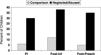
As described above, counterregulatory mechanisms result in downregulation of hypothalamic CRF in response to frequent elevations in glucocorticoids (Makino, Gold, & Schulkin, 1994) and downregulation of CRF receptors in the pituitary in response to chronic CRF drive (see for review, Heim et al., 2004). Thus, although hyperresponsiveness of the axis is often viewed as synonymous with acute stress, hypocortisolism is a likely consequence of chronic stress (Friese, Hesse, Hellhammer, & Hellhammer, 2005). This view is consistent with mounting evidence that children living under conditions of maltreatment tend to exhibit low early a.m. levels of cortisol and a relatively low pattern of cortisol production over the day (Gunnar & Vazquez, 2001). In our research network, we have observed low early a.m. cortisol levels among young children living in an orphanage in Russia (reviewed in Gunnar & Vazquez, 2001), toddlers within a month of adoption from Russian and Chinese orphanages (Bruce, Kroupina, Parker, & Gunnar, 2000), and both infants and preschoolers removed from their homes and placed in foster care (Dozier, Pelsos, Gordon, et al., in press; Fisher, 2005). Among the children recently removed from conditions of neglect and abuse, between 35 and 40% exhibit these abnormally low a.m. levels (see Figure 4). One of us (Fisher) has examined the abuse histories that differentiate preschool-aged children with low a.m. levels assessed within a month of foster placement. Neither physical nor sexual abuse predicted this pattern; instead the best was “failure to provide,” a measure of neglectful care. This finding is highly consist with evidence of low a.m. cortisol levels among institutionalized infants and toddlers, children whose care has been described as “institutional neglect.”

The percentage of neglected/abused children in three studies with extremely low early a.m. cortisol levels. PI, postinstitutionalized infants and toddlers (Bruce et al., 2000); Fost-Inf, infants in foster care and comparison infants (Dozier, Peloso, Gordon, et al., in press); Fost-Presch, preschoolers in foster care and community comparison preschoolers (Fisher, 2005).
As noted, it seems likely that these low a.m. levels reflect a transient downregulation of the axis in response to frequent or chronic CRF drive and elevated glucocorticoids. Abuse as well as neglect might have this effect. Two studies with rhesus infants also tend to support this argument. Sanchez and colleagues (McCormack, Maestripieri, Plotsky, & Sánchez, 2003), have studied the infants of rhesus mothers who spontaneously abuse their offspring. Abuse is most frequent in the first 2 months of life, followed by rejection and neglect in subsequent months. During the first month of life, early a.m. cortisol levels are elevated in these infants relative to infants of supportive mothers; however, beginning in the second month a.m. levels are suppressed below those of typically reared rhesus infants. A similar pattern has been noted for rhesus infants randomly assigned to a repeated, unpredictable separation paradigm (Sanchez, Lyon, et al., 2005; Sanchez, Noble, et al., 2005). These infants experience separations lasting between 30 min and 6 hr (unpredictably), multiple times per week from the time they are 3 to 6 months of age. These separations produce significant elevations in ACTH and cortisol. At 1 year of age, however, these infants exhibit low early a.m. cortisol levels that predict larger responses to acoustic startle when they are 2 years of age. However, these low a.m. levels seem to recover as the animals grow up. By 2 years of age differences in a.m. cortisol levels are not as robust as at earlier ages. Pharmacological probes of the HPA axis in these two rhesus paradigms confirm pituitary downregulation. Both spontaneously maltreated infants and infants subjected to repeated, unpredictable separations exhibit blunted responses to CRF challenge, suggesting chronic CRF drive and resulting downregulation of ACTH sensitivity to CRF.
These data raise the question of whether children experiencing chronic stressors will necessarily exhibit low cortisol levels. This seems unlikely. Rather, we might expect that acute psychosocial stressors could still produce large HPA axis responses in chronically stressed maltreated children. This argument is consistent with an analysis of ACTH responses of depressed abused 7- to 13-year-old children (Kaufman et al., 1997). Consistent with work on depressed adults, the researchers anticipated a blunting of the ACTH response to CRF among the depressed maltreated children. Instead, they noted large elevations. Upon close examination, ACTH responses were bimodal, with no overlap between those showing hyperactivation and normal to low activation in response to CRF. What distinguished the two groups was ongoing and severe emotional maltreatment in the families of the hyperresponders, including continuing exposure to violence, threats of abandonment and physical harm, and rejection of the child's attempts at emotional closeness.
Low a.m. cortisol levels may remit with improved care
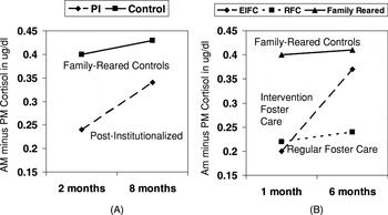
Several findings point to the possibility that with improved care, young children with low a.m. cortisol levels will begin exhibiting more normal patterns of diurnal cortisol production. First, although all young children examined in orphanages have thus far shown the blunted or suppressed pattern of early a.m. cortisol production (reviewed in Gunnar & Vazquez, 2001), in two studies of orphanage-reared children studied 3 or more years after adoption none of the children exhibit low a.m. cortisol levels (Gunnar, Morison, Chisholm, & Schuder, 2001; Kertes, Gunnar, & Madsen, 2006). Furthermore, in a small study of orphanage-reared toddlers studied first at 2 and then at 8 months postadoption, a.m. cortisol levels were low initially and the decrease from a.m. to p.m. was small, but then increased to approach the levels of family-reared toddlers (Bruce et al., 2000; see Figure 5A). These data suggest that we may be able to use a.m. cortisol and the diurnal decrease from a.m. to p.m. to track the impact of interventions in neglected, abused children. Consistent with this possibility, Fisher (2005) has shown that when children with low a.m. cortisol levels are placed in the treatment arm of the Early Intervention Foster Care program, they show increases in a.m. cortisol levels and larger diurnal decreases in a.m. to p.m. cortisol concentrations over time (see Figure 5B). This was not observed for children placed in regular foster care.

The increase in the diurnal change in salivary cortisol (μg/dl) from wakeup to bedtime between when children are first placed in supportive families and several months later: (A) data on children adopted internationally from institutions in Russia and China compared to children reared in their birth families in the United States (Bruce et al., 2000) and (B) data on preschoolers placed in either the intervention arm or regular foster care arm of the Early Intervention Foster Care Program compared to children in the community comparison group (Fisher, 2005).
Severe maltreatment may increase cortisol set points
There are now several studies of children who were severely maltreated in infancy and early childhood, removed from these maltreating environments, and assessed several years later when they were between approximately 7 and 13 years of age. In some, but not all of these studies, the children had chronic PTSD. Examining children with PTSD, De Bellis, Baum, et al. (1999) reported elevated 24-hr urinary cortisol and catecholamine levels. In a similar population, Carrion and colleagues (2002) noted elevated salivary cortisol levels. In both cases the maltreated children were compared to low-risk, family-reared children. Furthermore, neither study included maltreated children without behavioral or emotional problems. In contrast, Cicchetti and Rogosch (2001) collected salivary cortisol data on maltreated and control children during a 5-day summer camp. Overall, the maltreated and control children's cortisol levels did not differ. However, a subset of children with high (>1 SD) cortisol levels over the day were found. These children were more likely to have been severely and multiply abused. Severity of sexual abuse was also implicated. Studies of postinstitutionalized children also suggest elevated cortisol levels several years postplacement for children exposed to extreme deprivation (Gunnar et al., 2001), particularly those whose deprivation produced severe growth retardation (Kertes et al., 2006).
These studies of severely neglected and abused children stand in marked contrast to the data reviewed earlier on HPA axis activity among adults who were maltreated as children. In the adult studies, if anything, baseline levels of cortisol tend to be normal to low, consistent with reports on adults who develop PTSD following traumas experienced in adulthood (Yehuda, 2000). These are not the only data on abused children that differ from those obtained for adults with PTSD. Based on rodent stress models, hippocampal atrophy is expected due to either chronic increases in CRF or glucocorticoids. Adults with PTSD have often been shown to have smaller hippocampal volumes than controls (see for review, Bremner, 2002). This has also been noted for adult women who experienced abuse as children (Bremner et al., 2003). However, smaller hippocampal volumes in these studies were restricted to abuse survivors who developed either depression or PTSD. Notably, there is no evidence of hippocampal volume reduction in studies of maltreated children, even though all of the imaging studies have been conducted using maltreated children with chronic PTSD (Carrion et al., 2001; De Bellis, Keshavan, et al., 1999; Teicher et al., 1997). Similarly, rhesus monkeys reared in social isolation during infancy and assessed as young juveniles also failed to exhibit reduced hippocampal volumes relative to mother-reared rhesus juveniles despite marked disturbances in behavior (for a review, see Sanchez et al., 2001). Several researchers have suggested that hypocortisolism and hippocampal atrophy may take a number of years to develop and thus hypercortisolism in childhood and normal hippocampal volumes transition into hypocortisolism and hippocampal atrophy only with time or perhaps at puberty (De Bellis, 2001; Yehuda, Halligan, & Grossman, 2001). This possibility should alert researchers to look for dynamic changes in stress neurobiology related to development and ongoing physiological adaptations to previous disturbances. Any study of the impact of interventions will need to be designed to identify intervention-associated changes and differentiate them from endogenous change-inducing processes. This may be extremely difficult to accomplish when only one or a few indices of stress responding are used. Psychosocial challenge tests as well as pharmacological probes ultimately may be needed.
Summary
Although nonhuman primate studies have revealed few consistent long-term effects of early maternal deprivation on activity of the HPA axis, studies of survivors of child abuse reveal a number of alterations. The patterns noted depend on whether pharmacological challenges that act directly on the HPA axis or psychosocial challenges that require corticolimbic input are employed. They also depend on whether abused children are studied in childhood or as adults, and they likely also depend on the degree of ongoing trauma in the individual's life. Finally, they depend on the individual's clinical status. Age, time since rescue, current life stressors/traumas, and current and past psychopathology will need to be taken into account if preventive intervention researchers are to adequately incorporate stress-sensitive neurobiological measures.
Preventive Interventions
Many of the evidence-based practices for maltreated children emerged out of the field of family-based prevention science. Prevention science, in turn, grew out of the recognition that probabilistic developmental trajectories toward negative outcomes could be effectively averted through systematic and well-timed intervention efforts (Reid & Kavanagh, 1985). Parenting has been a major focus of preventive intervention efforts because it is malleable and effective in altering outcomes for young children (Reid & Eddy, 1997). Although efficacy trials to evaluate parenting interventions are beginning to include measures of stress neurobiology (e.g., Dozier's Attachment and Biobehavioral Catch-up, Dozier, Peloso, Sepulveda, et al., in press; Fisher's Early Intervention Foster Care, Fisher et al., 2005), the interventions themselves continue to be derived predominantly from psychological theories. Given our limited state of knowledge, moving beyond this point to use information on the impact of early experiences on stress neurobiology to more completely inform preventive interventions is probably somewhat beyond our grasp at this time. However, there are several approaches that may be useful.
Identifying therapeutic components of effective preventive interventions
As discussed earlier, many of the evidence-based interventions for high risk children focus on reducing externalizing problems through training caregivers, including birth, adoptive and foster parents, in effective behavior management techniques (e.g., Fisher et al., 2005). Some, as noted earlier, are focused on developing secure and supportive parent–child attachment relationships through training parents to be more accomplished at reading children's signals and responding appropriately and supportively to their needs (e.g., Cicchetti, 2005; Dozier, 2003). Although it would seem that the latter follow more directly from the early experience-stress literature, there is evidence that both alter activity of stress-sensitive systems (Dozier, Peloso, Sepulveda, et al., in press; Fisher, Gunnar, Chamberlain, & Reid, 2000). From an organizational perspective, both approaches likely address needs of children at different points in development (Dozier, Albus, Fisher, & Sepulveda, 2002). Establishing one or more secure attachment relationships is a critical developmental task of the infancy period, while learning to manage one's behavior in relation to rules and norms becomes important in the toddler and preschool years. Nonetheless, the early experience-stress research would seem to suggest that social regulation of the HPA axis is dependent on parental care, with the best evidence in humans suggesting that it is closely associated with sensitive, responsive care and relationship security.
Tracing association between preventive interventions to changes in parenting behaviors, child behaviors, and activity of stress-sensitive neurobiological systems may help to parse pathways through which successful interventions have their effects. It is certainly possible that by focusing on effective behavior management techniques, preventive interventions based on social learning theory may reduce noxious child behaviors, lead to reductions in hostile responses of parents and others to those behaviors, reduce child exposures to social threat, and prevent or ameliorate the development of hyperreactive, defensive stress neurobiology. It is also possible that such behavior management techniques, because they encourage and reward self-control, may help to foster more adequate corticolimbic pathways to stress and emotion regulation. However, a third possibility is that once the parent is assured of their competence in managing the child's noxious behaviors, this improves parent affect, including their warmth and sensitivity to the child, which in turn, promotes more a more secure parent–child attachment that then can serve as a better social regulator of the child's stress neurobiology. It is also possible that all of these pathways operate but are differentially salient at different points in development or for different children.
We have several preliminary pieces of evidence that suggest that changes in the neurobiology of fear and stress may track reductions in anxiety and increases in secure attachment behavior among children placed in an intervention that has its roots in social learning theory (Fisher et al., 2005). This intervention, which focuses on foster parents of preschool-aged children, is a comprehensive treatment model. Although foster parents are certainly trained in effective behavior management techniques, they are also provided training in appropriately and sensitively interpreting the children's behavior. Thus, although grounded in social learning theory, this intervention contains components that would also be found in interventions grounded in attachment and organizational approaches to preventive interventions. As noted, for children who enter foster care with low early a.m. cortisol levels, being assigned to the intervention as opposed to regular foster care arm of the study is associated with a normalization of the cortisol diurnal rhythm. More than that, however, improvements in the cortisol diurnal rhythm among these children tracked decreases in anxious, but not in externalizing behaviors. Furthermore, a pilot study of attachment behaviors displayed by the children over several months was conducted using a variant of the Stovall and Dozier (2000) attachment diary. Preschoolers in the intervention arm of the study showed a reduction in avoidant and increase in secure attachment behavior, while those in the regular foster care arm of the study, if anything, showed an increase in avoidant attachment behavior. Nonmaltreated comparison children showed stability in attachment patterns as would be expected as their family situation had not changed.
Of course, only home baseline cortisol levels were assessed in both the Fisher and Dozier preventive intervention studies. Improving the child's felt safety may reduce the chronic CRF drive on the pituitary and thus allow normalization of the diurnal cortisol rhythm. However, without additional probes of the HPA axis, this hypothesis cannot be verified. Furthermore, these studies did not involve any attempt to assess higher levels of the stress system, including higher levels of the HPA axis. Finally, such studies are only beginning to attempt assessment of corticolimbic pathways that may be involved in fear and stress regulation. It is certainly possible that different levels of stress neurobiology are responsive to different experiences. This suggests that examining pathways from preventive interventions to changes in stress neurobiology should include measures that assess different levels of organization of stress neurobiology (see also, Curtis & Cicchetti, 2003).
Potential neurobiological targets
One of the serious problems in studying the neurobiology of stress in preventive intervention designs with high-risk children involves ethical and practical limitations on the measures that can be employed. This is especially true if one wants to focus on preventive interventions with infants and young children. Functional imaging that allows assessment of corticolimbic pathways involved in processing threat stimuli is being employed with older children and adolescents (e.g., Nelson et al., 2003), but is not yet feasible with very young children. Structural imaging, including imaging of white matter tracts, may be feasible with young children, but typically it is difficult to get young children to lie still for long enough without medicating them, restricting the use of imaging procedures for research purposes. Using imaging to examine changes in corticolimbic networks involved in stress and emotions may however be highly useful in preventive interventions with older children and adolescents.
Electroencephalogram (EEG) measures are quite feasible with infants and young children, but are only beginning to be used in preventive intervention designs. EEG measures are being employed in a study of orphanage-reared children randomly assigned to continue in institutional versus move to therapeutic foster care (Zeanah et al., 2003). Consistent with evidence of reduced activity of regions in the PFC reported in a positron emission tomography study of postinstitutionalized children (Chugani et al., 2001), initial reports on the orphanage-reared children described underactivity in their EEGs compared to the EEGs of a family-reared comparison group (Marshall, Fox, & The Bucharest Early Intervention Project Core Group, 2004). A similar alteration in power across the EEG spectrum was noted in a large sample of adults who reported more early life stress (McFarlane et al., 2005). Although there have also been studies of differences in hemispheric development in abused children using EEG measures (Ito, Teicher, Glod, & Ackerman, 1998), to our knowledge there have been no published reports examining frontal EEG asymmetry in maltreated children. Given that associations between low parental responsiveness and right frontal EEG asymmetry have been reported (Hane & Fox, in press), EEG asymmetry measures may be a useful tool for preventive intervention researchers.
Fear-potentiated startle is another measure that may be used with even very young children, and could shed light on the development of stress neurobiology. Under conditions of threat, the eye-blink startle response is larger than under neutral or positive conditions. Fear-potentiated startle reflects activity of the amygdala and BNST (Davis, Walker, & Lee, 1997). Increased activity of extrahypothalamic CRF amplifies fear-potentiated startle responses (Lee, Schulkin, & Davis, 1994), and individuals who tend to show larger HPA responses to stressors exhibit larger startle reactions (Grillon et al., 2005).
As noted, assessments of the HPA axis in maltreated children have relied almost entirely on measures of cortisol (salivary or urinary). Measures of ACTH that require blood sampling have sometimes been collected, but pharmacological challenge tests have not been used with children prior to adolescents (e.g., De Bellis et al., 1994). Being limited to measures of cortisol seriously restricts the researcher's ability to assess changes in HPA axis activity. Combining assessments of cortisol with measures of other stress-sensitive systems, however, may provide useful information. There is increasing interest in examining the role of dehydoepiandrosterone (DHEA) in buffering or modulating effects of cortisol. DHEA and its sulfated version DHEA-S are androgens produced by the adrenal cortex that tend to have effects that oppose the actions of cortisol. The ratio of cortisol to DHEA may index the extent to which altered cortisol levels may affect physical and neurobiological development (Granger, Schwartz, Booth, Curran, & Zakaria, 1999). Recently, there has been increased evidence that low DHEA to cortisol ratios are associated with heightened fear-potentiated startle responses in adults (Grillon et al., 2005) and increased probability of developing depression in adolescents (Goodyer, Park, Netherton, & Herbert, 2001).
The relation of cortisol responses to responses of the SNS may also be relevant. The HPA axis and the sympathetic adrenomedullary (SAM) system are the two major arms of the stress system operating in the periphery of the body (Palkovits, 1987). Under conditions of threat, increases in both HPA and SAM activity are anticipated. Chronic stress may affect these systems differently. For example, in PTSD increased heart rate and sympathetic outflow are typically noted, whereas cortisol levels are normal or suppressed (Yehuda, 2000). Dissociation of the SAM and HPA systems may be an index of adverse experience and increased internalizing problems (Bauer, Quas, & Boyce, 2004). Measures of cardiac preejection period (PEP) can be used to assess sympathetic contributions to heart rate (Berntson, Cacioppo, Binkley, Uchino, & Quigley, 1994), and these measures are being employed in children (Alkon et al., 2003). It is also possible to indirectly measure sympathetic activity in saliva by assaying for α-amylase (AA; Granger et al., in press). AA is an enzyme produced locally in the oral mucosa that is sensitive to sympathetic stimulation. Increases in SNS (predominantly NE but perhaps also SAM) activity result in increased AA concentrations in saliva. Recently, salivary AA has been shown to be responsive to psychosocial stressors (i.e., the Trier Social Stress Test) in adults (Nater et al., 2005) and children (Kivlighan, Wewerka, Gunnar, & Granger, 2006).
There are more measures available than have been employed in studies of maltreated children and in the context of preventive intervention designs. Nonetheless, without theories about the effects that would be expected, a multisystems approach to measurement is not likely to provide clarity. This is especially true for developmental research as for many of these measures we have little normative developmental data. However, incorporated judiciously into preventive intervention designs, patterns of relations among measures (e.g., changes in DHEA to cortisol ratios; changes in frontal EEG asymmetry; alterations in cortisol and PEP or increased sympathetic–cortisol coregulation to psychosocial stressors) may help elaborate changes in neurobiology that support or perhaps herald improvement in child functioning.
Nonresponders and targeted interventions
Even preventive interventions with solid track records do not help all children. One hope for a psychoneurobiological approach to the effects of maltreatment and intervention efficacy is to develop models of children who do not respond to intervention efforts and to design interventions that are more effective for them. PTSD is challenging to diagnose in young children and few meet DSM-IV criteria for diagnosis (Scheeringa, Zeanah, Myers, & Putnam, 2005). Yet, traumatized children display high rates of functional impairment and respond poorly to interventions available in the community (Cicchetti, 2005; Scheeringa et al., 2005). PTSD may reduce responsiveness to preventive interventions that do not specifically address their disturbances in functioning. This is one place where the addition of neurobiological measures of stress and emotion may aid in improving preventive intervention designs. In addition, children who are hyper- or hyporesponsive to threat stimuli may require different types of intervention to improve their functioning. At least one study of children with disruptive behavior disorders has shown that those with low cortisol responsiveness to threat respond poorly to psychosocial intervention (van de Wiel, van Goozen, Matthys, Snoek, & van Engeland, 2004). Studies of typically developing children have also shown that those with low sympathetic tone respond poorly to authoritative patterns of parenting, requiring a close and secure relationship with the parent to develop patterns of moral or conscientious behavior (Fowles & Kochanska, 2000). Children who are hyperresponsive to threat, in contrast, may appear to be responding to treatments aimed at managing externalizing behavior problems, but the treatment may not be addressing their vulnerability to stress and risk of anxiety or depressive disorders (van Goozen, Matthys, Cohen-Kettenis, Buittelaar, & van Engeland, 2000).
Summary and Conclusions
Animal models of early experience and stress neurobiology are frequently invoked in discussions of how early abuse and neglect may increase the risk of psychopathology. Species differences and variations in the nature of adverse early experiences studied in animals and humans preclude direct translation of the animal studies to the human case. This, in turn, limits our ability to translate basic research on early experiences and stress into more effective preventive intervention designs for maltreated children. However, despite the great deal that is yet unknown, there appear to be striking continuities between the animal and human literatures. The most notable difference may reflect, at least partially, the timing of insults relative to neural maturity. In humans, it seems likely that experiences beginning in the prenatal period and extending through most of childhood may shape the development of stress neurobiology and risk for psychopathology. However, because the neurobiology of stress is more mature in the human than rodent at birth, it also seems likely that postnatal experiences will have their most long-term impacts on extrahypothalamic levels of stress system function, particularly networks involving corticolimbic pathways. Used alone, it is unlikely that noninvasive measures such as levels of salivary cortisol will be able to provide sufficient insight into the impacts of maltreatment and the efficacy of preventive intervention to be of much use. However, when combined with measures of other stress-responsive neural systems in designs that test specific hypotheses, assessments of stress neurobiology may prove to be useful tools in identifying (a) characteristics of maltreating and therapeutic environments that alter stress and defensive responding, (b) children who may benefit from different types of interventions, and (c) windows of opportunity to alter the developmental course of children subjected to abuse and neglect early in life.