Introduction
Suicidal behavior is a leading cause of morbidity and mortality in adolescence (Lubell et al. Reference Lubell, Kegler, Crosby and Karch2007). Despite progress in understanding risk factors for suicidal behavior (Brent et al. Reference Brent, Baugher, Bridge, Chen and Chiappetta1999), the pathogenesis is poorly understood, including possible alterations in neural circuitry that might predispose to suicidal behavior. Prevention of early-onset suicide attempt is identified as a mission of the US National Institute of Mental Health (NIMH). Progress in the prediction and prevention of suicidal behavior would be facilitated by identification of biomarkers for suicide risk, which could increase understanding of the pathogenesis of suicidal behavior in order to target and reverse such pathogenic processes.
Post-mortem studies have demonstrated abnormalities in the prefrontal cortex in adult victims of suicide (Mann et al. Reference Mann, Huang, Underwood, Kassir, Oppenheim, Kelly, Dwork and Arango2000). In addition, cognitive inflexibility and impaired executive function related to these brain regions have been shown to be characteristic of suicide attempters (Keilp et al. Reference Keilp, Sackeim, Brodsky, Oquendo, Malone and Mann2001; Jollant et al. Reference Jollant, Bellivier, Leboyer, Astruc, Torres, Verdier, Castelnau, Malafosse and Courtet2005). One neuroimaging study in adult suicide attempters (Oquendo et al. Reference Oquendo, Placidi, Malone, Campbell, Keilp, Brodsky, Kegeles, Cooper, Parsey, van Heertum and Mann2003b) reported lower glucose uptake in the prefrontal cortex and anterior cingulate gyrus in high versus low lethality suicide attempters. With regard to emotion processing, vulnerability to suicidal behavior has been associated with differences in response to both negative and positive emotion. Specifically, adult male suicide attempters showed greater activity in the right lateral orbitofrontal cortex [Brodmann area (BA) 47] and decreased activity in the right superior frontal gyrus (BA 6) to 100% intensity angry versus neutral faces relative to healthy and depressed non-attempter controls. In response to 50% intensity happy faces, attempters showed greater activity than healthy and non-attempter depressed controls in the right anterior cingulate gyrus (BA 32/10) (Jollant et al. Reference Jollant, Lawrence, Giampietro, Brammer, Fullana, Drapier, Courtet and Phillips2008). Together, these findings suggest that abnormal prefrontal cortical function during emotion processing may underlie vulnerability to suicidal behavior in depressed adults.
Regarding studies in adolescence, we reported in the same group of adolescents that suicide attempt may not be associated with abnormal activity in the neural circuitry supporting response inhibition. In fact, during response inhibition, adolescents with depression but no history of suicide attempt, relative to adolescents with depression and a history of suicide attempt, demonstrated a compensatory increase in activity in the right anterior cingulate gyrus (Pan et al. Reference Pan, Batezati-Alves, Almeida, Segreti, Akkal, Hassel, Lakdawala, Brent and Phillips2011). In parallel, there is evidence of greater activity during emotional processing in depressed, relative to healthy, adolescents in bilateral amygdalae and hippocampi (Lau et al. Reference Lau, Goldman, Buzas, Hodgkinson, Leibenluft, Nelson, Sankin, Pine and Ernst2010), and greater activity in the ventromedial prefrontal cortex and anterior cingulate when viewing fearful faces (Killgore & Yurgelun-Todd, Reference Killgore and Yurgelun-Todd2006). There are, however, no studies examining neural activity during emotion processing in adolescent suicide attempters. Research is therefore needed to determine the extent to which prefrontal cortical dysfunction during emotion processing may contribute to vulnerability to suicidal behavior in adolescents.
A complementary approach to regionally specific analyses assesses functional connectivity. This may be particularly salient in adolescence given prominent developmental changes in connectivity. For example, there is evidence that adults are more able to modulate activity in the anterior cingulate in response to fear and non-emotional cues than adolescents (Monk et al. Reference Monk, McClure, Nelson, Zarahn, Bilder, Leibenluft, Charney, Ernst and Pine2003) and that maturation of emotional processing is related to the progressive acquisition of greater functional activity within the prefrontal cortex in adolescence (Yurgelun-Todd & Killgore, Reference Killgore and Yurgelun-Todd2006). Fronto-limbic circuitry is implicated in pediatric depression (Hulvershorn et al. Reference Hulvershorn, Cullen and Anand2011). Abnormal functional connectivity of the anterior cingulate gyrus (BA 32), in particular, has been demonstrated during resting-state analysis with the amygdala, superior, medial and lateral prefrontal cortical regions, temporal regions, and the insula in adolescent depression (Cullen et al. Reference Cullen, Gee, Klimes-Dougan, Gabbay, Hulvershorn, Mueller, Camchong, Bell, Houri, Kumra, Lim, Castellanos and Milham2009). In adult depression, at rest and to emotional pictures, there is evidence of decreased functional connectivity between the anterior cingulate gyrus and limbic regions (Anand et al. Reference Anand, Li, Wang, Wu, Gao, Bukhari, Mathews, Kalnin and Lowe2005).
The aim of the present study was to measure neural activity during processing of emotional faces in adolescents with a history of depression and suicide attempt (ATT) versus age-matched adolescents with a history of depression but not suicide attempt (NAT) and age-matched healthy controls (HC), using functional magnetic resonance imaging (fMRI). We hypothesized that, like adult suicide attempters (Jollant et al. Reference Jollant, Lawrence, Giampietro, Brammer, Fullana, Drapier, Courtet and Phillips2008), ATT relative to the other two groups would show differential patterns of elevated prefrontal cortical activity to both angry and happy faces. We also examined functional connectivity by applying psychophysiological interaction (PPI) analysis (Friston et al. Reference Friston, Buechel, Fink, Morris, Rolls and Dolan1997) to fMRI data from the emotion-processing task. Existing data in depressed adults allowed us to hypothesize that both adolescent groups with a history of depression would show reduced functional connectivity among anterior cingulate gyri, prefrontal, temporal cortices and insulae to all face emotion conditions relative to HC.
Method
Participants
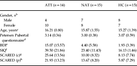
A total of 44 adolescents completed the study (Table 1), including those with: (1) lifetime history of suicide attempt and major depressive disorder (MDD) (ATT; n = 14); (2) history of MDD, but no history of suicide attempt (i.e. non-attempters; NAT; n = 15); and (3) healthy control participants without history of psychiatry disorder or suicide attempt (HC; n = 15).
Demographic information and clinical variablesa
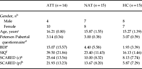
ATT, Adolescent suicide attempters; NAT, non-attempters with a history of major depressive disorder; HC, healthy controls; BDI, Beck Depression Inventory; SIQ, Suicidal Ideation Questionnaire; SCARED (c), Screen for Childhood Anxiety Related Emotional Disorders child version; SCARED (p), Screen for Childhood Anxiety Related Emotional Disorders parent version.
Data are given as mean (standard deviation) or as number of subjects.
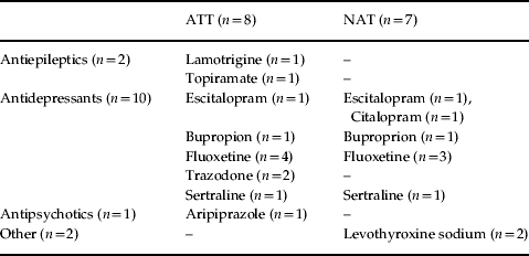
a All participants taking medication were taking an antidepressant. Two NAT had augmentation with levothyroxine. One ATT had augmentation with aripiprazole, and two ATT had augmentation with antiepileptic medications (see Table 2).
b ATT, NAT and HC did not differ significantly in gender ratio (p = 0.391).
c ATT, NAT and HC did not differ significantly in age (F 2,41 = 0.877, p = 0.506).
d ATT, NAT and HC did not differ significantly in Petersen Pubertal questionnaire (χ2 = 0.712, p = 0.701).
e ATT had significantly greater BDI scores than NAT and HC (χ2 = 13.92, p = 0.001).
f ATT had significantly greater SIQ scores than NAT and HC (χ2 = 20.53, p = 0.0001).
g ATT had significantly greater SCARED (c) than NAT and HC (χ2 = 14.63, p = 0.001).
h ATT had significantly greater SCARED (p) scores than NAT and HC (χ2 = 15.90, p = 0.0001).
ATT and NAT were recruited from existing studies and a clinic for depressed youth. HC were recruited from existing healthy control groups and advertisements in pediatric practices. Inclusion criteria for NAT and ATT included a lifetime history of MDD according to Diagnostic and Statistical Manual of Mental Disorders, Fourth Edition (DSM-IV) criteria. ATT had a history of at least one suicide attempt as defined by the Columbia Classification Algorithm of Suicide Assessment (Posner et al. Reference Posner, Oquendo, Gould, Stanley and Davies2007). All participants were right-handed.
Exclusion criteria included neurological disorders, head injury, Verbal Subtest of the Wechsler Intelligence Test score <80 (Wechsler, Reference Wechsler1999), use of sedative medication, drug or alcohol disorder or positive urine drug screen, pregnancy, magnetic resonance imaging (MRI) ineligibility, bipolar disorder or psychosis. Any ATT with suicide attempt causing neurological damage or long-term physiological effects was excluded.
DSM-IV criteria were assessed using the Schedule for Affective Disorders and Schizophrenia for School-Age Children-Present and Lifetime Version (Kaufman et al. Reference Kaufman, Birmaher, Brent, Rao, Flynn, Moreci, Williamson and Ryan1997). Suicide attempt was assessed at the time of the scan using the Suicide Intent Scale (Beck et al. Reference Beck, Schuyler, Herman, Beck, Resnick and Lettieri1974) and Suicide History Form (SHF; Oquendo et al. Reference Oquendo, Halberstam, Mann and First2003a). Depression, anxiety, suicidal ideation and pubertal status were assessed with the Beck Depression Inventory (BDI; Beck et al. Reference Beck, Ward, Mendelson, Mock and Erbaugh1961), Screen for Childhood Anxiety Related Emotional Disorders (SCARED; Birmaher et al. Reference Birmaher, Khetarpal, Brent, Cully, Balach, Kaufman and Neer1997), Suicidal Ideation Questionnaire (SIQ; Reynolds, Reference Reynolds1987) and the Petersen Development Scale (Petersen et al. Reference Petersen, Crockett, Richards and Boxer1988), respectively. After complete description of the study to all subjects, parental written informed consent and participant written informed assent were obtained.
A total of 51 participants were assessed. Exclusions included: two with claustrophobia, one with structural abnormality, one with marijuana dependence, two for younger age (10 years), and one due to scanner malfunction. No participants had to be excluded for movement (>3 mm). Median time since last attempt was 26.1 (s.d. = 29.1) months, and mean lethality of attempt from the SHF was 2.29 (s.d. = 1.20) (injury requiring medical intervention; Table S1). ATT had significantly higher SIQ, BDI and SCARED scores than NAT. Of the participants, three ATT and one NAT had a current depressive episode (BDI >20), and nine ATT and seven NAT were taking medication for depression. Medications included selective serotonin reuptake inhibitor (SSRI) antidepressant medication and augmentation (Table 2).
Medication
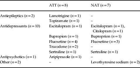
ATT, Adolescent suicide attempters; NAT, non-attempters with a history of major depressive disorder.
Paradigm
Participants completed two previously employed (Lawrence et al. Reference Lawrence, Williams, Surguladze, Giampietro, Brammer, Andrew, Frangou, Ecker and Phillips2004; Hassel et al. Reference Hassel, Almeida, Kerr, Nau, Ladouceur, Fissell, Kupfer and Phillips2008; Versace et al. Reference Versace, Thompson, Zhou, Almeida, Hassel, Klein, Kupfer and Phillips2010) 6-min event-related facial emotion-processing fMRI runs with 20 faces each of 100% intensity, 50% intensity and 0% intensity (neutral) faces totaling 60 randomized cues in each experiment of either (1) happy or (2) angry. Images were 10 individuals from a standardized series (Young et al. Reference Young, Perrett and Calder2002). Participants viewed each image for 2 s and determined the gender of the face with a button press (index finger for female, third finger for male). Task order was counterbalanced. The interstimulus interval included a baseline fixation cross and varied from 3 s to 8 s (mean = 4.9 s). These emotion-processing tasks have been associated with reliable activity within subcortical neural regions in healthy adults (Surguladze et al. Reference Surguladze, Brammer, Keedwell, Giampietro, Young, Travis, Williams and Phillips2005).
Data acquisition
Scans were acquired on a 3.0 T Siemens Allegra MRI scanner. Functional scans with full coverage of the adolescent brain of 34 contiguous 3-mm axial slices were acquired with a T2-weighted gradient echo planar imaging (EPI) sequence (repetition time: 2000 ms; echo time: 30 ms; field of view: 205 mm; matrix: 64 × 64; in-plane resolution: 3 × 3 mm). Stimuli were projected onto a screen approximately 55 cm from the subject with a rear screen projector. High-resolution T1-weighted magnetization prepared rapid gradient echo (MP-RAGE) structural images of 240 0.8-mm slices were acquired (repetition time: 1630 ms; echo time: 2.48 ms; inversion time: 800 ms; field of view: 200 mm; flip angle: 8°; matrix: 256 × 256).
Task performance
Task performance data were analysed using one-way analyses of variance (ANOVAs) to examine the main effect of group upon response time and numbers of correct gender responses in SPSS v. 20 (IBM, USA).
Imaging analyses
Data were pre-processed and analysed using Statistical Parametric Mapping software (SPM8; http://www.fil.ion.ucl.ac.uk/spm/). Data for each participant were first corrected for differences in acquisition time between slices; realigned using the first slice as a reference and unwarped to correct static inhomogeneity of the magnetic field and movement by inhomogeneity interactions. They were co-registered with the subject's anatomical image, segmented, normalized to standard Montreal Neurological Institute (MNI) template, re-sampled to 3 × 3×3 mm3 voxels, and spatially smoothed with a Gaussian kernel of 6 mm full-width at half-maximum (FWHM).
First-level fixed-effect models were constructed with 0%, 50% and 100% intensities of angry and happy facial emotions. Movement parameters from the realignment stage were entered as covariates of no interest. Trials were modeled using the canonical hemodynamic response function. Contrast maps from first-level analyses (i.e. happy condition: 100% happy face minus fixation, 50% happy face minus fixation, neutral face minus fixation; angry condition: 100% angry face minus fixation, 50% angry face minus fixation, neutral faces minus fixation) were then entered into the second-level analyses (random-effects) using a full factorial model. Separate models were computed for happy and angry conditions.
A second-level random-effects model was used for between-group comparison. We performed two separate 3 group (ATT, NAT, HC) × 3 condition (0%, 50% and 100% intensities) analyses of covariance (ANCOVAs) for angry and happy, covarying for age, to examine the group × condition interaction upon whole-brain activity. Specifically, the 3 × 3 ANCOVA was utilized to identify group differences of the effect of intensity difference. A 3 × 3 factorial design, examining differences between intensities, allowed for cancellation of other mental processes, such as gender identification, common to all groups. Although there was no significant difference in age between the three groups, age was added as a covariate in order to further control for potential effects of brain development (as in Ladouceur et al. Reference Ladouceur, Farchione, Diwadkar, Pruitt, Radwan, Axelson, Birmaher and Phillips2011; Pan et al. Reference Pan, Batezati-Alves, Almeida, Segreti, Akkal, Hassel, Lakdawala, Brent and Phillips2011). Age effect on emotion circuitry may differ differently across groups.
We maintained a cluster-level false-positive detection rate of p < 0.05 for this pattern of neural activity using a voxel-wise threshold of p < 0.05 with a cluster (k) extent empirically determined by Monte Carlo simulation implemented in AlphaSim (afni.nimh.nih.gov/afni/doc/manual/AlphaSim) (Gilbert et al. Reference Gilbert, Akkal, Almeida, Mataix-Cols, Kalas, Devlin, Birmaher and Phillips2005; Monk et al. Reference Monk, Telzer, Mogg, Bradley, Mai, Louro, Chen, McClure-Tone, Ernst and Pine2008; Dickstein et al. Reference Dickstein, Finger, Skup, Pine, Blair and Leibenluft2010; Pan et al. Reference Pan, Batezati-Alves, Almeida, Segreti, Akkal, Hassel, Lakdawala, Brent and Phillips2011; Kim et al. Reference Kim, Thomas, Rosen, Moscicki, Brotman, Zarate, Blair, Pine and Liebenluft2012) to account for spatial correlations between blood oxygen level-dependent (BOLD) signal changes in neighboring voxels. Our main findings survived small volume correction (SVC) with AlphaSim at p < 0.05.
Peak BOLD signal changes were extracted from regions with significant group × condition interaction in both the above 3 × 3 analyses. We utilized a standard peak voxel BOLD signal change to evaluate the peak magnitude for interpretation of the interaction. This is a more conservative approach, which has been employed previously (Hassel et al. Reference Hassel, Almeida, Kerr, Nau, Ladouceur, Fissell, Kupfer and Phillips2008; Pan et al. Reference Pan, Batezati-Alves, Almeida, Segreti, Akkal, Hassel, Lakdawala, Brent and Phillips2011). We then performed post-hoc tests on these extracted BOLD signal values to examine the extent to which pair-wise between-group differences in differences in activity contributed to the significant group × condition interactions, in the above analyses, using independent t tests and appropriate statistical thresholds to control for multiple tests. In these post-hoc tests for regions showing a significant group × condition interaction in the 3 × 3 ANCOVA, a significance threshold of p < 0.05/9=p < 0.006 was employed to control for the three independent between-group pair-wise tests for each of the three conditions in each region.
Functional connectivity analyses
PPI analyses were conducted to investigate the context-dependent contributions of seed region (right anterior cingulate gyrus, BA 32) activity in all the angry and happy conditions (intensities), and functional coupling with other brain regions in relation to task demands. The anterior cingulate seed region was chosen as it was activated in all groups to all conditions. PPI correlated the anterior cingulate seed region activation to other areas as a product of each condition. Motion parameters did not differ between groups, and no participant above 3 mm of motion was included in the analysis.
The PPI physiological variable was first computed by extracting the signal time course for all voxels in the right anterior cingulate gyrus region of interest (ROI) for each individual participant. The ROI was constructed using a mask from the Wake Forest University (WFU) PickAtlas with the anterior cingulate coordinates from the original whole-brain analysis. A total of six analyses were done for each participant (for 0%, 50% and 100% intensities minus baseline for happy and angry conditions). Analyses were Bonferroni corrected for multiple comparisons, with a statistical threshold of p = 0.05/6 (p < 0.008) for each 3 × 1 ANOVA. The PPI interaction variable was then computed as the product of the mean corrected right anterior cingulate gyrus activity and the vector coding for the experimental effect of each condition individually subtracted from baseline (1 for condition and −1 for baseline). A design matrix consisting of three regressors [(1) physiological variable, signal time course in the right anterior cingulate gyrus ROI; (2) psychological variable, representing the experimental task (condition – baseline); and (3) interaction variable] was then used to determine the PPI for each individual participant. A main effect of group analysis was then performed to examine the differences between the PPI and each group. Brain regions that received stronger right anterior cingulate gyrus influences during the angry and happy conditions subtracted from baseline were then determined by conducting a t test. We maintained p < 0.05 for this analysis using a voxel-wise threshold of p < 0.05 with a cluster (k) extent empirically determined by Monte Carlo simulation implemented in AlphaSim (Gilbert et al. Reference Gilbert, Akkal, Almeida, Mataix-Cols, Kalas, Devlin, Birmaher and Phillips2005; Monk et al. Reference Monk, Telzer, Mogg, Bradley, Mai, Louro, Chen, McClure-Tone, Ernst and Pine2008; Dickstein et al. Reference Dickstein, Finger, Skup, Pine, Blair and Leibenluft2010; Pan et al. Reference Pan, Batezati-Alves, Almeida, Segreti, Akkal, Hassel, Lakdawala, Brent and Phillips2011; Kim et al. Reference Kim, Thomas, Rosen, Moscicki, Brotman, Zarate, Blair, Pine and Liebenluft2012) to account for spatial correlations between BOLD signal changes in neighboring voxels.
Exploratory analyses
In exploratory analyses, we then examined potential relationships between extracted BOLD signals from the above neural regions and PPIs and: depression severity, anxiety, suicidal ideation, pubertal status, age, gender and medication status by class at the time of scanning. We employed Pearson correlation and independent t tests as appropriate in SPSS v. 20.
Results
Behavioral data
There was no significant effect of group upon task performance accuracy: percentage of accurate responses to angry faces [ATT 99.31 (s.d. = 0.9)%, NAT 99.04 (s.d. = 1.32) %, HC 99.25 (s.d. = 1.08) %, F 2,43 = 0.216, p = 0.806]; percentage of accurate responses to happy faces [ATT 99.5 (s.d. = 0.69) %, NAT 99.15 (s.d. = 1.56) %, HC 99.04 (s.d. = 1.32) %, F 2,43 = 0.534, p = 0.590]; percentage of total accurate responses [ATT 99.40 (s.d. = 0.53) %, NAT 99.09 (s.d. = 0.96) %, HC 99.15 (s.d. = 0.9) %, F 2,43 = 0.577, p = 0.567]. There was no significant effect of group upon reaction time: average reaction time to angry faces [ATT 1.01 (s.d. = 0.11) s, NAT 1.04 (s.d. = 0.17) s, HC 1.03 (s.d. = 0.11) s, F 2,43 = 0.098, p = 0.906]; average reaction time to happy faces [ATT 1.04 (s.d. = 0.12) s, NAT 1.02 (s.d. = 0.17) s, HC 1.02 (s.d. = 0.13) s, F 2,43 = 0.083, p = 0.921]; average total reaction time [ATT 1.03 (s.d. = 0.11) s, NAT 1.03 (s.d. = 0.16) s, HC 1.03 (s.d. = 0.11) s, F 2,43 = 0.001, p = 0.999].
Neuroimaging data
3 × 3 ANCOVAs
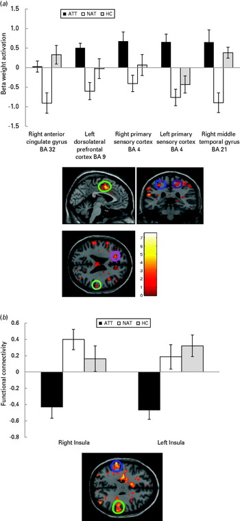
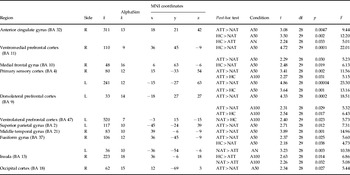
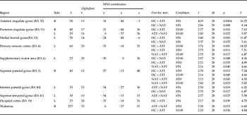
A 3 group × 3 condition ANCOVA for angry faces, covarying for age, revealed group × condition interactions in different dorsal anterior cingulate gyral, prefrontal, temporal, parietal and occipital cortical regions implicated in attention, emotion and visuospatial processing (Table 3, p < 0.05, corrected). A 3 × 3 ANCOVA for happy faces revealed group × condition interactions in dorsal anterior cingulate gyral, prefrontal, temporal, parietal, thalamic and occipital cortical regions (Table 4, p < 0.05, corrected). Post-hoc analyses (Tables 3 and 4, Fig. 1a) revealed that the following significant between-group comparisons contributed to these interactions.
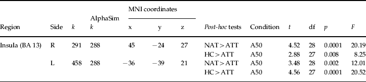
(a) Whole-brain 3 × 3 analysis of covariance (ANCOVA) identifying differences of the effect of intensity difference when viewing 50% intensity angry faces covarying for age in all groups. Suicide attempters (ATT) > depressed non-attempters (NAT) in the right anterior cingulate gyrus [Brodmann area (BA) 32, p = 0.0047], bilateral primary sensory cortices (BA 4, left, p = 0.00004, right, p = 0.002), left dorsolateral prefrontal cortex (BA 9, p = 0.0002) and the right middle temporal gyrus (BA 21, p = 0.001). ATT > healthy controls (HC) in the left primary sensory cortex (BA 4, p = 0.001). Values are means, with standard deviations represented by vertical bars. The brain image shown is the 3 × 1 contrast for 50% intensity angry faces. Sagittal image: green = right anterior temporal gyrus (BA 32). Coronal image: blue = left primary sensory cortex (BA 4); purple = right primary sensory cortex (BA 4). Horizontal image: pink = left dorsolateral prefrontal cortex (BA 9); light blue = right middle temporal gyrus (BA 21). (b) ATT show significantly lower functional connectivity between the right anterior cingulate gyrus and bilateral insulae when viewing 50% intensity angry faces than NAT (left, p = 0.002 and right, p = 0.0001) and HC (left, p = 0.0001 and right, p = 0.008). Values are means, with standard deviations represented by vertical bars. The left insula is shown at the top of the image (blue); the right insula is shown at the bottom (green).

Whole-brain 3 × 3 ANCOVA group (ATT, NAT, HC) × condition (A100, A50, AN) covarying for age

ANCOVA, Analysis of covariance; ATT, Adolescent suicide attempters; NAT, non-attempters with a history of major depressive disorder; HC, healthy controls; A100, Angry faces 100% intensity condition; A50, Angry faces 50% intensity condition; AN, Angry faces neutral condition; MNI, Montreal Neurological Institute; k, cluster; df, degrees of freedom; BA, Brodmann area; R, right; L, left.
Whole-brain 3 × 3 ANCOVA group (ATT, NAT, HC) × condition (H100, H50, HN) covarying for age

ANCOVA, Analysis of covariance; ATT, Adolescent suicide attempters; NAT, non-attempters with a history of major depressive disorder; HC, healthy controls; H100, Happy faces 100% intensity condition; H50, Happy faces 50% intensity condition; HN, Happy faces neutral condition; MNI, Montreal Neurological Institute; k, cluster; df, degrees of freedom; BA, Brodmann area; R, right; L, left.
Angry – ATT v. NAT
ATT showed significantly increased activity relative to NAT when viewing 50% intensity angry faces in the right anterior cingulate gyrus (BA 32, p = 0.0047), bilateral primary sensory cortices (BA 4, left, p = 0.00004, right, p = 0.002), the left dorsolateral prefrontal cortex (BA 9, p = 0.0002), and the right middle temporal gyrus (BA 21, p = 0.001). When viewing neutral faces presented in the angry run, ATT had significantly lower activity than NAT in the left fusiform gyrus (p = 0.003). (Fig. 1a)
Angry – ATT v. HC
ATT showed significantly greater activity than HC in the left primary sensory cortex (BA 4) to angry faces at 50% intensity (p = 0.001).
Angry – HC v. NAT
HC showed greater activity to angry faces at 50% intensity than NAT in the right anterior cingulate gyrus (p = 0.002) and right ventromedial prefrontal cortex (BA 11, p = 0.0001).
Happy – ATT v. HC
ATT showed significantly lower activity than HC when viewing 100% intensity happy faces (p = 0.001) in the left primary sensory cortex (BA 4). ATT showed less activity in the right anterior cingulate gyrus (BA 32, p = 0.0004) and left medial frontal gyrus (BA 10, p = 0.002) than HC to neutral faces presented in the happy run.
Functional connectivity analyses
PPI analyses using a right anterior cingulate seed region, selected because it was active in angry and happy conditions at all intensities in all groups, revealed a significant main effect of group in bilateral insulae during the 50% intensity angry condition (Table 5, Fig. 1b, p-corrected = 0.049). There were no significant findings in the other conditions. Post-hoc analyses revealed significantly reduced functional connectivity in ATT v. NAT (left, p = 0.002; right, p = 0.0001) and v. HC (left, p = 0.0001; right, p = 0.008) from a right anterior cingulate gyral seed to bilateral insulae to 50% intensity angry faces.
PPI activation with right anterior cingulate gyrus BA 32 as the seed region: 3 × 1 main effect of group (ATT, NAT, HC) × condition (A50)
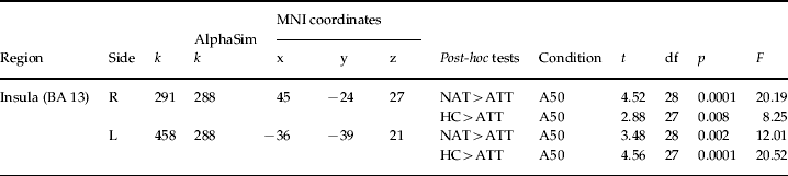
PPI, Psychophysiological interaction; BA, Brodmann area; ATT, Adolescent suicide attempters; NAT, non-attempters with a history of major depressive disorder; HC, healthy controls; A50, Angry faces 50% intensity condition; MNI, Montreal Neurological Institute; k, cluster; df, degrees of freedom; R, right; L, left.
Exploratory analyses
Exploratory analyses (Table S2) were performed to examine relationships between developmental variables (age, pubertal status), clinical variables (BDI, SCARED parent and child versions, SIQ, medication status), gender, and activity in ATT and NAT in regions showing significant differences in activity relative to HC, and to each other that emerged from the 3 × 3 interaction. A statistical threshold of p = 0.05/8 (p < 0.006) was employed to control for eight multiple tests (one for each of the exploratory variables) in each region. These analyses revealed no relationship between anxiety, medication status, gender, age, or pubertal status and activation. There was a positive correlation between activation in the right primary sensory cortex (BA 4) and BDI score in ATT to 50% intensity angry faces (r = 0.702, p = 0.005). In addition, in NAT, SIQ scores correlated negatively with activation in the left dorsolateral prefrontal cortex to 50% angry faces (r = –0.757, p = 0.001). This was repeated for the PPI extracted values, and no relationships between any of the exploratory variables and the PPI analyses were found.
Discussion
The specific aim of the study was to examine the extent to which ATT showed abnormal activity during processing of emotional faces compared with NAT and HC. We hypothesized that, like adult suicide attempters, ATT relative to the other two groups would show differential patterns of elevated prefrontal cortical activity to angry and happy faces (Jollant et al. Reference Jollant, Lawrence, Giampietro, Brammer, Fullana, Drapier, Courtet and Phillips2008). We further hypothesized that both adolescent groups with a history of depression would show reduced functional connectivity among anterior cingulate gyri, prefrontal and temporal cortices and insulae to all face emotion conditions relative to HC. Our 3 × 3 factorial design allowed for identification of group differences of the effect of intensity difference. In other words, we report on the group differences of a difference among the three intensities.
In support of our first hypothesis, when viewing 50% intensity angry faces ATT had significantly greater activity than NAT in different prefrontal cortical, anterior cingulate gyral, temporal, and sensory cortical regions, including the right dorsal anterior cingulate gyrus, bilateral primary sensory cortices, left dorsolateral prefrontal cortex, and the right middle temporal gyrus. ATT also showed significantly greater primary sensory cortical activity than HC to 50% intensity angry faces. Additionally, ATT showed reduced activity relative to the other groups in visual, sensory and prefrontal cortical regions and the anterior cingulate gyrus to intense happy faces and to neutral faces in both emotions. Largely in support of our second hypothesis, ATT, although not NAT, showed decreased functional connectivity from an anterior cingulate gyral seed region to bilateral insulae than other groups to 50% intensity angry faces.
Many of our significant findings were found in the dorsal anterior cingulate gyrus in ATT versus NAT and HC to 50% intensity angry faces. The dorsal anterior cingulate gyrus supports attention processing (Banich, Reference Banich2009), emotion processing, salience of emotion, and generation and regulation of emotional response (Etkin et al. Reference Etkin, Egner and Kalisch2011). Our findings regarding this region may therefore suggest that ATT attended more to 50% intensity angry faces than NAT. Furthermore, PPIs using this anterior cingulate cortical region as a seed region revealed diminished functional connectivity from this region to bilateral posterior insulae in ATT relative to NAT and HC. The posterior insula is implicated in determining stimulus intensity, response selection (Taylor et al. Reference Taylor, Seminowicz and Davis2009) and novelty seeking (Sugiura et al. Reference Sugiura, Kawashima, Nakagawa, Okada, Sato, Goto, Sato, Ono, Schormann, Zilles and Fukuda2000). Connections between the anterior cingulate and posterior insula, particularly the right posterior insula, are implicated in the anticipation of aversive experiences, especially in depressed individuals (Giesecke et al. Reference Giesecke, Gracely, Williams, Geisser, Petzke and Clauw2005). Furthermore, the posterior insula has a distinct pattern of connectivity, functioning as an input region for subcortical loci such as the amygdala and striatum (Craig, Reference Craig2011). In ATT, increased dorsal anterior cingulate gyral and other cortical activity, and reduced functional connectivity between this region and insulae, to 50% intensity angry faces may indicate inefficient strategies to regulate attention to, process the salience of, and select contextually appropriate behavioral responses to these stimuli. In contrast, the pattern of reduced anterior cingulate cortical activity in NAT relative to HC to 50% intensity angry faces may suggest abnormally diminished attention to displays of mild anger in NAT.
ATT also demonstrated greater activity than NAT to 50% intensity angry faces in: (1) the left dorsolateral prefrontal cortex, associated with motor planning and regulation of affect and action (Kaller et al. Reference Kaller, Heinze, Frenkel, Läppchen, Unterrainer, Weiller, Lange and Rahm2011), (2) the right middle temporal gyrus, supporting processing of familiar faces (Haxby et al. Reference Haxby, Hoffman and Gobbini2000); and (3) bilateral primary sensory cortices, that may underlie empathic responding to emotional stimuli (Nummenmaa et al. Reference Nummenmaa, Hirvonen, Parkkola and Hietanen2008), and greater activity than HC in the left primary sensory cortex to these faces. NAT also showed significantly less activity than HC in the right ventromedial prefrontal cortex, a region implicated in emotion valuation (Grabenhorst & Rolls, Reference Grabenhorst and Rolls2011) to 50% intensity angry faces. These findings may indicate greater attention to, processing of, or empathic response toward, 50% intensity angry faces in ATT than NAT and HC, and abnormally reduced attention to these faces in NAT.
Many of our findings related to 50%, rather than to 100%, intensity angry faces. Milder intensity facial expressions are more likely than 100% ‘prototypical’ intensity facial expressions to be representative of social displays of emotion (Calder et al. Reference Calder, Young, Rowland and Perrett1997; Surguladze et al. Reference Surguladze, Brammer, Keedwell, Giampietro, Young, Travis, Williams and Phillips2005). Our findings therefore suggest that ATT were distinguished from other groups by neural activity to socially relevant, rather than to prototypical, displays of anger. Greater attention to, and processing of, these pertinent social displays of disapproval may contribute to vulnerability to suicidal behavior in adolescents with a history of depression.
Additional findings indicate significantly reduced activity in anterior cingulate gyral, primary sensory, prefrontal, and visual cortical regions in ATT than NAT or HC to ambiguous neutral faces in the angry run and intense happy and neutral faces in the happy run. ATT had less activity than HC in the left primary sensory cortex to 100% intensity happy faces, less activity in the right anterior cingulate gyrus and left medial frontal gyrus than HC to neutral faces presented in the happy run, and less activity in the left fusiform gyrus than NAT to neutral faces in the angry run. This suggests that ATT may have attended less or demonstrated reduced neural response to these non-angry stimuli than other groups. The diminished regional activation in ATT compared with HC in right BA 32 and left BA 10 to neutral faces in the happy run may further indicate functional abnormalities of the anterior cingulate gyrus or its connectivity in ATT compared with HC. Furthermore, the finding of reduced primary sensory cortical activity to 100% intensity happy faces in ATT relative to HC may suggest abnormally reduced empathic response to these prototypical displays of social approval and reward in ATT (Nummenmaa et al. Reference Nummenmaa, Hirvonen, Parkkola and Hietanen2008).
Exploratory analyses revealed a positive correlation between activation in the right primary sensory cortex (BA 4) and BDI score in ATT to 50% intensity angry faces, indicating ATT with more depressive symptoms had more activation in this area. Given the role of the primary sensory cortex in empathic responding to sensory stimuli (Nummenmaa et al. Reference Nummenmaa, Hirvonen, Parkkola and Hietanen2008), this finding suggests greater empathic response to 50% intensity angry faces in ATT with greater severity depression. Surprisingly, in NAT the SIQ score correlated negatively with activation in the left dorsolateral prefrontal cortex to 50% intensity angry faces, indicating NAT with greater morbid ideation had less activity in this region. Given that ATT had greater activity in this region than NAT to 50% intensity angry faces, this finding is difficult to interpret, but suggests different strategies for processing 50% intensity angry faces in ATT compared with NAT with higher ideation.
We have previously shown that suicide attempt during adolescence may not be associated with abnormal activity in the neural circuitry supporting response inhibition, but rather that NAT, relative to ATT, demonstrated a compensatory increased recruitment of the right anterior cingulate gyrus (Pan et al. Reference Pan, Batezati-Alves, Almeida, Segreti, Akkal, Hassel, Lakdawala, Brent and Phillips2011). It is intriguing that the opposite pattern, ATT demonstrating greater activity in this area relative to NAT, was shown during 50% intensity angry face processing. Together, our findings suggest that greater recruitment of the anterior cingulate gyrus with emotion (mild angry face emotion processing), rather than functional abnormalities in this region with cognition (response inhibition), may indicate risk for suicide attempt in adolescence.
It was necessary to recruit ATT and NAT taking medication for depression. Exploratory analyses did not, however, show any significant relationships in NAT and ATT between medication status and activity to angry or happy faces in those regions showing between-group differences. Future studies should aim to recruit medicated and unmedicated ATT and NAT. Given the continued development of cingulo-frontal circuitry throughout adolescence (Monk et al. Reference Monk, McClure, Nelson, Zarahn, Bilder, Leibenluft, Charney, Ernst and Pine2003), future longitudinal studies should also examine the developmental trajectories and functional connectivity of these regions during emotion processing.
There were other limitations to the present study. Our main findings from whole-brain analysis survived SVC with AlphaSim at p < 0.05. AlphaSim is a method of correction for multiple comparisons employed in other software packages [e.g. NIMH's Analysis of Functional NeuroImages (AFNI)] and has been used in previous neuroimaging studies of pediatric populations (Gilbert et al. Reference Gilbert, Akkal, Almeida, Mataix-Cols, Kalas, Devlin, Birmaher and Phillips2005; Monk et al. Reference Monk, Telzer, Mogg, Bradley, Mai, Louro, Chen, McClure-Tone, Ernst and Pine2008; Dickstein et al. Reference Dickstein, Finger, Skup, Pine, Blair and Leibenluft2010; Pan et al. Reference Pan, Batezati-Alves, Almeida, Segreti, Akkal, Hassel, Lakdawala, Brent and Phillips2011; Kim et al. Reference Kim, Thomas, Rosen, Moscicki, Brotman, Zarate, Blair, Pine and Liebenluft2012). Future studies may focus on region of interest analyses as new data about neural circuitry underlying adolescent suicide become available. Our study only examined two emotions. Our a priori hypothesis involved angry and happy faces because of previous use in adults with a history of suicide attempt (Jollant et al. Reference Jollant, Lawrence, Giampietro, Brammer, Fullana, Drapier, Courtet and Phillips2008). Inclusion of other emotions was limited by task duration, but should be a focus of future studies. ATT were more symptomatic than NAT, although there were few significant relationships between measures of depression, anxiety and suicidal ideation and patterns of neural activity in either ATT or NAT. It is not possible to be certain whether the differences between the groups are due to suicide attempt history or illness severity. However, NAT differed more than ATT from HC, despite ATT having greater symptomatology. This is the opposite of what would be expected if the differences were secondary to illness severity. Nonetheless, current depression severity in NAT is much closer to that of HC than ATT, and current symptoms could still be a confound.
Our study is the first to examine the functional integrity of neural circuitry in adolescents with a history of suicide attempt during emotion processing. Our findings suggest that functional abnormalities in neural circuitry implicated in emotional processing, especially in processing mild intensity angry faces, may underlie risk for suicide attempt in adolescence. A goal for future studies is thus to determine the extent to which functional abnormalities in neural circuitry supporting emotional processing and other cognitive processes relevant to suicidal behavior may yield potential biomarkers of suicide risk in adolescence.
Supplementary material
For supplementary material accompanying this paper visit http://dx.doi.org/10.1017/S0033291712002966.
Acknowledgements
All work was carried out within the Department of Psychiatry of the University of Pittsburgh. The neuroimaging data were collected at the Brain Imaging Research Center (University of Pittsburgh & Carnegie Mellon University).
We thank the participants who made this work possible.
This research was supported by the American Foundation for Suicide Prevention, the Klingenstein Third Generation Foundation Fellowship for Adolescent Depression [NIMH/National Institute of Child Health (NICHD) 1K23MH082884-01 to L.A.P.], National Institutes of Health grants (MH66775, MH65368, MH56612 and MH18951 to D.A.B.) and NIMH MH076971 (to M.L.P.).
Declaration of Interest
None.