Introduction
Low-cost, fast, portable, sensitive, and selective devices are essential to improve point-of-care diagnostics and on-site environmental monitoring.Reference Martinez, Phillips, Whitesides and Carrilho 1 Paper is an attractive substrate for constructing such devices as (1) it is extremely cheap and commercially available; (2) liquids can be transported by capillary forces, thus it does not require external power; (3) it is easily patterned; and (4) it consists of porous cellulosic fiber networks that make it biofriendly (see the Introductory article in this issue). Biological entities (enzymes, antibodies, bacteriophages—viruses that infect bacteria, and aptamers—single stranded oligonucleic acids that bind to specific targets) are known to be powerful recognition elements for the construction of sensors, as they naturally exhibit high selectivity for target analytes. Therefore construction of bioactive paper (i.e., paper-based material that incorporates biologically active entities) should lead to extremely efficient, cheap sensors.
At present, lateral flow immunoassays based on nitrocellulose (e.g., pregnancy test kit) are one of the only commercially available rapid testing technologies available, but these are generally not quantitative and can detect only one analyte at a time. Recently, a global research effort has started involving the use of cellulose substrates for production of bioactive paper sensors. 2 Aspects such as control of liquid flow in paper, development of multianalyte detection capability, new methods for signal generation, and improved methods for biomolecule immobilization have received renewed attention.
In this review, we focus on techniques for the immobilization of biomolecules (enzymes, antibodies, bacteriophages, and aptamers) onto cellulosic paper substrates and provide a few examples of their application for on-site environmental monitoring. The aim of this review is to outline current methods for immobilizing such biomolecules in a manner that is compatible with the requirements for large-scale printing, and to highlight remaining challenges. For a more extensive description, readers should refer to the review written by Pelton.Reference Pelton 3
Biomolecule immobilization on paper
Immobilization is performed with the goal of retaining active biomolecules in a defined region with maximal density and minimal leaching. In the case of paper-based sensors, an added requirement is the need for immobilization by a printing method that allows patterning of biomolecules. Biomolecule immobilization on paper is normally done by one of four different methods: physical immobilization, covalent and affinity-based attachment, immobilization on carriers, and encapsulation.
Physical immobilization
Physical immobilization occurs when biomolecules spontaneously adhere onto the surface. The process is driven mainly by Van der Waals and electrostatic interactions. Generally, solutions of biomolecules can be easily deposited via contact printing or printed by non-contact methods (i.e., inkjet printing) onto paper. However, commonly used filter paper surfaces are slightly anionic, thus biomolecules that are not highly cationic are weakly adsorbed and can be easily washed off during assays, especially if these utilize reaction conditions involving high ionic strength or non-physiological pH values.
Protein loading can be improved by increasing the strength of interactions via pre-coating paper with cationic polymers that will adhere to cellulose and provide a highly cationic charged surface, or in some cases, by grafting hydrophobic molecules onto cellulose fibers, which was shown to improve adsorption of horseradish peroxidase, which has an isoelectric point of 7.2 (i.e., pH at which an enzyme carries no net electrical charge).Reference Di Risio and Yan 4 To enhance electrostatic adsorption, polyelectrolyte multilayers (i.e., polyelectrolytes of opposite charges that are consecutively adsorbed by layer-by-layer self-assembly) have been used for immobilization of enzymesReference Xing, Eadula and Lvov 5 and, more recently, bacteriophagesReference Larsson, Puttaswamaiah, Ly, Vanerek, Hall and Drolet 6 onto paper. However, this method requires a multistep coating/printing method to adsorb the various layers, making for more complicated manufacturing.
A challenge with physical adsorption is control of biomolecule orientation, which is critical for preserving bioactivity. For example, bacteriophages are viruses composed of a capsid (head) and a tail by which they interact with bacteria. Therefore, oriented immobilization of bacteriophages by their heads is required so that their tails remain active. In order to immobilize bacteriophages on paper, Anany et al. characterized the surface charges of the head and tail and found, in accordance with previous reports, that heads had a net negative charge and tails a net positive charge.Reference Anany, Chen and Pelton 7 Thus, a cellulose membrane coated with cationic polyvinylamine was used to immobilize a cocktail of phages that could detect and infect Listeria monocytogenes or Escherichia coli. Coated membranes bound significantly more phages than unmodified ones, and phages immobilized on those membranes were shown to effectively reduce the population of the target bacteria in ready-to-eat and raw meat. This bioactive paper membrane is a promising technology for reducing bacterial contamination in food samples.
A second challenge when using paper that is modified to increase electrostatic or hydrophobic interactions is the increase in nonspecific binding. This can be reduced by further treatment with commonly used blocking agents (casein, bovine serum albumin, Tween 20) following the adsorption of the species of interest,Reference Cheng, Martinez, Gong, Mace, Phillips, Carrilho, Mirica and Whitesides 8 but again this adds complexity to the manufacturing process and can also add cost to the final bioactive paper test strip. Thus, while certainly the simplest method for biomolecule immobilization, physical adsorption often requires significant surface modification and provides minimal control over biomolecule orientation, suffers from leaching and long-term degradation and denaturation of biomolecules, and potential interference from nonspecifically adsorbed species.
Covalent and affinity-based attachment
Direct covalent attachment requires chemical coupling between biomolecules and paper. Cellulose substrates are not highly reactive, as they only possess a few carboxyl and hydroxyl groups, thus the surface needs to be activated prior to bioconjugation. This approach involves multiple chemical steps, which are generally not compatible with manufacturing low-cost devices.
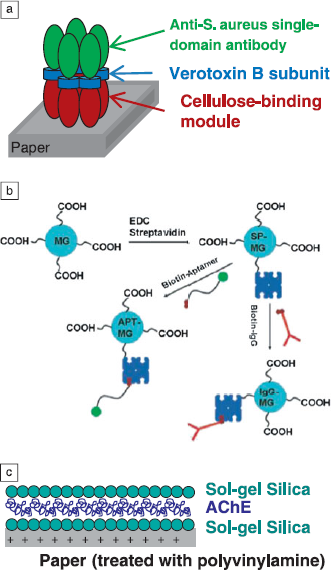
An alternative route is the use of biochemical coupling where biomolecules express cellulose-binding modules (CBM), which spontaneously attach to cellulose. The fusion of CBM and antibodies (Ab) has been described.Reference Lewis, Keshavarz-Moore, Windust, Bushell and Parry 9 To construct CBM-Ab fusions that would irreversibly bind to cellulose with high affinity for a target, Hussack et al. engineered bispecific pentameric antibodies: five single-domain antibodies and five CBM linked by a subunit (see Figure 1 a).Reference Hussack, Luo, Veldhuis, Hall, Tanha and MacKenzie 10 The multimerization simultaneously increased the affinity for cellulose and for the antibody targets. The constructed molecule was bifunctional: It bound cellulose and identified Staphylococcus aureus to a limit of detection of 105 cfu/mL. Genetically modified bacteriophages that express CBM on the head of the phage have also been reported for oriented immobilization onto cellulose beads.Reference Tolba, Minikh, Brovko, Evoy and Griffiths 11
(a) Schematic representation of a bispecific pentamer. Adapted with permission from Reference 10. © 2009 Cambridge University Press. (b) Schematic representation of DNA aptamers or antibodies coupling on carboxylated microgels. Reprinted with permission from Reference 13. © 2013 American Chemical Society. (c) Schematic representation of AChE immobilized onto paper between two sol-gel silica layers. Adapted with permission from Reference 17. © 2013 American Chemical Society.

A drawback of CBM as an immobilization technology is the need for extensive protein engineering and incompatibility with nonprotein biorecognition elements (i.e., aptamers), making this approach of potentially limited versatility and potentially also incompatible with low-cost and large-scale printing. Interestingly, it has been reported that small DNA aptamers can be designed that selectively bind to cellulose, thus one can imagine the design of more complex bifunctional aptamers that would bind to cellulose, and detect and signal specific analytes.Reference Boese and Breaker 12
Immobilization on carriers
In this approach, biomolecules are first immobilized onto a biocompatible carrier and then layered onto paper. For example, Su et al. covalently coupled DNA aptamers and antibodies with carboxylic poly(N-isopropylacrylamide) (PNIPAM) colloids, as shown in Figure 1b.Reference Su, Ali, Filipe, Li and Pelton 13 They demonstrated that when the microgels were left to air-dry after deposition, they remained where deposited and were not dispersed with a water front, probably due to mechanical entrapment within the cellulosic network. In another report, biotinylated DNA aptamers immobilized on streptavidin coated polystyrene beads that were deposited onto paper showed minimal bead displacement after elution.Reference Liu, Xiang, Lu and Crooks 14 This method has the advantage that the carriers are usually water-soluble and thus can be formulated into water-based inks. However, the need to couple the biological agent onto the carrier adds complexity to the manufacturing process.
Entrapment
Entrapment of biomolecules within inorganic materials obtained by the sol-gel process has been widely used for bio-applications, as entrapped biomolecules retain activity for long periods.Reference Avnir, Coradin, Lev and Livage 15 An inorganic network is formed around biomolecules, tight enough to avoid leaching but sufficiently loose for small molecule diffusion and local motions required for substrate binding. Using co-immobilization of acetylcholinesterase (AChE) and gold nanoparticles in sol-gel based silica materials, Luckham et al. constructed a paper-based sensor for AChE inhibitors such as neurotoxins and organophosphates, with a shelf-life of at least three months.Reference Luckham and Brennan 16 However, the biomolecule doped sol was coated on paper by dip-casting, as the material gelled too quickly to allow for printing (<1 h). To overcome this issue, Hossain et al. used a sandwich approach, where AChE was deposited between two layers of silica that were printed at lower pH to extend the gelation time (see Figure 1c).Reference Hossain, Luckham, Smith, Lebert, Davies, Pelton, Filipe and Brennan 17 Piezo inkjet printing also required the addition of both surfactant and glycerol to lower the surface tension and increase viscosity, respectively, producing biocompatible inks that allowed for large-scale printing of biomolecules onto paper.Reference Hossain, Luckham, Smith, Lebert, Davies, Pelton, Filipe and Brennan 17
A final entrapment-based method is the encapsulation of biomolecules into polymer microcapsules, usually made from a semi-permeable polymeric material. While this is a well-established method for protein immobilization, it has only recently been applied to the production of bioactive paper through the application of screen printing of laccase-containing poly(ethyleneimine) microcapsules onto paper.Reference Savolainen, Zhang, Rochefort, Holopainen, Erho, Virtanen and Smolander 18 While this is a versatile method, it can suffer from a loss of enzyme activity with time, biomolecule leaching and dehydration, and rupture of capsules, which can affect the shelf-life of sensors.
Bioactive paper for environmental sensing
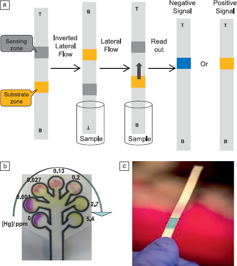
Bioactive paper has been widely utilized in two key areas: point-of-care diagnostics and on-site environmental monitoring. Herein, we focus on the latter, and describe recent reports on detection of organophosphate pesticides, heavy metals, and bacteria in food and water. One of the earliest reports on environmental monitoring with paper-based sensors was by Hossain et al., who developed a reagentless bioactive paper sensor for organophosphate and carbamate pesticide detection, which are environmental pollutants that affect both animals and humans and are widely used in the developing word.Reference Hossain, Luckham, McFadden and Brennan 19 The assay relied on colorimetric detection of acetylcholinesterase (AChE) activity, using the chromogenic indicator indophenyl acetate (IPA), the hydrolysis of which is catalyzed by AChE to generate a yellow-to-blue color change. In the presence of nanomolar levels of pesticides, AChE activity is inhibited, resulting in a decrease of the blue intensity. To construct the paper-based sensor, AChE and IPA were immobilized between two silica layers, as described previously, and printed into a sensing zone (AChE and a cationic capture polymer of polyvinylamine) and a substrate zone, as shown in Figure 2 a. The assay involved lateral flow of the sample to the enzyme side of the strips, followed by incubation and lateral flow from the opposite end to move IPA to the enzyme zone. The achieved limits of detection (LOD) were below 10 nM for the four pesticides (bendiocarb, carbaryl, malathion, paraoxon) tested.
(a) Schematic representation of the detection of organophosphate and carbamate pesticides with inverted lateral flow. Adapted with permission from Reference 19. © 2013 American Chemical Society. (b) Multiplexed patterned paper sensor for heavy metal detection showing a dose-dependent decrease of color intensity at each Hg (II) concentration. Reprinted with permission from Reference 20. © 2013 American Chemical Society. (c) Bioactive paper showing positive response for coliform and E. coli detection.

Based on the concept of colorimetric detection of enzymatic activity, the authors reported a bioactive paper sensor for the environmental detection of metals.Reference Hossain and Brennan 20 The sensitivity of the sensor was Hg(II)>Ag(I)>Cu(II)>Pb(II) = Cr(VI)>Ni(II), with LOD ranging from 0.001 to 0.230 ppm. The assay involved simply dropping the sample on the enzyme zone, followed by lateral flow of substrate to produce a color change, which allowed the design of a multiplexed sensor based on inhibition of immobilized β-galactosidase by the metal ions (see Figure 2b). More recent reports have used simple chromogenic dyes to detect metals upon chelation (as was also reported by Hossain et al.), but in general, the detection limits were not as good as was reported for the enzyme inhibition assay.Reference Ratnarathorn, Chailapakul, Henry and Dungchai 21
Bacterial contamination has huge public health implications; therefore, its detection is receiving a lot of attention. Two recent publications from the Henry groupReference Jokerst, Adkins, Bisha, Mentele, Goodridge and Henry 22 and our groupReference Hossain, Ozimok, Sicard, Aguirre, Ali and Brennan 23 reported the use of colorimetric paper sensors for bacterial detection via enzymatic activity. Both devices detected β-galactosidase that was present in the bacteria, which is commonly used as an indicator for total coliform presence, using a colorimetric substrate printed onto paper. Our sensor was multiplexed to also detect bacterial β-glucuronidase activity, commonly used as an indicator for fecal coliform/E. coli presence (see Figure 2c). Within 30 minutes, without culturing, the LOD were 104 cfu/mL and 2.5 × 10³ cfu/mL for E. coli BL21 and E. coli 0157:H7, respectively. LOD values were further improved using a capture zone to preconcentrate product ions and an immunomagnetic preconcentration step (E. coli antibodies bound to magnetic particles) to concentrate 10 mL of sample to 0.1 mL for testing, resulting in LOD values of 20 cfu/mL and 5 cfu/mL for E. coli BL21 and E. coli 0157: H7, respectively. Henry’s group also reported detection of active enzymes characteristic of Salmonella species and Listeria monocytogenes, but with relatively high detection limits.
Conclusions
At present, most bioactive paper sensors utilize physisorbed biomolecules that are manually deposited onto paper. Emerging technologies based on affinity interactions, use of carriers or entrapment, and the development of bio-inks that are suitable for printing will be key for further developing this field. Current paper-based sensors for environmental diagnostics are mostly enzyme-based devices. However, both aptamer and bacteriophage sensors are emerging technologies for cell detection, and in the case of phages cannot only detect cells but also kill them. Structure switching signaling DNA aptamersReference Carrasquilla, Li and Brennan 24 and RNA aptamersReference Carrasquilla, Lau, Li and Brennan 25 immobilized with silica sol-gel based materials were also recently reported. We expect the development of new bioactive paper sensors using such biorecognition elements in the near future. Although none of the paper-based assays described herein are commercially available, extensive validation studies and scale-up of printing are under way.
Acknowledgments
The authors thank the Natural Sciences and Engineering Research Council of Canada and the Sentinel—Canadian Network for the Development and Use of Bioactive Paper for funding.