Introduction
Maya blue is a greenish-blue pigment that is found widely in the artifacts of the Mayan cultural heritage (Van Olphen, Reference Van Olphen1966; Yacamán et al., Reference Yacamán, Rendón, Arenas and Serra Puche1996; Vázquez de Ágredos Pascuala et al., Reference Vázquez de Ágredos Pascuala, Doménech Carbó and Doménech Carbó2011). It is famous for its fantastic hue and exceptional stability (Gettens, Reference Gettens1962; Shepard, Reference Shepard1962; Giustetto et al., Reference Giustetto, Wahyudi, Corazzari and Turci2011b; Cavalcanti et al., Reference Cavalcanti, Souprayen, Guillermin, Rodrigues, Fonseca and Jaber2022). After decades of investigation, it is now clear that the main components of Maya blue are plant-derived indigo and the clay mineral palygorskite. In addition, a few studies have reported the discovery of sepiolite in Maya blue specimens (Gettens, Reference Gettens1962; Shepard, Reference Shepard1962; Giustetto et al., Reference Giustetto, Wahyudi, Corazzari and Turci2011b). Therefore, sepiolite may also be present as a substrate of Maya blue. Experimental studies have shown that palygorskite and sepiolite can be combined with indigo under heated conditions (usually <190°C) to form pigments resistant to acid. However, the interactions between clay minerals indigo in Maya blue pigments, as well as their effects on the properties of the pigments, have not been understood fully so far.
Previous studies have generally linked the hue and stability of Maya blue to the structure of palygorskite or sepiolite (Gettens, Reference Gettens1962; Kleber et al., Reference Kleber, Masschelein-Kleiner and Thissen1967). Palygorskite and sepiolite are typical fibrous clay minerals (Serratosa, Reference Serratosa1979; Guggenheim and Krekeler, Reference Guggenheim and Krekeler2011). They have a similar structure in which two tetrahedral sheets sandwich an octahedral sheet to form a 2:1 layer (Suárez and García-Romero, Reference Suárez and García-Romero2011). The tetrahedra extend in two dimensions to form continuous tetrahedral sheets. Discontinuous octahedra are formed as the apical oxygen of the tetrahedra reverses periodically. As a result, palygorskite and sepiolite have a large number of channels and tunnels in their structures. The dimensions in the channels/tunnels of palygorskite and sepiolite are 3.7×6.4 Å and 3.7×10.6 Å, respectively (Ruiz-Hitzky et al., Reference Ruiz-Hitzky, Aranda, Álvarez, Santarén and Esteban-Cubillo2011). Some exchangeable cations (e.g. Na+ and Ca2+) and various water molecules are also present in these channels/tunnels (Esteban-Cubillo et al., Reference Esteban-Cubillo, Pina-Zapardiel, Moya, Barba and Pecharromán2008; Ruiz-Hitzky et al., Reference Ruiz-Hitzky, Aranda, Álvarez, Santarén and Esteban-Cubillo2011; Suárez and García-Romero, Reference Suárez and García-Romero2012). Ideally, four and eight zeolitic water molecules are present in one cell of palygorskite and sepiolite, respectively. Coordinated water molecules are present at the edges of the channels/tunnels and coordinate with octahedral cations (generally Mg2+) using oxygen atoms.
Based on the structural characteristics of palygorskite and sepiolite, several hypotheses for the formation and stabilization of Maya blue have been proposed. It is generally believed that indigo molecules can be inserted into clay mineral channels/tunnels under moderate heating while zeolitic water molecules are removed (Littmann, Reference Littmann1982; Caliandro and Dooryhee, Reference Caliandro and Dooryhee2019). Finally, the inserted indigo molecules form hydrogen bonds with coordinated water on both channel/tunnel sides to form a stable pigment (Ovarlez et al., Reference Ovarlez, Giulieri, Chaze, Delamare, Raya and Hirschinger2009; Giulieri et al., Reference Giulieri, Ovarlez and Chaze2012; Yamamoto and Koga, Reference Yamamoto and Koga2019). Earlier studies suggested that indigo molecules may be present on the external surface of clay minerals, in surface grooves, at the entrance of tunnels, and within tunnels (Mulders and Oelkers, Reference Mulders and Oelkers2020; Walczyk et al., Reference Walczyk, Karcz, Kryściak-Czerwenka, Napruszewska, Duraczyńska, Michalik, Olejniczak, Tomczyk, Klimek, Bahranowski and Serwicka2020a). Several possible interactions can also occur between indigo molecules and minerals, including hydrogen bonding, van der Waals forces, and direct bonding between cations (in tetrahedra or octahedra) and indigo molecules (Sánchez del Río et al., Reference Sánchez del Río, Boccaleri, Milanesio, Croce, van Beek, Tsiantos, Chyssikos, Gionis, Kacandes, Suárez and García-Romero2009; Giustetto et al., Reference Giustetto, Seenivasan, Bonino, Ricchiardi, Bordiga, Chierotti and Gobetto2011a; Bemardino et al., Reference Bemardino, Constantino and de Faria2018; Grazia et al., Reference Grazia, Buti, Amat, Rosi, Romani, Domenici, Sgamellotti and Miliani2020). It has also been suggested that iron oxide nanoparticles and dehydroindigo are responsible for the greenish-blue hue of Maya blue (Manciu et al., Reference Manciu, Reza, Polette, Torres and Chianelli2007; Sánchez del Río et al., Reference Sánchez del Río, Boccaleri, Milanesio, Croce, van Beek, Tsiantos, Chyssikos, Gionis, Kacandes, Suárez and García-Romero2009; Rondão et al., Reference Rondão, Séixas de Melo, Bonifácio and Melo2010; Doménech-Carbó et al., Reference Doménech-Carbó, Holmwood, Di Turo, Montoya, Valle-Algarra, Edwards and Doménech-Carbó2019). The widely accepted view is that indigo molecules enter the channels/tunnels while zeolitic water is removed under heating. Ultimately, the inserted indigo molecules form hydrogen bonds with the coordinated water on both sides of the channel/tunnel to form a stable pigment.
The proposed hypotheses of clay mineral–indigo interactions primarily stem from the association of palygorskite with indigo. There have been few studies on the interaction of sepiolite with indigo, as well as on the structure and properties of the corresponding Maya blue-like pigment (Sánchez del Río et al., Reference Sánchez del Río, Doménech, Doménech-Carbó, Vázquez de Agredos Pascual, Suárez and García-Romero2011). Despite the structural similarities between sepiolite and palygorskite, they also have significant differences. For example, sepiolite has a larger channel/tunnel size than palygorskite and generally a magnesium content (Ovarlez et al., Reference Ovarlez, Giulieri, Delamare, Sbirrazzuoli and Chaze2011; Ruiz-Hitzky et al., Reference Ruiz-Hitzky, Aranda, Álvarez, Santarén and Esteban-Cubillo2011; Yalçin and Bozkaya, Reference Yalçin and Bozkaya2011). Indeed, the structure and property differences between palygorskite-based Maya blue pigment and the sepiolite-based analog have been reported previously (Ovarlez et al., Reference Ovarlez, Giulieri, Delamare, Sbirrazzuoli and Chaze2011; Suárez and García-Romero, Reference Suárez and García-Romero2011; Doménech-Carbó et al., Reference Doménech-Carbó, Valle-Algarra, Doménech-Carbó, Domine, Osete-Cortina and Gimeno-Adelantado2013). Sánchez del Río et al. (Reference Sánchez del Río, Martinetto, Dooryhée, Reyes-Valerio and Suárez2006) found that sepiolite-based Maya blue pigment exhibited poorer acid resistance than palygorskite-based pigment. Previous reports have indicated that sepiolite-based Maya blue pigments exhibited inferior chemical stability yet superior photostability compared with palygorskite-based Maya blue pigments.
The structure and properties of sepiolite may influence the interaction between sepiolite and indigo, thereby controlling the color and stability of sepiolite-based Maya Blue pigments. In a previous study, Li et al. (Reference Li, Zhuang, Li, Yuan, Deng and Guo2022) applied thermal treatment to sepiolite and found that the effect of zeolitic and coordinated water in sepiolite on pigment color and stability is different. However, the influence of the chemical composition of the sepiolite on its interaction with indigo and the pigment properties is not fully uncovered. Acid treatment of the sepiolite (Li et al., Reference Li, Zhuang and Yuan2023) leads preferentially to the dissolution of the octahedral sheets at their edges (including the channel edges) more than the tetrahedral sheets, however, thus degrading the chemical stability of the pigment. The effect of alkali treatment of sepiolite, however, has not been investigated fully and may lead to a different outcome with respect to the interaction between the sepiolite and indigo.
Generally, the stability of sepiolite differs in neutral and alkaline media compared with that in acidic media (Jozefaciuk and Bowanko, Reference Jozefaciuk and Bowanko2002; Martinez-Ramirez et al., Reference Martinez-Ramirez, Puertas and Blanco-Varela2018). Sepiolite releases more Si4+ and less Mg2+ under alkaline conditions compared with acidic conditions (Mulders and Oelkers, Reference Mulders and Oelkers2020; Walczyk et al., Reference Walczyk, Karcz, Kryściak-Czerwenka, Napruszewska, Duraczyńska, Michalik, Olejniczak, Tomczyk, Klimek, Bahranowski and Serwicka2020a). Alkali activation enhances the activity of sepiolite material by partially breaking the Si-O-Si and Si-O-M bonds (Wang et al., Reference Wang, Wang, Kang and Wang2015). Previous studies have shown that strong base catalysts can be prepared by substituting some of the Mg2+ ions located at the edges of sepiolite channels/tunnels with alkaline ions (Li+, Na+, K+, Cs+) (de la Fuente et al., Reference de la Fuente, Juárez, de Rafael, Villamiel and Olano1999; Corma et al., Reference Corma, García, Leyva and Primo2003). NaOH treatment has been shown to cause partial decalcification of loughlinite (Na-sepiolite), accompanied by the formation of amorphous Mg(OH)2 within the structural channels/tunnels (Walczyk et al., Reference Walczyk, Karcz, Kryściak-Czerwenka, Napruszewska, Duraczyńska, Michalik, Olejniczak, Tomczyk, Klimek, Bahranowski and Serwicka2020a; Walczyk et al., Reference Walczyk, Michalik, Napruszewska, Kryściak-Czerwenka, Karcz, Duraczyńska, Socha, Olejniczak, Gaweł, Klimek, Wójcik-Bania, Bahranowski and Serwicka2020b). Loughlinite has a structure similar to sepiolite, where two Na+ ions replace the Mg2+ at the end of the octahedral complex (Fahey et al., Reference Fahey, Ross and Axelrod1960; Preisinger, Reference Preisinger1961; Akyuz and Akyuz, Reference Akyuz and Akyuz2010; Akyuz et al., Reference Akyuz, Akyuz and Akalin2010; Giustetto et al., Reference Giustetto, Wahyudi, Corazzari and Turci2011b). Therefore, the effect of NaOH treatment on the microstructure of sepiolite may influence the coordinated water and micropores, thereby affecting pigment properties.
The objective of the present study was to explore the impact of alkali modification of sepiolite on the properties of sepiolite-based Maya blue pigments and the interactions between sepiolite and indigo.
Materials and methods
Materials
Sepiolite (Sep) and indigo (97%) were purchased from Shanghai Aladdin Bio-Chem Technology Co. The chemical composition of Sep was reported previously (Li et al., Reference Li, Zhuang, Li, Yuan, Deng and Guo2022; Li et al., Reference Li, Zhuang and Yuan2023) and the cation exchange capacity was 8.143 cmol+ kg–1. The chemical compositions (mass%), as measured by X-ray fluorescence (XRF), was 55.61% SiO2, 22.04% MgO, 1.9% Al2O3, 0.59% Fe2O3, 0.52% K2O, 0.33% CaO, 0.27% Na2O, 0.08% TiO2, 1.19% others, and 17.47% loss on ignition. Concentrated nitric acid (HNO3, 68 wt.%) and dimethyl sulfoxide (DMSO, 99.9 wt.%) were obtained from the Guangzhou Chemical Reagent Factory.
Alkali treatment of sepiolite
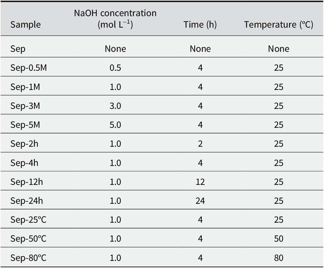
The alkali treatment procedure for sepiolite was carried out by adding 3.0 g of sepiolite to 60 mL of NaOH solution of varying concentrations. The modified sepiolite was separated from suspension by centrifugation, washed with deionized water, freeze-dried, and ground with an agate mortar. The influence of NaOH concentration, reaction time, and temperature on the structure and properties of sepiolite and sepiolite-based pigments was also determined (Table 1).
Summary of the alkali treatment of sepiolite

Preparation of hybrid pigments
Sepiolite materials (including raw sepiolite and alkali-modified sepiolite) and indigo (2% of sepiolite material in mass) were mixed and ground in a Fritsch PULVERISETTE 6-type ball mill at 350 rpm for 3 min. Then, the indigo–sepiolite mixtures were heated at 180°C for 4 h in an oven. The final hybrid pigments were obtained and marked following the template of In-Sep-X, where X represents the NaOH concentration, reaction time, and temperature. For example, In-Sep represents hybrid pigments prepared from indigo and raw sepiolite, and In-Sep-1M indicates the hybrid pigments prepared from indigo and 1 mol L–1 of NaOH-modified sepiolite (Table 1).
Characterization
The UV-Vis absorbance spectra, reflectance spectra, and CIE parameters were obtained using an Ideaoptics (Shanghai, China) spectrometer FX2000L. The Ideaoptics HL-2000 light source (λ=380–2000 nm) was used for reflectance spectra. The CIE 1976 color space system was used to evaluate the color of the pigments.
Before the reflectance and CIE tests, the samples were lightly pressed into pellets using a 5 mm die set and a metal collar to be placed in a Quick Press from Tianjin Optical Instrument Factory (HF-2). L* represents the lightness, a* indicates green (−) or red (+), and b* refers to blue (−) or yellow (+) (Zou and Yeo, Reference Zou and Yeo2024). The total change in color was calculated as
 $ \Delta {E}^{\ast }=\sqrt{{\left({a}_1^{\ast }-{a}_0^{\ast}\right)}^2+{\left({b}_1^{\ast }-{b}_0^{\ast}\right)}^2+\left({L}_1^{\ast }-{L}_0^{\ast}\right)} $
, where subscripts 1 and 0 refer to the sample and reference, respectively. The color saturation was calculated as
 $ {S}_{ab}=\frac{\sqrt{a^{\ast 2}+{b}^{\ast 2}}}{\sqrt{L^{\ast 2}+{a}^{\ast 2}+{b}^{\ast 2}}}\times 100\% $
. Photostability was evaluated under visible light for 180 h using a CST (Dongguan, China) SL21-63.9-WDG type LED light. The working distance between the light and the samples was 10 cm, corresponding to a diameter of 2.5 cm of the working area, with an illuminance of 100 kLux. Hence, the fading test applied for the pigments gave a total light dose of 18,000 kLux h for 180 h. These light doses are the equivalent of ~30 years in a common museum gallery illuminated at 200 Lux (10 h of light exposure per day, 6 days per week, and 50 weeks per year). A similar calculation can be found in the literature (Zhuang et al., Reference Zhuang, Jaber, Rodrigues, Rigaud, Walter and Zhang2019; Li et al., Reference Li, Zhuang, Li, Yuan, Deng and Guo2022; Zhuang et al., Reference Zhuang, Li, Li and Yuan2022). The chemical stability of pigments was assessed using chemical attacks by concentrated HNO3 (68%) and DMSO. HNO3 attack was performed as follows: 0.04 g of pigment was added to 2 mL of concentrated HNO3 solution, and the dispersion was shaken for 15 min; the dispersion was then centrifuged immediately at 6000 rpm (2415 × g) and washed. DMSO extraction was completed as follows: 0.04 g of pigment was added to 5 mL of DMSO and shaken for 24 h; the final pigment and corresponding solution were obtained after centrifugation. All the resulting pigments were dried at 60°C for 24 h.
The Brunauer-Emmet-Teller (BET) surface area and micropore measurements were performed using a Micromeritics ASAP-2020 instrument (N2 adsorption at 77 K). The samples were pre-degassed at 60°C under vacuum on the apparatus. The degassing process was terminated when the vacuum pressure decreased to 3 μm Hg. Samples prepared as KBr pellets were analyzed by Fourier-transform infrared (FT-IR) spectroscopy and the spectra were collected using a Bruker VERTEX 70v type FT-IR spectrometer in the range 4000–400 cm–1 in vacuum. The final FT-IR spectra were obtained after 64 scans with a resolution of 4 cm–1. Transmission electron microscopy (TEM) was performed using an FEI Tecnai Spirit G2 F30 transmission electron microscope at a voltage of 200 kV. The samples were dispersed in ethanol with ultrasound for 10 min; the dispersions were then dropped on copper supports with a carbon grid.
Results and Discussion
Pigment properties and stability
The reflection spectra and CIE parameters characterize accurately the features and variations in the color of the sepiolite–indigo hybrid pigments. The reflection spectra of the sepiolite–indigo hybrid pigments (Fig. 1a), consistent with previous literature reports (Grazia et al., Reference Grazia, Buti, Amat, Rosi, Romani, Domenici, Sgamellotti and Miliani2020; Li et al., Reference Li, Zhuang, Li, Yuan, Deng and Guo2022; Li et al., Reference Li, Zhuang and Yuan2023), show a central reflection band at 426 nm and a shoulder at 503 nm for In-Sep that indicated a blue-green hue. Compared with In-Sep, the pigments prepared from alkali-treated sepiolite exhibited increased reflectance and red shift at both the primary (426 nm) and secondary (503 nm) reflection bands, with a notable enhancement in reflectance at the shoulder band of 503 nm. This suggests increased brightness and a change in hue. Additionally, In-Sep-80°C showed a notable shift of the 503 nm reflection center to 540 nm, and In-Sep-5M exhibited a different reflectance ratio at both the primary and secondary reflection centers compared with other samples. This suggests that the structural changes in those two sepiolite materials impact the properties of the hybrid pigments significantly.
Reflectance spectra and colors of hybrid pigments: (a) at different alkali concentrations and temperatures; and (b) after visible light exposure for 180 h.

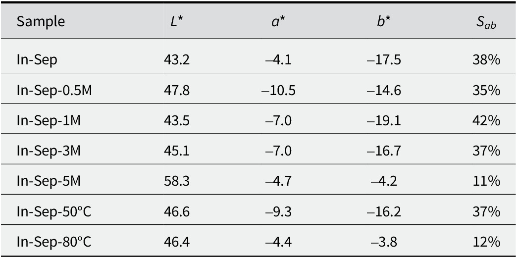
Based on the analysis of the reflection spectra, the CIE parameters of samples under various alkaline conditions were compared (Table 2; see also Table S1 in the Supplementary material). In-Sep exhibited a blue-green hue with L*=43.2, a*=–4.1, and b*=–17.5. After alkali treatment, the L* values of the hybrid pigments range from 43.5 to 58.3, indicating an increase in brightness, consistent with the reflection spectra results. The hybrid pigments treated with alkali were categorized into two groups based on the similarity of their CIE parameters. Group I included In-Sep-0.5M, In-Sep-1M, In-Sep-3M, and In-Sep-50°C, while Group II included In-Sep-5M and In-Sep-80°C. Compared with In-Sep, the notable change in Group I was the larger |a*| values, indicating a more pronounced green hue. Group II showed similar a* values but significantly smaller |b*| values, suggesting a reduced blue hue for these pigments. They also exhibited larger L* values and a significantly smaller Sab (below 27%). Comparing the two groups indicated that appropriate alkali treatment enhances the green hue and brightness of the pigment, whereas severe alkali treatment is detrimental to the pigment’s application.
CIE parameters and color saturation of hybrid pigments at different alkali concentrations and temperatures

Several studies have demonstrated the characteristic resistance of Maya blue to organic solvents (acetone, chloroform, methanol), acids (hydrochloric acid, sulfuric acid, nitric acid), and bases (sodium hydroxide, potassium hydroxide) (Van Olphen, Reference Van Olphen1966; Hubbard et al., Reference Hubbard, Kuang, Moser, Facey and Detellier2003; Domènech et al., Reference Domènech, Doménech-Carbó, Sánchez del Río, Goberna-Ferrón and Lima2009; Yamamoto and Koga, Reference Yamamoto and Koga2019). The parameter ΔE∗, representing color difference, is employed frequently to characterize numerically changes in color under various conditions (Szadkowski et al., Reference Szadkowski, Kuśmierek, Kozanecki, Nowakowska, Rogowski, Maniukiewicz and Marzec2023). To characterize the stability of the pigments after alkali treatment, a comparison was made of the ΔE* values for visible light exposure, HNO3, and DMSO erosion.
After visible light exposure for 180 h (Fig. 1b), In-Sep exhibited the primary and shoulder reflections at 432 nm and 530 nm, respectively. The reflectance bands of hybrid pigments shifted to slightly longer wavelengths and increased in intensity, particularly the shoulder bands. This phenomenon indicates that visible light altered the color of pigments and is consistent with the digital photo results. Furthermore, hybrid pigments via alkali exhibited poor light resistance, with larger ΔE* values from 7.93 to 12.9, compared with 3.21 for In-Sep (Fig. 2a). These ΔE* value changes suggest that photostability is compromised even under mild alkali conditions, such as in the case of In-Sep-0.5M.
Color differences in alkali-treated hybrid pigments: (a) after visible light exposure for 180 h, (b) after HNO3 attack for 15 min, and (c) after DMSO erosion for 24 h.

The hybrid pigments show positive resistance to HNO3 attack (Fig. 2b). When dark blue indigo powder comes into contact with concentrated nitric acid, it converts into a yellowish-red precipitate, indicating the decomposition of indigo into isatin (C8H5NO2) (Sánchez del Río et al., Reference Sánchez del Río, Martinetto, Dooryhée, Reyes-Valerio and Suárez2006; Giustetto et al., Reference Giustetto, Wahyudi, Corazzari and Turci2011b). However, the alkali-treated hybrid pigments retained their blue hue. In-Sep has large ΔE* values of 23.1, whereas others ranged from 13.5 to 6.33, denoting that alkali treatment effectively conserves the chemical integrity of the hybrid pigments. The DMSO results of alkali-treated hybrid pigments were similar to the HNO3 attack, which showed smaller ΔE* values ranging from 5.03 to 14.6, compared with 24.6 for In-Sep (Fig. 2c). These results were consistent with the UV-visible absorbance spectra of DMSO extracts of hybrid pigments at different alkali conditions (see Fig. S2 in the Supplementary material). Notably, In-Sep-0.5M exhibited the minimum value of 5.03, demonstrating that the enhancement of DMSO-resistant stability is very sensitive to slight changes in alkali conditions. Despite the decline in photostability, hybrid pigments have better chemical stability following alkali treatment. Alkali treatment improves the chemical stability of the pigments.
Structure of pigments
Based on the analysis of the properties and stability of pigments, representative samples under various alkaline conditions were selected. Alkali treatment altered the specific surface area and micropore structure of both the sepiolite materials and the resulting hybrid pigments (Table 3; see also Figs S3 and S4 in the Supplementary material). The specific surface area of Sep was 352 m2 g–1, and the specific surface area values of the alkali-treated samples varied significantly, gradually decreasing with increasing alkali treatment conditions, with Sep-80°C and Sep-5M decreasing to as low as 160 and 129 m2 g–1, respectively. The specific surface area is related to the crystallinity of the samples and affects the accessibility of N2 in the structural channels/tunnels (Suárez and García-Romero, Reference Suárez and García-Romero2012). The further decrease in the BET surface area of the sepiolite materials indicated structural evolution. The micropore surface area and micropore volume of the samples also decreased following the alkali treatment, with Sep-5M having the smallest values of 164 m2 g–1 and 33 mm3 g–1. The micropore size distribution of the sepiolite materials was primarily in the range of 0.50–0.54 nm (Fig. 3a). Sep-0.5M had the largest dV/dw of 4.54 cm3 g–1 nm–1 at 0.50 nm, indicating that alkali treatment increased pore volume. Notably, the pore volume for Sep-80°C and Sep-5M decreased significantly, with the maximum pore volume for Sep-5M shifting to 0.95 nm. With increasing alkali treatment conditions, the specific surface area decreased substantially, and the micropore surface area, micropore volume, and micropore rate all dropped several times. This suggests that alkali treatment disrupts structural channels/tunnels of sepiolite, resulting in a decrease in both specific surface area and micropore volume.
Summary of surface properties of sepiolite materials and alkali treatment hybrid pigments

Horvath-Kawazoe differential pore volume vs pore-width plots of sepiolite materials and alkali- treated hybrid pigments.

Like alkali-treated sepiolite, the hybrid pigments exhibited a gradual decrease in specific surface area and micropore volume. In-Sep had a surface area of 250 m2 g–1 and a micropore volume of 52 mm3 g–1, while In-Sep-5M had the smallest specific surface area (27 m2 g–1) and micropore volume (8 mm3 g–1). Before pigment preparation, Sep-5M had a surface area of 129 m2 g–1 and a micropore volume of 33 mm3 g–1 (Table 3). The hybrid pigments differed in micropore distribution from pristine sepiolite materials (Fig. 3b), with the characteristic sepiolite micropore distribution at 0.50–0.54 nm disappearing, being primarily distributed within the range of 0.72–1.2 nm. This indicated that the micropores in the sepiolite material were occupied by indigo. Therefore, after the preparation of the hybrid pigment, indigo further blocks the structural tunnels of the sepiolite material.
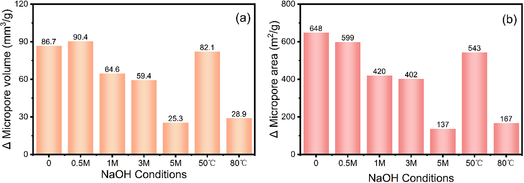
The differences in micropore volume and specific surface area between sepiolite materials and hybrid pigments under various NaOH conditions decreased as the alkalinity increased, and these differences were classified into two groups based on the observed variations. Significant differences were observed in In-Sep-0.5M, In-Sep-1M, In-Sep-3M, and In-Sep-50°C, whereas smaller differences were seen between In-Sep-80°C and In-Sep-5M. The difference in micropore volume between Sep and In-Sep was 86.7 mm3 g–1, while that between Sep-0.5M and In-Sep-0.5M was 90.4 mm3 g–1, indicating that mild alkaline treatment increased the microporosity of sepiolite slightly. As Sep-0.5M, Sep-1M, Sep-3M, and Sep-50°C had certain specific surface areas and pore volumes (Table 3 and Fig. 4), the indigo entered and clogged the pore structures after the pigments were formed. Meanwhile, Sep-80°C and Sep-5M already had very small specific surface areas and pore volumes, offering fewer structural pathways to accommodate indigo molecules. This suggests that indigo enters the structural channels/tunnels of sepiolite and demonstrates how the pore structure of the sepiolite material changes after alkaline treatment.
Difference of micropore volume and area between sepiolite materials and hybrid pigments at various NaOH conditions.

The X-ray diffraction (XRD) patterns of raw sepiolite exhibited the typical diffraction peaks of sepiolite, with the (110) reflection appearing at °2θ=7.38, corresponding to a d 110 value of 11.97 Å. After alkali treatment, all samples still revealed the typical reflections of sepiolite (Fig. 5a), consistent with previous reports (Suárez and García-Romero, Reference Suárez and García-Romero2012; Fitaroni et al., Reference Fitaroni, Venâncio, Tanaka, Gimenez, Costa and Cruz2019; Walczyk et al., Reference Walczyk, Michalik, Napruszewska, Kryściak-Czerwenka, Karcz, Duraczyńska, Socha, Olejniczak, Gaweł, Klimek, Wójcik-Bania, Bahranowski and Serwicka2020b). The XRD patterns of Sep-80°C and Sep-5M showed a slight decrease in the (110) reflection intensity and an increase of corresponding full width at half-maximum (see Fig. S5 in the Supplementary material), indicating that higher NaOH concentrations or treatment at higher temperatures result in a decrease in sepiolite crystallinity. In addition, Sep-80°C and Sep-5M exhibited d 110 values of 12.20 and 12.15 Å, slightly larger than that of Sep (d 110=11.97 Å). This phenomenon indicates that a slight structural evolution occurred in sepiolite under these alkaline conditions (Suárez and García-Romero, Reference Suárez and García-Romero2012; Taimur and Yasin, Reference Taimur and Yasin2017). When incorporated with indigo, these sepiolite materials maintained their structure in hybrid pigments, as revealed by the XRD patterns of pigments (Fig. 5b) being comparable to related sepiolite materials.
XRD patterns of: (a) sepiolite material and (b) hybrid pigments under different conditions.

Consistent with previous research (Li et al., Reference Li, Zhuang and Yuan2023), the morphology of sepiolite remained unchanged before and after pigment preparation (Fig. 6). The TEM results of sepiolite material before and after pigment preparation were compared here (Fig. 6; see also Fig. S6 in the Supplementary material). Sepiolite exhibited a typical fibrous shape (Tiemblo et al., Reference Tiemblo, García, Hoyos, Mejía and de Francisco2015; Zhou et al., Reference Zhou, Yan, Zhang, Tan, Wang, Zhou and Pu2016). After alkali treatment, the sepiolite fibers did not undergo drastic changes, but there was a gradual trend of breaking and shortening with increasing severity of the alkali treatment. Sep-5M, which underwent the most severe alkali treatment, showed fibers significantly shorter than Sep but still maintained its fibrous morphology (Martinez-Ramirez et al., Reference Martinez-Ramirez, Puertas and Blanco-Varela2018; Walczyk et al., Reference Walczyk, Michalik, Napruszewska, Kryściak-Czerwenka, Karcz, Duraczyńska, Socha, Olejniczak, Gaweł, Klimek, Wójcik-Bania, Bahranowski and Serwicka2020b). In addition to changes in fiber morphology, alkali treatment also affected the elemental composition of sepiolite (Fig. 7). Three typical samples from the present experiment were selected for comparative analysis of typical Mg, Si, and Na elemental changes. The decreasing Mg/Si ratio indicated a slight dissolution of both Mg2+ and Si4+, with Si4+ dissolving more than Mg2+. The Na/Si and Na/Mg ratios increased gradually; in Sep-5M, the Na/Si ratio was eight times that of Sep (0.140), and the Na/Mg ratio was nine times that of Sep (0.293), indicating that Na+ replaced Mg2+ during the alkali-treatment process (de la Fuente et al., Reference de la Fuente, Juárez, de Rafael, Villamiel and Olano1999; Walczyk et al., Reference Walczyk, Michalik, Napruszewska, Kryściak-Czerwenka, Karcz, Duraczyńska, Socha, Olejniczak, Gaweł, Klimek, Wójcik-Bania, Bahranowski and Serwicka2020b), further causing a reduction in coordinated water associated with Mg2+.
TEM images of sepiolite materials and pigments: (a) Sep, (b) Sep-5M, (c) In-Sep, and (d) In-Sep-5.

The average elemental ratios of sepiolite materials derived from the EDS results.

Interaction between alkali-modified sepiolite and indigo
The FT-IR absorption spectra of Sep, Sep-0.5M, Sep-24h, Sep-80°C, and Sep-5M within the range 4000–400 cm–1 showed changes in the microstructure of the sepiolite after alkaline treatment (Fig. 8). The stretching vibrations of sepiolite hydroxyl groups appeared in the 4000–3000 cm–1 range, while the bending vibration modes were found in the 1700–1600 cm–1 range. Specifically, the bands at 3685, 3627, and 3565 cm–1 were attributed to the stretching vibrations of Mg3OH (Casal et al., Reference Casal, Merino, Serratosa and Ruiz-Hitzky2001), Al2-OH, and coordinated water (Fernandes et al., Reference Fernandes, Manjubala and Ruiz-Hitzky2011), respectively. The absorption bands at 1660 and 1628 cm–1 belonged to the bending vibrations of zeolitic water and coordinated water, respectively (Serna and Vanscoyoc, Reference Serna and Vanscoyoc1979; Casal et al., Reference Casal, Merino, Serratosa and Ruiz-Hitzky2001; Frost et al., Reference Frost, Locos, Ruan and Kloprogge2001; Bukas et al., Reference Bukas, Tsampodimou, Gionis and Chryssikos2013). Silicate absorption features were observed in the 1400–400 cm–1 range, corresponding mainly to Si-O bonding in tetrahedral sheets and Mg-O stretching vibrations. The band at 1210 cm–1 is related to the asymmetric stretching vibration of Si-O-Si linkages between inverted tetrahedra at the joints of alternating ribbons. The absorption bands at 1021 and 979 cm–1 corresponded to in-plane stretching vibrations of Si-O, while the absorption at 1078 cm–1 is associated with out-of-plane stretching of Si-O. The absorption band at 781 cm–1 is due to O-Si-O and/or Si-O-Si bending vibrations, and the absorptions at 694 and 650 cm–1 stem from the translation and bending of hydroxyls in Mg3OH groupings. The absorption at 470 cm–1 corresponded to the bending of Si-O-Si, while the absorptions at 440 and 425 cm–1 were due to Si-O-Mg vibrations (Yariv, Reference Yariv1986; Vicente-Rodriguez et al., Reference Vicente-Rodríguez, Suarez, Bañares-Muñoz and Lopez-Gonzalez1996; McKeown et al., Reference McKeown, Post and Etz2002; Bukas et al., Reference Bukas, Tsampodimou, Gionis and Chryssikos2013; Lescano et al., Reference Lescano, Castillo, Marfil, Barbosa and Maiza2014; Madejová et al., Reference Madejová, Gates and Petit2017; Walczyk et al., Reference Walczyk, Michalik, Napruszewska, Kryściak-Czerwenka, Karcz, Duraczyńska, Socha, Olejniczak, Gaweł, Klimek, Wójcik-Bania, Bahranowski and Serwicka2020b).
FT-IR spectra of raw sepiolite and alkali-treated samples under various conditions: (a) 3800 to 1550 cm–1, and (b) 1530 to 400 cm–1.

With intensified alkali treatments, the absorption intensity at 3685 cm–1 initially increased and then decreased, indicating changes in the environment surrounding Mg3-OH due to alkali treatment. Sep-0.5M and Sep-24h exhibited a significant decrease in absorption intensity at 3627 and 3565 cm–1, suggesting that alkali treatment causes the loss of octahedral aluminum and magnesium cations, thereby reducing coordinated water associated with magnesium ions. Notably, for Sep-80°C, the absorption intensity at 3627 cm–1 weakened, and the absorption band at 3565 cm–1 decreased significantly, indicating a further reduction in coordinated water. In particular, Sep-5M lost the absorption bands at 3627 and 3563 cm–1 and exhibited a new absorption band at 3608 cm–1, suggesting that water coordinated to magnesium ions at the edges of the structural channels/tunnels was removed (Fernandes et al., Reference Fernandes, Manjubala and Ruiz-Hitzky2011). Another similar feature of coordinated water disruption was that the absorption intensity at 1660 and 1628 cm–1 for Sep-0.5M, Sep-24h, and Sep-80°C weakened compared with Sep, while Sep-5M generated a new absorption at 1654 cm–1. In summary, alkali treatment reduces the amount of coordinated water, which decreases further with more severe alkali treatments conditions.
After alkali treatment, from Sep-0.5M, Sep-24h, and Sep-80°C to Sep-5M, a gradual broadening and decrease in intensity of the absorption bands corresponding to the stretching vibrations of Si-O were observed. With a further increase in the concentration of NaOH (Sep-5M, 5 mol L–1), the absorptions at 1021 and 979 cm–1 were replaced by 1014 cm–1, and the intensity of the absorption band at 1210 cm–1 gradually decreased, indicating that the silicon-oxygen tetrahedron is sensitive to the etching effect of alkali. The increasing disorder within the silicate layers was also reflected in the decreasing intensity and broadening of the 781 cm–1 absorption band. In contrast, the absorption bands at 694 and 650 cm–1 were only slightly affected, suggesting that although coordinated water was disrupted, the octahedral sheet retained its identity. In summary, the Si-O in the tetrahedral structure of sepiolite is eroded by alkali, and the erosion increases with more severe alkali treatments. Notably, compared with Sep, the absorption band at 1401 cm–1, attributed to carbonates, increased gradually after alkali treatment. New absorption bands attributed to carbonates were generated at 868 and 1450 cm–1. In addition, Sep-5 produced a broad absorption band between 1400 and 1600 cm–1 (Fig. 8) (Gunasekaran et al., Reference Gunasekaran, Anbalagan and Pandi2006; Olszówka et al., Reference Olszówka, Karcz, Bielańska, Kryściak-Czerwenka, Napruszewska, Sulikowski, Socha, Gaweł, Bahranowski, Olejniczak and Serwicka2018). Many researchers have reported the adsorption of CO2 by sepiolite under certain temperatures and pressures, as well as the transformation between Mg(OH)2 and MgCO3 under specific temperature and pressure conditions (Siriwardane and Stevens, Reference Siriwardane and Stevens2009; Jeon et al., Reference Jeon, Choi, Yun and Lee2014; Cecilia et al., Reference Cecilia, Vilarrasa-García, Cavalcante, Azevedo, Franco and Rodríguez-Castellón2018; Chouikhi et al., Reference Chouikhi, Cecilia, Vilarrasa-García, Besghaier, Chlendi, Franco Duro, Rodriguez Castellon and Bagane2019; Gómez-Pozuelo et al., Reference Gómez-Pozuelo, Sanz-Pérez, Arencibia, Pizarro, Sanz and Serrano2019). Therefore, it is worth considering whether the changes in the absorption bands attributed to carbonates can prove the formation of Mg(OH)2 after alkali treatment.
The FT-IR spectral features for indigo and hybrid pigments appear in the range 4000–1250 cm–1 (Fig. 9a). The changes in indigo within In-Sep were consistent with previous literature (Tatsch and Schrade, Reference Tatsch and Schrade1995; Sánchez del Río et al., Reference Sánchez del Río, Boccaleri, Milanesio, Croce, van Beek, Tsiantos, Chyssikos, Gionis, Kacandes, Suárez and García-Romero2009; Amat et al., Reference Amat, Rosi, Miliani, Sgamellotti and Fantacci2011; Li et al., Reference Li, Zhuang, Li, Yuan, Deng and Guo2022; Zhuang et al., Reference Zhuang, Li, Li and Yuan2022). The red shift of the C=O stretching band indicated strong guest–host interactions and the formation of hydrogen bonds (Tilocca and Fois, Reference Tilocca and Fois2009). Although the adsorption at 1626 cm–1 corresponding to the C=O stretching overlapped with the bending vibration of coordinated water, the bands related to benzene rings (νCC and δCH) at 1483, 1462, 1317, and 1299 cm–1 shifted to 1488, 1463, 1321, and 1302 cm–1, indicating the formation of indigo–sepiolite interaction. As the alkalinity of the treatment conditions intensified, the characteristic absorption bands of indigo in the hybrid pigments became progressively less pronounced when compared with In-Sep. Absorption bands attributable to indigo were observed in In-Sep-0.5M and In-Sep-80°C but were absent in In-Sep-5M. Several fundamental changes are presented as follows: (1) with increasing alkali treatments, the characteristic absorption of indigo decreases; (2) hybrid pigments possess new bands at 1401 and 1385 cm–1, corresponding to 1411 and 1392 cm–1 of indigo, instead of 1411 cm–1 as in In-Sep; (3) strong alkali treatments produce a broad absorption band between 1350 and 1550 cm–1, especially notable for In-Sep-5M; (4) In-Sep-5M shows a distinct FT-IR spectrum with bands at 3610, 3449, and 1637 cm–1. The Raman spectroscopy data exhibit similar variations, with the absorption band centered at 1500 cm–1 changing in response to alterations in the alkaline treatments, particularly notable for In-Sep-80°C and In-Sep-5M (Fig. 9b). According to the literature, the band in this region may correspond to a central C=C stretching vibration, C=O vibration, and N-H deformation of indigo molecules (Bemardino et al., Reference Bemardino, Constantino and de Faria2018; Ricci et al., Reference Ricci, Lofrumento, Becucci and Castellucci2018). These results indicate that the alkali treatment of sepiolite alters indigo–sepiolite interaction by destroying the pore structure and varying the coordinated water.
(a) FT-IR and (b) Raman spectra of hybrid pigments after various alkali treatments.

The specific surface area and pore volume of sepiolite material decreased after alkali treatment, therefore. The EDS data indicated an increase in Na content and decreases in the Mg and Si contents within the samples, suggesting that Na+ ions replaced Mg2+ ions. Although the d 110 diffraction did not increase from 12.27 Å (sepiolite) to 12.90 Å (loughlinite) as reported in previous studies (Caillerie and Fripiat, Reference Caillerie and Fripiat1992; Giustetto et al., Reference Giustetto, Wahyudi, Corazzari and Turci2011b), slight changes were observed in the diffraction of the (110) crystal planes. Such observations combined with FT-IR and Raman analyses prove further that alkali treatment erodes the tetrahedral Si-O of sepiolite and disrupts the octahedral Mg2+, altering the microstructure of sepiolite. Alkaline treatment affects the micropores and coordinated water of sepiolite, which involves the process of indigo molecules entering the sepiolite structural channels/tunnels and interacting with the coordinated water (Fig. 10). Although this reduces the light stability of the hybrid pigment, it enhances the greenish hue and chemical stability of the pigment.
Suggested schematic diagram of the interaction between the indigo molecule and alkali-treated sepiolite.

Summary and conclusions
In this study, an alkali pre-treatment was first carried out on sepiolite, after which a series of hybrid pigments was prepared from the pre-treated sepiolite and indigo. A comprehensive study was then conducted on the color properties, stability, structure, and sepiolite–indigo interaction of the pigments. The results demonstrated that the alkali treatment damaged the micropore structures and coordinated water of sepiolite by dissolving the Si4+ ions in the tetrahedral sheet and the channel/tunnel edge Mg2+ ions in the octahedral sheet, and varied the micropore structure through the precipitation of Mg2+ ions in the channels/tunnels. This alteration subsequently affected the entry of indigo into the sepiolite channels/tunnels, which led to the interaction between indigo and coordinated water through the carbonyl group. Although the photostability of the hybrid pigments was not improved after the alkali treatment of sepiolite, the chemical stability and green hue were enhanced significantly. Therefore, appropriate alkali treatment is a viable strategy to achieve better chemical stability and saturated hue while maintaining a certain level of photostability for preparing pigments.
Supplementary material
The supplementary material for this article can be found at http://doi.org/10.1017/cmn.2025.2.
Author contributions
In this work, Li Li: Data curation, Formal analysis, Investigation, Methodology, Writing – original draft. Guanzheng Zhuang: Conceptualization, Investigation, Validation, Writing – review & editing. Yanfu Wei: Funding acquisition, Writing – review & editing. Mohammad Fahimizadeh: Writing – review & editing. Peng Yuan: Supervision, Funding acquisition, Writing – review & editing.
Acknowledgements
None.
Financial support
This work was supported by National Natural Science Foundation of China (grant nos. 52161145405 and 42102027) and the Science and Technology Development Fund, Macau SAR (0094/2022/A2).
Competing interests
The authors declare none.
Data Availability Statement
All data generated or analysed during this study are included in this published article and its supplementary information files.