Percutaneous coronary intervention for stenosis of coronary artery after Kawasaki disease presents various challenges. The diameters of reference vessels and femoral artery in children are smaller, and the morphological changes are different from adults with atherosclerotic disease. Herein, we describe our successful experience with a severe coronary artery stenosis at the proximal portion of left anterior descending treated with cutting balloon followed by drug-coated balloon dilation.
Case report
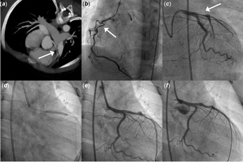
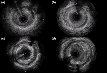
A 6-year-old boy was admitted to our hospital because of a positive treadmill test displaying a horizontal ST segment depression exceeding 2 mm in leads III and augmented Vector Foot (AVF), with the ST/HR index (the ratio of the difference between exercise and rest ST depression and the difference between exercise and rest heart rate) = 2.8 μv/beat/minute during the recovery period. He developed aneurysmal dilation of the right coronary artery and the left anterior descending due to Kawasaki disease at 4 months of age, which were treated with warfarin and aspirin, but he did not follow-up regularly. Coronary CT could not depict the proximal right coronary artery due to important calcification and showed a moderate narrowing at the proximal of left anterior descending coronary artery (Fig 1a). Coronary angiography was then performed under general anaesthesia via right femoral artery and detected a total occlusion of the proximal right coronary artery with bridge collaterals formation (Fig 1b). Additionally, a severe stenosis at the proximal portion of left anterior descending was also delineated (Fig 1c). After obtaining his parents informed consent, we decided to perform percutaneous revascularisation for the stenotic lesion of the left anterior descending considering that collateral arteries had developed well for the distal segment of right coronary artery. A 6-French EBU 3.0 guiding catheter (Medtronic, Minneapolis, MN) was inserted from the right femoral artery and 5000u of heparin was administered intravenously. A Sion guide wire (Asahi Intecc Co., Ltd., Aichi, Japan) was successfully introduced into the distal left anterior descending. Intravascular ultrasound imaging (IVUS, UltraCross 3.2Fr 30 MHz catheter, Boston Scientific Co., Natick, MA, USA) showed an eccentrically fibrous intimal thickening at the culprit lesion with homogeneous high intensity. The minimum lumen diameter was 1.82 mm × 1.8 mm, and the minimal lumen area was 2.59 mm2 (Fig 2a). Then this lesion was pre-dilated in turn with two cutting balloons (2.5 mm × 6 mm, 3.0 mm × 6 mm) (Boston Scientific Co., Natick, MA, USA) gradually inflated to 6 atm. An optimal angiographic result was achieved, the minimal lumen diameter was 2.5 mm × 3.24 mm, and the minimal lumen area was improved to 6.48 mm2 (Fig 2b). Finally, we performed drug-coated balloon angioplasty with a 3.0 mm × 20 mm paclitaxel-coated balloon (3 mcg/mm2, B. Braun, Melsungen, Germany) at 7 atm for 60s (Fig 1d). The final angiography showed a well-dilated culprit lesion with no apparent dissection and cracks (Fig 1e). The subsequent intravascular ultrasound revealed the minimal lumen diameter was 2.68 mm × 3.09 mm, and the minimal lumen area was 6.52 mm2 (Fig 2c). The procedure concluded free of complications. After the procedure, a repeat treadmill test showed definite improvement in exercise performance, ST-T changes, and the ST/HR index = 1.7 μv/beat/minute. The patient was commenced on dual antiplatelet therapy with aspirin 100 mg once daily and ticagrelor 45 mg twice daily post procedure. No thrombus and restenosis at the culprit site were detected by follow-up angiography 6 months later, and the minimal lumen area was 6.84 mm2 (Figs 1f and 2d).
The CT and coronary angiography before and after percutaneous coronary intervention (PCI). ( a ) CT before PCI depicted moderate stenosis at the proximal of the left anterior descending coronary artery (LAD, White arrow). ( b ) Right coronary angiography before PCI showed complete occlusion of the right coronary artery with bridge collaterals formation (White arrow). ( c ) Coronary angiography before PCI developed localised tight stenosis just at the proximal portion of the LAD (White arrow). ( d ) Drug-coated balloon (DCB) angioplasty with a 3.0 mm × 20 mm SeQuent Please was performed. ( e ) Left coronary angiography immediately after DCB angioplasty showed a well-dilated LAD. ( f ) Coronary angiography at 6-month follow-up showed no thrombus or restenosis in the LAD.

Intravascular ultrasound images before and after catheter intervention. ( a ) Intravascular ultrasound (IVUS) image of the stenotic lesion in the proximal left anterior descending coronary artery (LAD) showed an eccentrically fibrous intimal thickening with homogeneous high intensity before percutaneous coronary intervention (minimal lumen area was 2.59 mm2). ( b ) IVUS image at the stenosis site after the cutting balloon angioplasty (minimal lumen area was 6.48 mm2). ( c ) IVUS image at the stenosis site after the drug-coated balloon angioplasty (minimal lumen area was 6.52 mm2). ( d ) IVUS image at the stenosis site 6 months later (minimal lumen area was 6.84 mm2).

Discussion
Coronary artery complications are the most important sequelae of Kawasaki disease. The normal vascular structure can be disrupted by pan-vasculitis, which leads to coronary artery dilation, thrombosis, intimal proliferation, calcification, and finally narrowing of the lumen (negative remodeling). Reference Fukazawa, Kobayashi and Ayusawa1 Despite the importance of management of ischaemic heart disease to improve symptoms and prevent or treat cardiovascular events, current experiences for managing young children and adolescent with coronary sequelae of Kawasaki disease are extremely limited and are mostly extrapolated from adults with atherosclerotic coronary disease, with marked difference in the choice of intervention between heart centres. Reference Muta and Ishii2,Reference Dionne, Bakloul and Manlhiot3 According to the JCS/JSCS guidelines, patients with evidence of inducible myocardial ischaemia on testing should undergo invasive coronary angiography. And elective percutaneous coronary intervention is recommend for a localised stenosis >75% with the symptoms of myocardial ischaemia just like our case. Reference Fukazawa, Kobayashi and Ayusawa1 If the localised lesion is caused primarily by intimal hypertrophy without severe calcification, plain old balloon angioplasty is recommended for treatment. However, conventional balloon angioplasty alone is not an effective treatment for revascularisation in patients with coronary sequelae of Kawasaki disease, because such lesions are stiff and there is a relatively high rate of restenosis because intimal thickening caused by Kawasaki disease can progress with ageing after the procedure.
Although the early results after stenting are good, some complications can develop in the late period, such as aneurysm, in-stent reocclusion, or malposition of the stent. Reference Fukazawa, Kobayashi and Ayusawa1 These issues should be carefully considered in children as the long-term outcome remains unknown. Moreover, the diameters of reference vessels in children are small and thus the lumen loss after implantation occupies a higher percentage, which could lead to smaller final minimum lumen diameter and higher rates of in-stent restenosis. Therefore, “stent-less intervention” seems to be a better strategy for children.
Drug-coated balloons are a novel therapeutic strategy for coronary artery disease with the advantage of rapid release of anti-proliferative medicine at the local site to inhibit intimal hyperplasia and without the need for permanent implants at the stenotic lesions. Recently, drug-coated balloons have been proven effective in several large and adequately designed trials for the treatment of small-vessel disease, usually defined as lesions in vessels ≤2.75 or <3.0 mm. Reference Jeger, Eccleshall and Wan Ahmad4 There are only a few case reports on use of drug-coated balloon for treating coronary sequelae of Kawasaki disease in adults, two cases were combined with rotational atherectomy by Shiraishi and Shi, respectively, another was combined with laser excimer angioplasty and cutting balloon by Kawamura. Reference Shiraishi, Matsubara and Yanagiuchi5–Reference Kawamura, Komiyama, Fukamizu, Shibui, Ashikaga and Sakurada7 They all showed good results, and rotational atherectomy followed by drug-coated balloon dilation might contribute to deferring repeat intervention. Reference Shiraishi, Matsubara and Yanagiuchi5 To our knowledge, our case is the first to describe successful percutaneous revascularisation using drug-coated balloon for coronary sequelae of Kawasaki disease in paediatric. In our patient, the proximal left anterior descending showed intimal proliferation without obvious calcification and atherosclerotic change, so rotational atherectomy or laser excimer was not attempted. Although cutting balloon was associated with some procedural advantages compared with conventional balloon, but the efficacy of cutting balloon for coronary sequelae of Kawasaki disease has not been investigated. Furthermore, cutting balloon angioplasty did not reduce recurrent coronary restenosis. Reference Albiero, Silber and Di Mario8 Taking the high risk of restenosis into consideration, we used cutting balloons for pre-dilation of the stenotic lesion and followed by a paclitaxel-coated balloon. The results of exercise test after the procedure and 6 months follow-up angiography are favourable.
In adult patients, the use of paclitaxel-coated stents and balloons seems associated with increased mortality in the treatment of peripheral arterial disease but not in coronary artery disease. Reference Jeger, Eccleshall and Wan Ahmad4 In paediatric patients, some studies had reported good results on the use of drug-coated balloons for vascular intervention in pulmonary artery or hepatic vein, Reference Cohen, Glickstein and Crystal9,Reference Parra and Brandao10 but there are limited published case reports on the use of drug-coated balloons for coronary intervention. Paediatric patients have a smaller volume of distribution compared to adults and thus the plasma level of paclitaxel could be higher following device placement. Our case shows relatively good outcomes after the use of paclitaxel-coated balloons for coronary intervention in a 6-year-old child, but longer follow-up on our patient and additional cases are needed to provide more information on this topic.
Our case demonstrates the clinical feasibility of drug-coated balloon dilation in de novo lesions of small coronary vessel in paediatric patients due to Kawasaki disease, which provide initial evidence for a possible new treatment of coronary sequelae of Kawasaki disease. Large-scale clinical trials and long-term follow-up studies are needed to assess the safety and efficacy of drug-coated balloons in this patient population.
Acknowledgements
None.
Financial support
This research received no specific grant from any funding agency, commercial, or not-for-profit sectors.
Conflicts of interest
None.