Birth weight is an important predictor of the future health condition of an individual.Reference Carlsson, Persson and Alvarsson 1 Low birth weight is an important public health issue in developing countries.Reference Longo-Mbenza, Vangu Ngoma and Mbungu Fuele 2 The prevalence of low birth weight in Zimbabwe is 10%, higher in female individuals than in male individuals. 3 Low birth weight is associated with an increased risk for coronary artery disease,Reference Kent, Sonnet, Perrie, Natasha, Job and George 4 hypertension,Reference Barker and Osmond 5 and heart failure.Reference Barker, Osmond, Golding, Kuh and Wadsworth 6 Babies that are growth restricted prenatally are more likely to have hearts with detrimental structural and functional changes.Reference Kent, Sonnet, Perrie, Natasha, Job and George 4 , Reference Geelhoed and Jaddoe 7
Research has shown that prolonged strenuous exercise produces a transient decrease in cardiac function, both diastolic and systolic function – a phenomenon referred to as exercise-induced cardiac fatigue. This has been demonstrated in both well-trained and untrained participants.Reference Dawson, George, Shave, Whyte and Ball 8 , Reference Oxborough, Birch, Shave and George 9 Low birth weight individuals may be particularly prone to exercise-induced cardiac fatigue because of the changes in structure and function as highlighted above.
Exercise-induced hypertension is used as a predictive tool for future hypertension and in asymptomatic normotensive individuals.Reference Singh, Larson and Manolio 10 , Reference Allison, Cordeiro, Miller, Daida and Gau 11 Similar methodology may be useful in using exercise-induced diastolic dysfunction among young adults to predict future heart failure in asymptomatic individuals.Reference Dawson, George, Shave, Whyte and Ball 8 , Reference Oxborough, Birch, Shave and George 9 Abnormalities in left ventricular diastology, as defined by the mitral inflow E/A ratio, among asymptomatic older individuals have been associated with subsequent development of heart failure.Reference Kane, Karon and Mahoney 12
The purpose of the study was to determine whether low birth weight predicts development of left ventricular diastolic dysfunction after prolonged exercise for 75 minutes in young black adults.
Materials and methods
A case–control study was conducted in the Department of Physiology, University of Zimbabwe College of Health Sciences. All participants volunteered and gave informed consent to take part in this study. Participants were required to produce their neonatal Ministry of Health and Child Welfare birth cards, which documents birth weight. Ethical permission was granted by Joint Parirenyatwa Hospital and College of Health Sciences Research Ethics Committee and the Medical Research Council of Zimbabwe.
A total of 23 eligible participants were randomly selected. A self-administered questionnaire was used to obtain variables such as age, history or clinical evidence of diabetes or cardiovascular diseases, and any form of diseases in the participant that can affect cardiovascular function. Weight and height of the participants were measured with the participants wearing light clothing and without shoes using an analogue scale, SECA model 770, on day 1 of the study protocol. 24 hours before the exercise test participants were advised to abstain from caffeine and taurine products such as coffee and energy drinks that may influence their cardiovascular perfomance during test.
Participants initially underwent graded exercise testing – Trojan Stamina 315 model – according to the Bruce protocol to determine maximal aerobic power, VO2max on day 1 of the study protocol. These results were used to establish running velocities, eliciting heart rate 80% VO2max, high intensity, during prolonged exercise sessions, which were conducted on the following day in a controlled laboratory setting. Graded treadmill exercise testing was performed to exhaustion.
Participants completed a high-intensity, 80% VO2max,Reference Banks, Sasson, Busato and Goodman 13 , Reference Banks, Sasson, Esfandiari, Busato and Goodman 14 exercise challenge involving 75 minutes of running on a treadmill – Trojan Stamina 315 model – at room temperature. To minimise changes in the hydration state and cardiac-loading conditions, participants were encouraged to consume 250 ml of water every 20 minutes. Post-exercise measures were taken within 5 minutes after exercise completion.Reference Banks, Sasson, Busato and Goodman 13 , Reference Banks, Sasson, Esfandiari, Busato and Goodman 14 Echocardiography was performed by a trained sonographer blinded to participant birth weight, using a portable ultrasound device – M-Turbo, SonoSite Inc, Bothell, Washington, United States of America. Pulse-wave Doppler flow velocities were recorded from the transthoracic apical window at the site of mitral valve leaflet tip coaptation. Diastolic function was determined before and after exercise as the ratio of the early mitral inflow velocity, E wave, to late mitral inflow velocity, A wave), represented as the unitless E/A ratio.Reference Dawson, George, Shave, Whyte and Ball 8 , Reference Kane, Karon and Mahoney 12
Statistical methods
Low birth weight was defined as weight between 1900 and 2499 g at term. Normal birth weight was defined as weight between 2500 and 4000 g at term; no participants had birth weights outside these ranges. Exercise-induced cardiac fatigue was defined as ∆E/A ratio ⩾0.40.Reference Dawson, George, Shave, Whyte and Ball 8 Independent samples two-tailed t-test at 5% level of significance was used to compare the various means of metabolic parameters between the two grouping variables – that is, low birth weight and normal birth weight. Odds ratios were calculated using the Pearson χ 2 test in SPSS at 95% confidence interval. Participants’ demographic characteristics and all additional data are reported as mean±standard deviation. SPSS software version 16.0, SAS Institute, Cary, North Carolina, United States of America, was used to analyse the data. Tables were generated using SPSS 16.0 and Microsoft Excel 2010.
Results
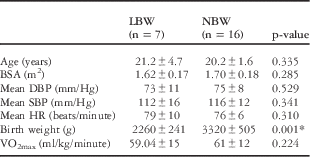
A total of 23 young adult participants, 9 (39%) female individuals and 14 (61%) male individuals, with a mean age of 20.7±3.3 years were recruited; of them, seven had low birth weight, three female individuals and four male individuals, and 16 had normal birth weight, six female individuals and 10 male individuals. Table 1 shows detailed demographic characteristics of the study sample.
Demographics of study sample

HR=heart rate; LBW=low birth weight; NBW=normal birth weight; SD=standard deviation; BSA=body surface area; DBP=diastolic blood pressure; SBP=systolic blood pressure.
Formulae, VO2max=14.8−(1.379×T)+(0.451×T2)−(0.012×T3) in male individuals and VO2max=4.38×T−3.9 in female individuals.
*p<0.05; significant difference between NBW and LBW individuals.
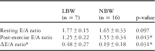
According to Table 2, of the 23 participants studied, seven (30%) developed a ∆E/A ratio ⩾0.40, five low birth weight and two normal birth weight individuals. There was a significant association between low birth weight and exercise-induced cardiac fatigue (odds risk ratio 4.64; 95% confidence interval 1.19–18.1, p<0.05).
Haemodynamic measurements

∆E/A ratio – change in the E/A ratio from rest to post-exercise. Exercise-induced cardiac fatigue was defined as ∆E/A ratio ⩾0.40.8 Of the 20 participants who were studied, only seven developed EICF(exercise induced cardiac fatigue), five low birth weight and two normal birth weight individuals. There was a significant association between low birth weight and EICF (the χ 2 test p<0.05, odds risk ratio 4.64 95% confidence interval 1.19–18.1).
*p<0.05; significant difference between NBW and LBW individuals.
Comment
This study is the first to elucidate the relationship between birth weight and exercise-induced cardiac fatigue. Compared with young adults with normal birth weight, those with low birth weight had greater diastolic dysfunction after prolonged aerobic exercise, as evidenced by mitral inflow E/A ratio. This supports the hypothesis that low birth weight individuals have permanent structural and functional cardiac impairments due to intrauterine growth retardation, making them more prone to exercise-induced cardiac fatigue.Reference Kent, Sonnet, Perrie, Natasha, Job and George 4 , Reference Geelhoed and Jaddoe 7
Prior study on exercise-induced cardiac fatigue has focused primarily on trained athletes participating in prolonged endurance.Reference Dawson, George, Shave, Whyte and Ball 8 , Reference Oxborough, Birch, Shave and George 9 The strenuous exercise and high motivation of athletes because of the competitive nature of these events provide a unique model in which to view cardiac dysfunction. The students in this study appeared to experience evidence of cardiac fatigue after a shorter amount of time and at lower exercise intensities compared with competitive and recreational endurance athletes.Reference Dawson, George, Shave, Whyte and Ball 8 , Reference Oxborough, Birch, Shave and George 9
Low birth weight individuals showed even greater rates of exercise-induced cardiac fatigue. This may be explained by epigenetic modification that occurs in response to intrauterine hypoxia and lack of nutrition, leading to a decrease in protection against injury in the left ventricular myocardium,Reference Kent, Sonnet, Perrie, Natasha, Job and George 4 besides structural and functional impairments in utero.Reference Kent, Sonnet, Perrie, Natasha, Job and George 4 , Reference Geelhoed and Jaddoe 7
Furthermore, the mechanisms of exercise-induced cardiac fatigue involve transient myocardial stunning and a decrease in sensitivity of myocardium to catecholamines, reducing the effect of adrenaline in improving both systolic and diastolic function during exercise.Reference Dawson, George, Shave, Whyte and Ball 8 , Reference Oxborough, Birch, Shave and George 9 These factors may be impaired to some extent in low birth weight individuals, thus making them more prone to exercise-induced cardiac fatigue. In fact, low birth weight is associated with chronic symphathoexcitation later in life, as measured by high noradrenaline “spillover” in low birth weight animals and children.Reference Rebbecca, Reynolds, Walker, Phillips, Dennison and Scott 15 , Reference Ijzerman, Stehouwer and de Geus 16 This also arises from the restricted growth leading to stress-induced increases in the circulating levels of cortisol and other glucocorticoids, and this prenatal excessive glucocorticoid activity might increase the hypothalamic–pituitary–adrenal axis with the resultant high symphathoexcitation.Reference Rebbecca, Reynolds, Walker, Phillips, Dennison and Scott 15 Hence, if there is high sympathetic discharge at rest, β receptor sensitivity to catecholamines, which are already abundant, may easily and quickly decrease during exercise.
Limitations
The use of E/A ratio of transmitral valve flow by pulse-wave Doppler as an index of diastolic function is load dependent. Participants were asked to drink water during exercise, because post-exercise E/A changes may be influenced by sweating and changes in preload. Other sonographic measures of diastology were not systematically recorded, nor were natriuretic peptide levels obtained, both of which may have provided additional information about cardiac structure and function. Although the sample size was small, it was adequate to show statistically significant differences in the two groups of interest. Further studies with a larger sample size and more comprehensive physiological testing may provide enhanced understanding of subclinical cardiac changes in low birth weight individuals.
Conclusions
Low birth weight is associated with exercise-induced cardiac fatigue with diastolic dysfunction in young black adults. Hence, low birth weight may be a risk factor for the development of diastolic dysfunction and heart failure in low birth weight individuals later in life.
Acknowledgement
The authors thank Mr E. Nhandara and the Physiology Department technical staff, including Mr G. Musapatika, for their assistance.
Financial Support
The study was funded by the National Institute of Neurological Disorders and Stroke (NINDS), Office of the Director – National Institutes of Health (OD) and National Heart, Lung, and Blood Institute (NHLBI) through a Medical Education Partnership Initiative (MEPI) Grant to the University Of Zimbabwe College Of Health Sciences (1 R24 TW008881 and 1 R24 TW008905).
Conflicts of Interest
None.
Ethical Standards
The authors assert that all procedures contributing to this work comply with the ethical standards of the Medical Research Council of Zimbabwe (MRCZ) on human experimentation and with the Helsinki Declaration of 1975, as revised in 2008, and have been approved by the Joint Parirenyatwa Hospital and College of Health Sciences Research Ethics Committee (JREC).