Introduction
More than a decade ago, Villa & Wise (Reference Villa and Wise1998) reported “thoracosphaerid fragments” within the Quaternary drill core CRP-1 recovered from the McMurdo Sound, western Ross Sea, Antarctica. The fragments were found only within a carbonate-rich unit referred to as lithostratigraphic unit 3.1 (LU 3.1) (Cape Roberts Science Team 1998). Villa & Wise (Reference Villa and Wise1998) identified and described these remains as “Thoracosphaera spp.”, i.e. as belonging to various species of the calcareous dinoflagellate genus Thoracosphaera. Several of the fragments and a putative complete cyst were depicted (Villa & Wise Reference Villa and Wise1998, pl. 1, Reference Bellemofigs a–e, pl. 2, figs c–h). The finding of these remains had been considered as remarkable as they indicate the first occurrence of calcareous dinoflagellates in Quaternary sediments of the Antarctic, south of the modern Antarctic Divergence (i.e. 77°S).
As the specimens described by Villa & Wise (Reference Villa and Wise1998) are mainly fragmentary and do not in our opinion allow an unequivocal systematic determination, we re-analysed two samples from LU 3.1 (CRP-1 core) for their microfossil content. Specific aims were to look for complete cysts of calcareous dinoflagellates and to further investigate the systematic affinity of the reported “thoracosphaerid fragments”.
Material, methods and locality
Two samples from the lower part of LU 3.1 of core CRP-1 were chosen for this study, i.e. CRP-1 (33.72) and CRP-1 (33.69). The drill site of CRP-1 is located at 77.008°S, 163.755°E, 16 km offshore from Cape Roberts, McMurdo Sound, western Ross Sea. For further details on core CRP-1 see Taviani & Claps (Reference Taviani and Claps1998). Each of the two samples was processed in two different ways according to the two fossil groups of interest, i.e. calcareous dinoflagellates and foraminifera.
To detect calcareous dinoflagellate cysts, c. 0.5 g of each sample was suspended in a weak solution of water and ammonia and repeatedly frozen and thawed to achieve complete disintegration of the sediment. Subsequently, the material was washed through 90 and 20 μm sieves. The residue was dried and the individual fractions, particularly the 20–90 μm fraction, were checked for calcareous dinoflagellate cysts and other microfossils under a binocular microscope. For faunal analyses of benthic foraminifera c. 1 g of each sample was wet sieved over a 63 μm mesh and oven dried. We have identified at least 300 foraminifera specimens from the oven dried residues or an aliquot, obtained with a microsplitter. Selected specimens of all foraminifera and calcareous dinoflagellate taxa were put on aluminum stubs, sputter-coated with gold, and photographed using a scanning electron microscope (SEM) (Zeiss Supra35VP and Philips XL 30 ESEM) at the Evolutionary Biology Centre (Microscopy Unit) of Uppsala University and the Alfred Wegener Institute of Polar and Marine Sciences in Bremerhaven, respectively.
Systematics of foraminifera follow the classifications of Loeblich & Tappan (Reference Loeblich and Tappan1988) and Sen Gupta (Reference Sen Gupta1999). Those of calcareous dinoflagellates follow Elbrächter et al. (Reference Elbrächter, Gottschling, Hildebrand-Habel, Keupp, Kohring, Lewis, Meier, Montresor, Streng, Versteegh, Willems and Zonneveld2008). All illustrated specimens are deposited in the palaeontological collections of the Museum of Evolution, Uppsala University, Uppsala, Sweden (acronym PMU).
Results
In accord with earlier investigations, the sediments of the two analysed samples of LU 3.1 were carbonate-rich and yielded a wide variety of macro- and microfossils (Cape Roberts Science Team 1998, Taviani & Claps Reference Taviani and Claps1998, Taviani et al. Reference Taviani, Beu and Lombardo1998, Webb & Strong Reference Webb and Strong1998, Scherer et al. Reference Scherer, Bohaty, Dunbar, Esper, Flores, Gersonde, Harwood, Roberts and Taviani2008). Because both samples revealed an almost identical calcareous fauna, they are discussed together here. The > 90 μm fraction is dominated by foraminifera and fragments of macrofossils, i.e. mainly molluscs (gastropods and bivalves) and bryozoans, but also echinoid spines and ossicles, holothuroid spicules, diverse calcareous sponge spicules, a variety of ostracod taxa, and serpulid worms have been identified. The 20–90 μm fraction consists mainly of biogenic fragments, sponge spicules, juvenile, unilocular, and broken foraminiferal tests, as well as rare cysts of calcareous dinoflagellates.
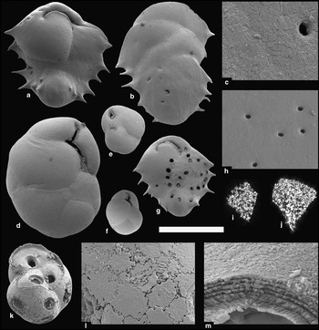
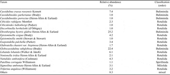
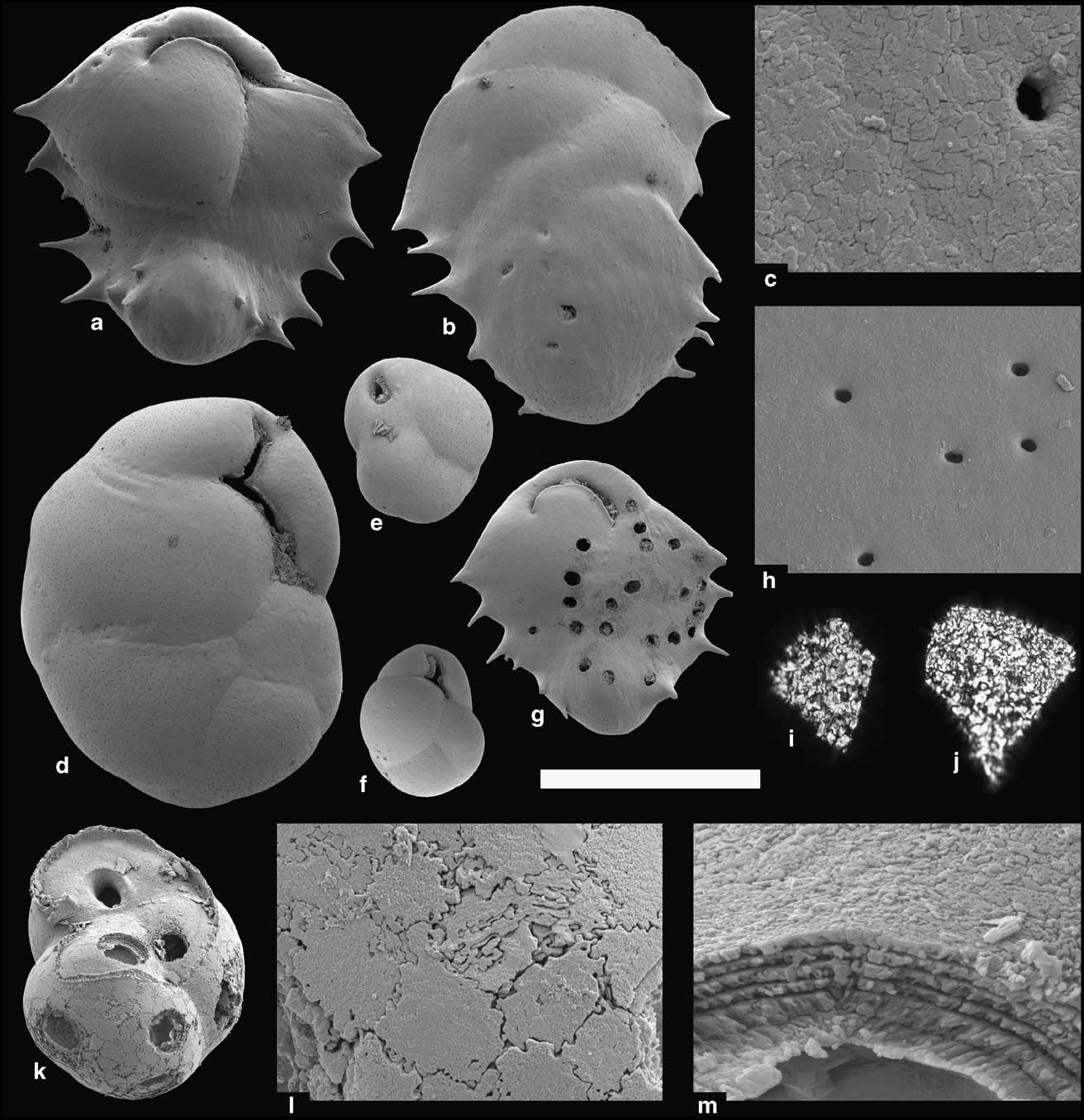
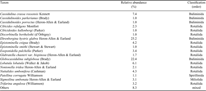
Our faunal analyses of the benthic foraminifera in the two LU 3.1 samples are in agreement with previous analyses (Webb & Strong Reference Webb and Strong1998), revealing a strong dominance by two species, Ehrenbergina hystrix glabra Heron-Allen & Earland (25.2%; Fig. 1a, b & g) and Globocassidulina subglobosa (Brady) (22.4%; Fig. 1e) (Table I). The majority of calcareous taxa exhibit a bilamellar wall type with 57% belonging to the order Buliminida and > 30% to the order Rotaliida (Table I). Observed corrosion and fragmentation of the buliminid taxa, such as E. hystrix glabra, Cassidulina crassa rossenis Kennett (Fig. 1d & f), Cassidulinoides spp. and G. subglobosa, was high, much higher than in lagenids or large-sized species of the orders Miliolida or Rotaliida (e.g. Cibicides spp., Trifarina angulosa Williamson). Various corroded test surfaces of buliminid and rotaliid specimens revealed a taxa-typical ultrastructure of large cleaved crystals separated from each other by jigsaw-puzzle-like structures (Fig. 1k & l). In contrast, pristine specimens show smooth surfaces (Fig. 1h), with the irregular sutures only visible under polarized light (Fig. 1i & j).
Selected foraminifera from samples CRP-1-33.69 (a–j) and CRP-1-33.72 (k–m). a. & b. Large specimens of Ehrenbergina hystrix glabra Heron-Allen & Earland (PMU 25724, PMU 25725), c. outer test surface of E. hystrix glabra (PMU 25726), d. large specimen of Cassidulina crassa rossensis Kennett (PMU 25727), e.Globocassidulina subglobosa (Brady) (PMU 25728), f. juvenile form of C. crassa rossensis (PMU 25729), g. multiple bored test of E. hystrix glabra (PMU 257230) (borings represent the trace fossil Oichnus coronatus Nielsen & Nielsen), h. outer test surface of C. crassa rossensis (detail of d.), i. & j. test fragments of C. crassa rossensis (i.; PMU 25731) and E. hystrix glabra (j.; PMU 25732) in polarized light, k.–m. multi-chambered test fragment of an unidentified bilamellar foraminifera (PMU 25733) showing multiple borings (Oichnus spp.), l. ultrastructure with crystal units separated by “interdigitated sutures” (detail of k.), m. laminar test wall (detail of k.). Scale bar equals 400 μm (a., b. & d.–g.), 100 μm (i. & j.), 40 μm (k.), 20 μm (h.), 8 μm (l.), and 4 μm (c. & m.).
Occurrence of benthic foraminifera in analysed CRP-1 samples. Species with a relative abundance < 1% are summarized as ‘others’.
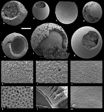
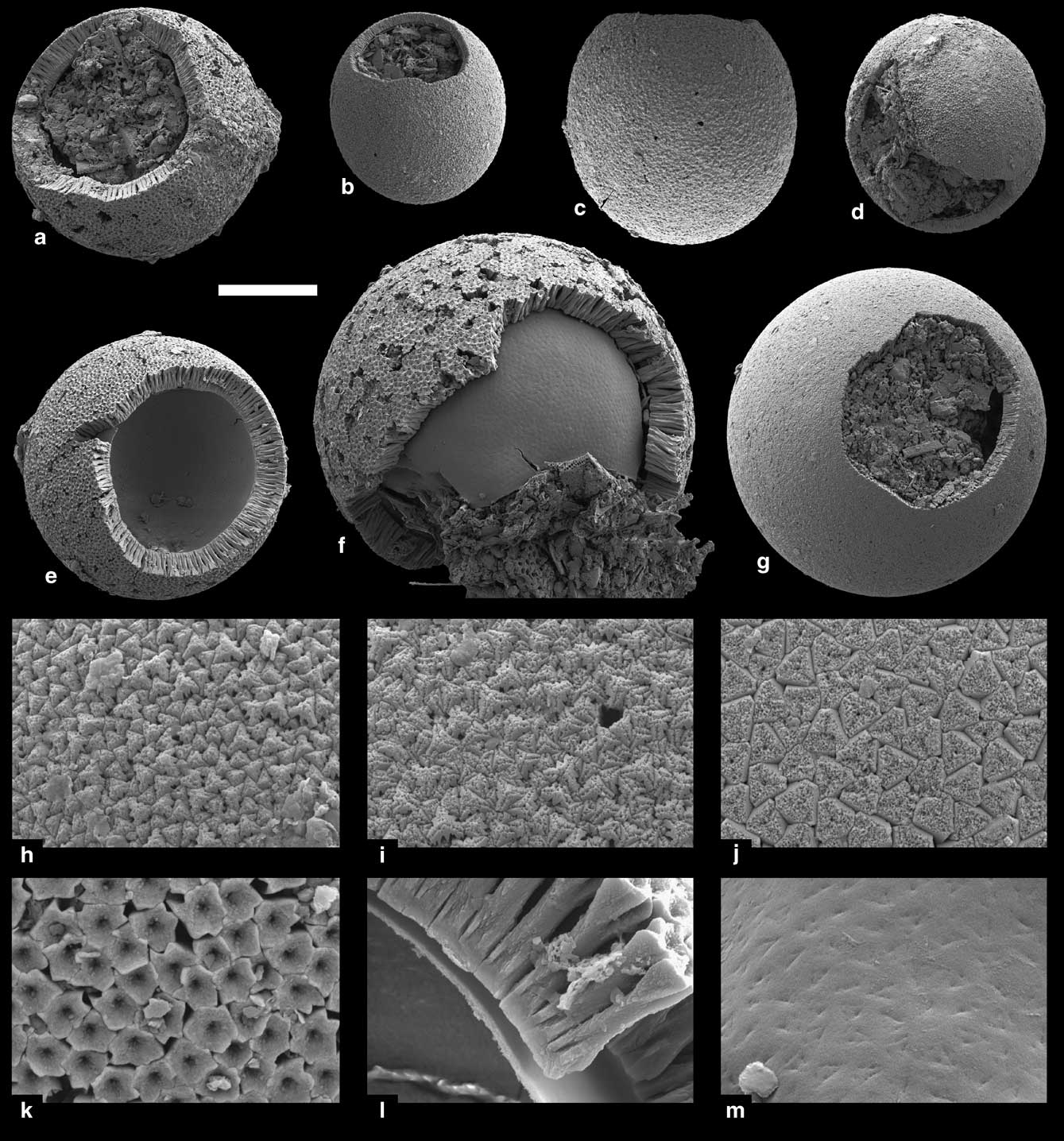
Intensive search for calcareous dinoflagellate cysts in the two studied samples resulted in the recovery of 20 cysts of Caracomia arctica (Gilbert & Clark) Streng, Hildebrand-Habel & Willems (Figs 2 & 3). As the studied samples were not prepared quantitatively, no absolute abundances can be calculated. However, the general abundance of “cysts per picking tray” appears to be similar to other high latitude Pleistocene localities, for which extreme low absolute abundances of less than 100 cysts per gram sediment were calculated (Streng et al. Reference Streng, Hildebrand-Habel and Willems2002). The cysts are spherical to slightly ovaloidal in shape ranging from 37–85 μm in maximum diameter (for further details on cysts see Systematic palaeontology).
Caracomia arctica forma rossensis f. nov. (a., e., f. & k.–m.) and Caracomia arctica forma arctica Autonym (b.–d. & g.–j.) from lithostratigraphic unit 3.1 (sample CRP-1-33.72; early Pleistocene). a. Holotype with subangular mesoepicystal archeopyle (PMU 25720), b.–d. ovaloidal cysts with densely packed wall-forming crystals (PMU 25734, PMU 25735, PMU 25736), e. paratype with secondarily slightly enlarged archeopyle (PMU 25721), f. relatively large paratype showing loose coalescence of wall-forming crystals and inner organic layer exposed (PMU 25722), g. large, spherical cyst with subangular mesoepicystal archeopyle (PMU 25737), h.–j. outer cyst surfaces showing different degrees of corrosion (details of d., c. & g.; note central canals of crystals visible in j.), k. outer cyst surface with funnel-shaped crystal terminations (detail of e.), l. wall cross-section showing radially arranged wall-forming crystallites and organic inner layer (detail of e.), m. outer surface of organic layer with imprints of proximal tips of wall-forming crystals (detail of f.). Scale bar equals 20 μm for whole cysts (a.–g.) and 3 μm for close-ups (h.–m.).
Discussion and implications
A recent palaeoenvironmental interpretation of LU 3.1 based on micropalaeontology, isotope and palaeomagnetism evidence (Scherer et al. Reference Scherer, Bohaty, Dunbar, Esper, Flores, Gersonde, Harwood, Roberts and Taviani2008) confirmed the initial interpretation by Villa & Wise (Reference Villa and Wise1998) of deposition during relatively warmer conditions and also agrees with our results on the presence of C. arctica. In fact, the discovery of C. arctica in LU 3.1 has two main implications: 1) it permits inferences about the life cycle of this particular species, and 2) it provides constraints on the palaeoenvironmental interpretation of LU 3.1.
Although C. arctica is considered as a modern species (Streng et al. Reference Streng, Hildebrand-Habel and Willems2002, Vink Reference Vink2004, Elbrächter et al. Reference Elbrächter, Gottschling, Hildebrand-Habel, Keupp, Kohring, Lewis, Meier, Montresor, Streng, Versteegh, Willems and Zonneveld2008), its motile stage and life cycle are still unknown, and thereby ambiguity exists about the organism′s metabolism and the function of the cyst. However, all modern calcareous cyst producing dinoflagellates are considered to be photosynthetic, i.e. they live in the photic zone and need sunlight for their metabolism (Zonneveld et al. Reference Zonneveld, Meier, Esper, Siggelkow, Wendler and Willems2005 and references therein). The same most probably applies to C. arctica. As C. arctica can occur at high latitudes beyond the polar circles, i.e. at latitudes where for extended periods of time no sunlight is available, the organism must be specially adapted to cope with such extreme conditions. Studies have shown that survival times of more than two years are not uncommon among resting cysts of marine organic-walled dinoflagellates (Lewis et al. Reference Lewis, Harris, Jones and Edmonds1999) and the same is probably true for calcareous cysts. Thus, cysts of C. arctica might be best interpreted as resting cysts that enable the organism to endure adverse conditions, such as polar winters, or generally speaking, times in which no sunlight is available for metabolism. The low cyst abundance in the sediments of LU 3.1 might suggest a low cyst production rate, which also points towards a function as a resting rather than a vegetative cyst (but see Gilbert & Clark Reference Gilbert and Clark1983, who argued that the cysts of C. arctica might have been vegetative). Similar to other dinoflagellate taxa, germination of C. arctica is probably triggered by increasing temperature and/or light availability, e.g. in the course of polar summers.
Irrespective of the functional interpretation of the cyst of C. arctica, the presence of the cysts has implications for the interpretation of the palaeoenvironment of LU 3.1. Supposed extant occurrences of monospecific associations of C. arctica are known from two localities in the South Atlantic Ocean (Streng et al. Reference Streng, Hildebrand-Habel and Willems2002, Vink Reference Vink2004). Both localities are under the influence of eutrophic, cold currents (the Falkland Current and Antarctic Circumpolar Current), having a combined temperature range from -2°C (winter minimum temperature, potential of sea ice coverage) to 10°C (maximum summer temperature) with a salinity range of 34–35 psu. This contrasts with present-day conditions in the Ross Sea, which are characterized by multiannual fast ice, seasonal sea ice and summer surface water temperatures not above -1.6°C (Davey et al. Reference Davey, Barrett, Cita, van der Meer, Tessensohn, Thomson, Webb and Woolfe2001, Scherer et al. Reference Scherer, Bohaty, Dunbar, Esper, Flores, Gersonde, Harwood, Roberts and Taviani2008). Thus, judging from the known ecological preferences of C. arctica, significantly warmer and more open marine conditions may have prevailed during the deposition of LU 3.1. This agrees with palaeoenvironmental reconstructions based on diatom associations, which indicate warmer than present-day water temperatures and an open water setting with little or no summer ice (Scherer et al. Reference Scherer, Bohaty, Dunbar, Esper, Flores, Gersonde, Harwood, Roberts and Taviani2008). Pending further detailed information on the ecological preferences of C. arctica, the presence and absence of the species in sediments around Antarctica could possibly be used to assess fluctuations of seawater temperatures, productivity and sea ice coverage during the Pleistocene. However, despite the potential of C. arctica as a proxy for high latitude oceanographic conditions, it should be stressed that C. arctica is typically an extremely rare species (see Results; Streng et al. Reference Streng, Hildebrand-Habel and Willems2002, but see also Gilbert & Clark Reference Gilbert and Clark1983) and it is unlikely that its cysts would be detected using standard nannoplankton sample preparation techniques, i.e. smear slides. To recover specimens of C. arctica, cysts have to be concentrated by sieving and subsequently be hand-picked under a binocular microscope (see above).
Based on our results and observations on the microfossil content of LU 3.1, the dinoflagellate nature of the described and illustrated fragments of “Thoracosphaera spp.” of Villa & Wise (Reference Villa and Wise1998) cannot be confirmed. We consider the ultrastructure of the “Thoracosphaera spp.” fragments to be identical to that of bilamellar foraminifera (see e.g. Towe & Cifelli Reference Towe and Cifelli1967, Bellemo Reference Bellemo1974, or Nomura Reference Nomura1984 for details on foraminiferan ultrastructure). Bilamellar foraminifera are the most common group of foraminifera in the analysed samples of LU 3.1 (> 87%) and show frequent corrosion and a high degree of fragmentation in combination with abundant bioerosion (see Results, Fig. 1g & k). Furthermore, described morphology, surface structures, and sizes of “Thoracosphaera spp.” do not match any known calcareous dinoflagellate species, but match well with test characters of the most common foraminifera. This also applies to the putative complete cyst of Villa & Wise (Reference Villa and Wise1998, pl. 1, fig. e), which in our opinion represents a proloculus of a bilamellar foraminifera (e.g. G. subglobosa). For these reasons, we interpret the specimens illustrated and described by Villa & Wise (Reference Villa and Wise1998) as fragments of bilamellar foraminifera.
Systematic palaeontology
Family Thoracosphaeraceae Schiller, 1930
Remarks: Calcareous dinoflagellates are dinoflagellates that have the ability to form calcareous cysts during their life cycle. Based on rRNA sequencing data, calcareous dinoflagellates are today considered to belong to a monophyletic clade within the Peridiniales (Gottschling et al. Reference Gottschling, Keupp, Plötner, Knop, Willems and Kirsch2005). Accordingly, they have been recently united within the single family Thoracosphaeraceae (Elbrächter et al. Reference Elbrächter, Gottschling, Hildebrand-Habel, Keupp, Kohring, Lewis, Meier, Montresor, Streng, Versteegh, Willems and Zonneveld2008), contrasting the previous polyphyletic concept of Fensome et al. (Reference Fensome, Taylor, Norris, Sarjeant, Wharton and Williams1993) (see Elbrächter et al. Reference Elbrächter, Gottschling, Hildebrand-Habel, Keupp, Kohring, Lewis, Meier, Montresor, Streng, Versteegh, Willems and Zonneveld2008 for details). The calcareous cysts, which are typically spherical in shape, have in natural environments a reported size range from c. 9–132 μm in diameter, with Thoracosphaera heimii (Lohmann) Kamptner being the smallest and Orthopithonella? globosa (Fütterer) Lentin & Williams being the largest species known (Fütterer Reference Fütterer1977, Reference Fütterer1984, respectively). Most cysts, however, have diameters of 25–70 μm with most modern oceanic species being predominantly in the lower half of that range (Streng, observation 1999–2003). The cyst wall may have one, two, or three (only one Cretaceous taxon) calcareous layers. Two-layered cysts were quite common during Cretaceous and Palaeogene times, and diminished during the Neogene. The youngest reported double-layered cyst is Pirumella parva Fütterer, Reference Fütterer1984 from the early Pleistocene (Fütterer Reference Fütterer1984). Individual calcite crystals whose morphological long axes are either radially or obliquely oriented construct each layer. The crystallographic c-axis of the radially arranged wall-forming crystals, however, can be perpendicular or parallel to the cyst surface. Thus, three different cyst ultrastructures are distinguished in calcareous dinoflagellates: a radial, a tangential, and an oblique ultrastructure (see Elbrächter et al. Reference Elbrächter, Gottschling, Hildebrand-Habel, Keupp, Kohring, Lewis, Meier, Montresor, Streng, Versteegh, Willems and Zonneveld2008 for details). Pores and pore-like structures that penetrate the cyst wall perpendicularly have been described for various species of calcareous dinoflagellates (e.g. Streng et al. Reference Streng, Hildebrand-Habel and Willems2002, Reference Streng, Hildebrand-Habel and Willems2004a). Pore diameters range from 0.2 to c. 1 μm. Due to their mineralized walls, cysts of calcareous dinoflagellates have a high preservation potential (for complementary reading see Streng et al. Reference Streng, Hildebrand-Habel and Willems2004a, Elbrächter et al. Reference Elbrächter, Gottschling, Hildebrand-Habel, Keupp, Kohring, Lewis, Meier, Montresor, Streng, Versteegh, Willems and Zonneveld2008, Versteegh et al. Reference Versteegh, Servais, Streng, Munnecke and Vachard2009).
Genus Caracomia Streng, Hildebrand-Habel & Willems, Reference Streng, Hildebrand-Habel and Willems2002
Caracomia arctica (Gilbert & Clark, Reference Gilbert and Clark1983) Streng, Hildebrand-Habel & Willems, Reference Streng, Hildebrand-Habel and Willems2002
Diagnosis: See Streng et al. (Reference Streng, Hildebrand-Habel and Willems2002).
Discussion: Caracomia arctica is the only real cold water calcareous dinoflagellate species recognized so far that can clear the hurdle of physical limitations on carbonate secretion in polar environments (Streng et al. Reference Streng, Hildebrand-Habel and Willems2002, Reference Streng, Hildebrand-Habel and Willems2004b, Vink Reference Vink2004). It is known from the late mid Miocene (Hildebrand-Habel & Streng Reference Hildebrand-Habel and Streng2003, Streng et al. Reference Streng, Banasová, Reháková and Willems2009) where it also can be found in more temperate environments. Since the late Pliocene, however, its distribution seems to be restricted to cold waters of both hemispheres where it occurs in monospecific associations reflecting the rapid cooling trend in the late Pliocene and the onset of the Pleistocene glaciations (Gilbert & Clark Reference Gilbert and Clark1983, Streng et al. Reference Streng, Hildebrand-Habel and Willems2004b). In the northern hemisphere, C. arctica has been reported from early Pleistocene sediments as far north as 85°50′N (Mudie Reference Mudie1985, see also Gilbert & Clark Reference Gilbert and Clark1983). Gilbert & Clark (Reference Gilbert and Clark1983) explained the high latitude occurrence by relative productive surface waters with thin and/or seasonal sea ice. The most southern occurrence on the southern hemisphere was, until the discovery of the specimens described herein, specimens from surface sediment samples of Maud Rise at 64°31′S (Streng et al. Reference Streng, Hildebrand-Habel and Willems2002). The presence of C. arctica in early Pleistocene sediments of LU 3.1 at 77°S suggests an explanation similar to that of Gilbert & Clark (Reference Gilbert and Clark1983) for the Northern Hemisphere, i.e. reduced seasonal sea ice coverage favouring increased primary production rates (see Discussion and implications).
Caracomia arctica is quite variable in its morphology, which is reflected in its division into four distinct forms (see Streng et al. Reference Streng, Banasová, Reháková and Willems2009 for summary). Among the cysts of C. arctica found in LU 3.1, two different morphotypes are evident, one of which is interpreted as a new and fifth form.
Caracomia arctica forma arctica Autonym (Fig. 2b–d & g–j)
Material: Seven cysts: four cysts from sample CRP-1-33.72, and three cysts from sample CRP-1-33.69.
Description: Cyst characters and cyst dimensions generally agree with previous descriptions of specimens from the Southern Hemisphere (Streng et al. Reference Streng, Hildebrand-Habel and Willems2002; see also Vink Reference Vink2004 for light optical images) and won′t be repeated here. Cysts from LU 3.1 are spherical to slightly ovaloidal in shape and measure 37–66 μm in maximum diameter (mean 47 μm, n = 7). Cyst walls are 1.8–3.1 μm thick, positively correlated to cyst size, measuring 5–6% of maximum cyst diameter (n = 6).
Discussion: See discussion of Caracomia arctica forma rossensis f. nov. below.
Caracomia arctica forma rossensis f. nov. (Fig. 2a, e, f, k–m & Fig. 3)
Material: Thirteen cysts: three cysts from sample CRP-1-33.72, and ten cysts from sample CRP-1-33.69.
Etymology: Named after the type locality, Ross Sea.
Holotype: Specimen PMU 25720, Fig. 2a, sample CRP-1-33.72; early Pleistocene (Marine Isotope Stage 31); Ross Sea, Antarctica.
Paratypes: Topotypic specimens PMU 25721 and PMU 25722 (Fig. 2e & f), and specimen PMU 25723 from sample CRP-1-33.69 (Fig. 3).
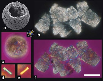
Single cyst of Caracomia arctica forma rossensis f. nov. from lithostratigraphic unit 3.1 (sample CRP-1-33.69; early Pleistocene) (PMU 25723) in the SEM and under the light microscope. a. Whole cyst in the SEM; note that specimen is not sputter coated, b. & f. specimen crushed under crossed nicols, without (b.) and with gypsum plate (f.) employed, c. whole cyst under the light microscope with crossed nicols and gypsum plate showing negative optic sign, d. & e. an individual wall-forming crystal tilted 45° counter-clockwise and clockwise against the light path. Scale bar equals 50 μm for a.–c. & f.; picture width equals 10 μm in d. & e.

Diagnosis: Thick-walled form of C. arctica with funnel-shaped distal crystal terminations.
Description: Cysts are all spherical in shape, measuring 51–85 μm in diameter (n = 12). The single-layered calcareous wall is 5.5–8.8 μm thick, with thickness positively correlated to cyst size, measuring c. 9–11% of cyst diameter (n = 10). Radially arranged, rod-shaped wall-forming crystals are densely packed proximally but less so distally. The individual crystals slightly increase in diameter towards their distal and proximal ends. Distal crystal tips are characterized by an irregular hexagonal outline and a central funnel-shaped depression. All crystals appear to have a central, axially running fine canal that terminates in the centre of the funnel-shaped depression (Fig. 2k). A robust inner organic layer is preserved in all but one cyst. It is c. 200 nm thick and has a granular ultrastructure with the individual granules measuring 30–40 nm. Occasionally, imprints of proximal crystal tips are preserved on its outer surface (Fig. 2m). Most specimens represent dehisced cysts displaying a large, subangular mesoepicystal archeopyle. Other specimens show irregular openings, potentially resulting from secondary enlargement of the archeopyle during deposition.
The crystallographic c-axis of the individual wall-forming crystals corresponds to their morphological long axis, characteristic for the radial ultrastructure of the cyst wall. Whole cysts show under the light microscope in crossed nicols and with gypsum plate a faint extinction cross with interference colours that indicate a negative optic sign. Interference colours are yellow/orange in the first and third quadrant, whereas the second and forth quadrant show a blue colour (Fig. 3c). The faint interference colours in whole cysts is due to the relatively thick walls and large diameter of the cysts. Isolated wall-forming crystals from artificially crushed specimens, however, show clear interference colours when tilted 45° against the light path (Fig. 3d & e).
Occurrence: Early Pleistocene (Marine Isotope Stage 31); Ross Sea, Antarctica.
Discussion: The new form is separated from other forms of C. arctica by the habitus of the wall-forming crystals, i.e. the distal crystal terminations, which are developed as funnel-shaped depression. It is most similar to C. arctica f. arctica, from which it is additionally distinguished by the thickness of the cyst wall. In relation to the cyst size, cysts of C. arctica f. rossensis have a thicker cyst wall than those of C. arctica f. arctica (compare descriptions above). Although cysts of C. arctica f. rossensis tend to be bigger than the LU 3.1 specimens of C. arctica f. arctica, size cannot be used as a distinguishing character. Cyst diameters of C. arctica f. arctica of up to 105 μm have been reported from the Arctic Ocean (Gilbert & Clark Reference Gilbert and Clark1983).
Also distinctive for C. arctica f. rossensis seems to be a weak coalescence of the individual wall-forming crystals. This results in wide interspaces between wall-forming crystals and the inclination of discrete or bunches of crystals (Figs 2a, 2f & 3a). Interspaces and inclined crystals are, however, not considered as cyst characters but are interpreted to be secondary, i.e. taphonomically induced.
The fine central canals, as seen in the wall-forming crystals of C. arctica f. rossensis, are also present in the crystals of C. arctica f. arctica. Here, the canals only become visible if the outer cyst surface has been affected by corrosion (Fig. 2j).
Acknowledgements
Funding by the Swedish Research Council (Vetenskapsrådet) to MS is acknowledged. Gary Wife (Uppsala) helped with SEM photography of the uncoated specimen. We thank S. Meier (Kiel), an anonymous reviewer, and the editor for their critical comments and efforts on an earlier version of the manuscript.