Introduction
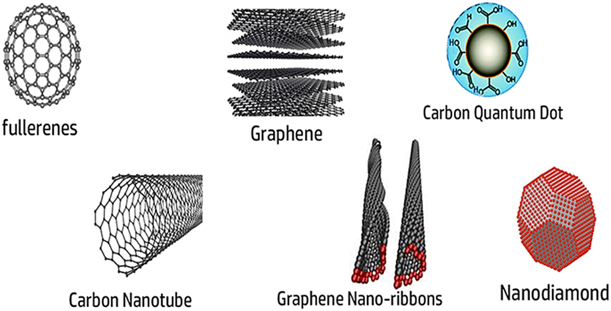
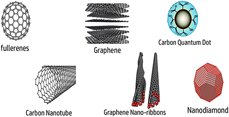
Extensive research outcomes for the last few decades in the field of nanotechnology have reached at such a level that it offers the scope of building up the nanostructures with required size and shape by controlling it at an atomic level, in order to get the desired properties. That changes the landscape of various fields including healthcare [Reference Duncan and Gaspar1, Reference Mahon, Salvati, Baldelli Bombelli, Lynch and Dawson2, Reference Liu, Cui and Losic3, Reference Goenka, Sant and Sant4]. One such nanoscale materials is carbon-based nanomaterials (CBNs) which has been receiving much interest due to their exceptional electrical, thermal, mechanical and biological properties [Reference Baughman5, Reference Zhang6, Reference Kam, O’Connell, Wisdom and Dai7]. These have been captivating researchers with their diverse forms such as single and multi-walled carbon nanotubes (CNTs), graphene, fullerenes, nano-horns, nano-fibres, nano-diamonds, carbon quantum dots (C-Dots) and so forth (Fig. 1). These nanomaterials offer exclusive and unique properties which are suitable for various biomedical applications that expand from the development of novel therapeutics to diagnostic techniques. Moreover, the chance of lower toxicity of CBNs as compared to several other nanoscale materials increases its prospects in several applications like bioimaging, development of diagnostic kits, fluorescence labelling, drug delivery and adjuvant therapy.
Various types of CBNs, showing the structural and morphological diversity. Each and every class of CBNs show unique physicochemical properties which are different from other.

Intravenously administered nanoscale materials enter the circulatory system and immediately encounter the blood which is a complex biological fluid. Interactions of nanoscale materials with various components of blood, trigger cascade of events like platelet aggregation, thrombosis, haemolysis, or complement activation. These parameters are all-together known as hemocompatibility. Hence, biomedical applications of CBNs deem to be minute probing of their hemocompatibility. Hemocompatibility test aims to identify adverse blood–nanomaterial interactions, which may stimulate immunogenic responses or destroy the cellular components of blood. These are very fundamental prerequisites that need to be addressed prior to intravenous administration of any nanoscale materials. Hence, activation of the complement system, effects on blood cells (e.g., RBCs, WBCs, and platelet) and interference in the coagulation process ultimately decide the scope of the nanoscale materials for the real-life applications.
For the last few decades, there have been many experimental works that illustrated the various kinds of CBNs and blood interactions by focusing on varieties of blood components. Individual research groups have carried out studies by focusing on a particular domain of hemocompatibility study. For instance, interactions of the CBNs with the complement system of the blood have been a matter of interest for several groups. Contemporarily, some researchers have also carried out studies on the behavior of blood cells such as white blood cells (WBCs), red blood cells (RBCs), and platelets after the interaction with CNBs. However, only a few reports have attempted to provide a complete depiction about the hemocompatibility by considering all the parameters and possibilities of interactions in a systematic way for better understanding and clarifications [Reference Ilinskaya and Dobrovolskaia8, Reference Sobot, Mura, Couvreur, Kobayashi and Müllen9]. In this review, we have tried to combine these recent experimental findings in order to fulfil the gap in the research outcomes of various research groups. Various parameters have been considered and combined herein to get a better interpretation of the blood and CNBs interactions. We have also exemplified various ways of surface modification of the CNBs in order to alleviate the adverse effects and to increase the efficiency and performance, which is essential for the biological applications [Reference Yang, Wan, Zhang, Zhang, Lee and Liu10, Reference Singh, Singh, Kulkarni, Sonkar, Grácio and Dash11, Reference Awasthi, Singh, Singh, Dash and Srivastava12, Reference Yang, Thordarson, Gooding, Ringer and Braet13, Reference Schulz-Dobrick, Sarathy and Jansen14, Reference Neouze and Schubert15, Reference Ruckenstein and Li16].
Response of blood plasma and complement system in presence of CBNs
CNTs
Among the various allotropes of the carbon, CNTs are one of the extensively studied nanoscale materials which were first brought into the limelight by Iijima in 1991 [Reference Iijima17]. For the synthesis of the CNTs, arc-discharge evaporation method and chemical vapour deposition (CVD) techniques are widely used. CVD is mostly employed as it is economical compared to other techniques. Although different types of CVD techniques are used, most of them use metals as a catalyst in order to promote the nucleation which is required for the formation of CNTs. Metals like Molybdenum, Cobalt, Nickel, Iron, etc., can serve as mono and bi-metallic catalyst for the formation of CNTs. Thus, predominantly CVD techniques have been used to synthesize single and double walled nanotubes by various research groups.
In the first report, the as-synthesized CNTs were characterized by Transmission electron microscope (TEM) which showed coaxial tubes of graphitic sheets of size 2–50 nm. From the TEM images, it was observed that each CNT is comprised of hexagonal carbon-atoms that were arranged in a helical fashion. These are hollow cylindrical structured nanoscale materials of a single layer or multilayer compositions that are commonly coined as single-walled nanotubes (SWCNTs) and multi-walled nanotubes (MWCNTs), respectively. It has gained huge attention from the scientific communities owing to its exceptional properties of very high surface area as well as huge aspect ratio (length to diameter proportion of up to 132,000,000:1). Generally, SWCNTs are having the diameter ranges from 1 to 2 nm whereas MWCNTs are anywhere between 10 and 100 nm based on the number of layers. However, the length of both these types of CNTs varies from 50 nm to 1 cm depending on the synthesis methods. Both the types of CNT are made up from smooth graphene sheets comprising of sp 2 bound carbon atoms which wounds up in the form of thin and hollow tubes. The presence of sp 2 bound aromatic rings in their pristine form makes CNTs highly hydrophobic. But this hydrophobic surface may effectively be converted into hydrophilic or a charged surface by functionalizing them with some other molecules. It would be mentioned here that ease of surface functionalization and high surface area have made CNTs particularly attractive in various biomedical applications like the development of gene and drug delivery cargos, cancer therapy, and tissue engineering. However, protein binding and its selectivity are highly influenced by surface features of functionalized CNTs such as surface charge, hydrophobicity and hydrophilicity of the functional groups present on the CNTs [Reference Bussy, Methven and Kostarelos18]. Several studies have been carried out to the find out the effect of CNTs on the blood complement system, which is a vital part of the innate immune system that plays an essential role in protection against microorganisms, altered cells and toxic synthetic materials (including nanoscale materials). The complement system is comprised of a group of soluble proteins present in the blood serum that interact with one another in a highly regulated manner, in order to eliminate the pathogens. Essentially, complement proteins are proteases that are themselves activated by proteolytic cleavage.
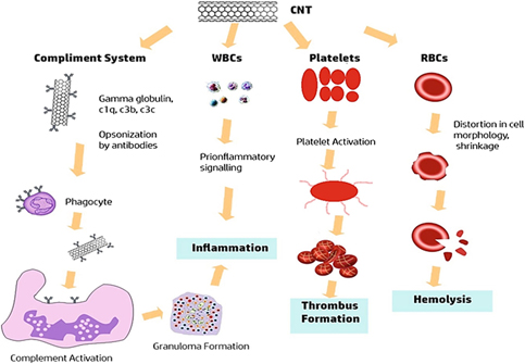
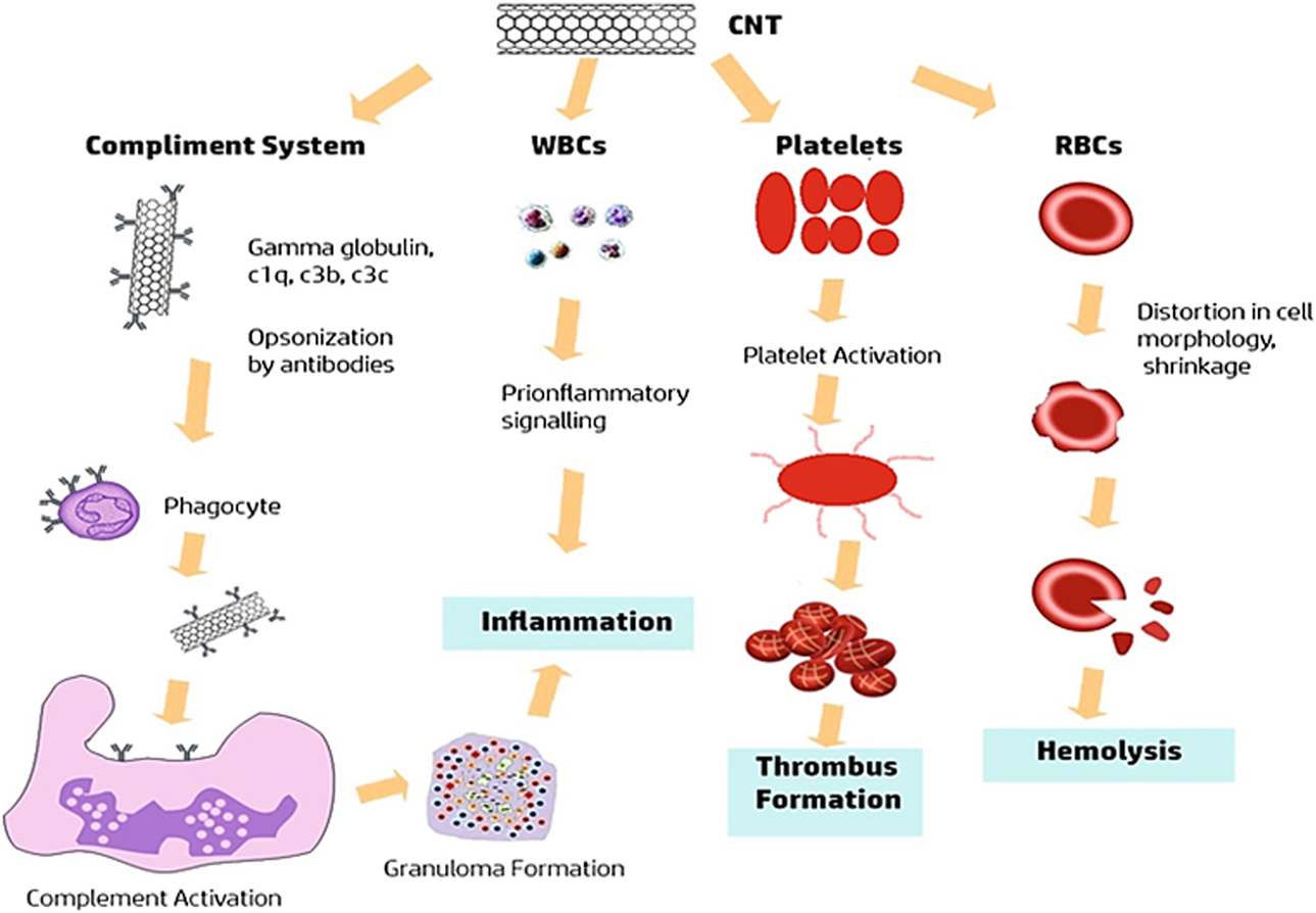
The dissociation of one protein leads to dissociation of the next complement protein as well as leads to the generation of bioactive protein fragments. It plays a vital role in the immune system by eliciting the function of phagocytic cells (e.g., Macrophage and Natural Killer cells) and antibodies in the case of opsonisation. It also eradicates the invading elements (e.g., micro-organisms or foreign materials including nanoscale materials) and altered cells (e.g., apoptotic or necrotic cells) present in the host. Thus, activation of the complement systems may trigger several immune responses such as phagocytosis, inflammation and membrane attack complex (MAC) formation (Fig. 2).
Interaction of CNTs with various blood components and possible biological responses, showing the cascades of events may occur due to the presence of CNTs including complement activation, haemolysis and thrombus formation.
Activation of the complement system occurs by three pathways, viz. the classical, lectin and alternate pathway. All three pathways produce the same end product that is MAC that kills the target cells (i.e., pathogen) by integrating into the bi-lipid layer of the pathogen. However, the starting point of each pathway is different. For example, the classical pathway starts while the C1 component (‘C’ stand for complement protein) that binds to antigen–antibody complexes. The C1 component comprises C1q, C1r, and C1s subunits where different subunits perform different functions. For instance, C1q is responsible for the binding of the C1 protein with antigen–antibody complex and C1r as well as C1s subunits cause the proteolysis of other complement proteins such as C4 and C2 which results in C3 convertase (i.e., C4b2a) that subsequently catalyses C3 complements proteins into C3a and C3b. These cascades of reaction form C5b that ultimately binds to C6, C7, C8, and C9 to form the C5b-9 (i.e., membrane attack complex, MAC). In the lectin pathway, mannose-binding lectin (MBL) interacts with neutral sugar residues and binds to its targets that ultimately lead to the activation of the complement systems. In the alternate pathway, complement activation begins by attachment of C3b to the pathogen’s surface and the rest of the events are similar to the classical pathway [Reference Salvador-Morales, Flahaut, Sim, Sloan, Green and Sim19].
Recent reports stated that complement activation may be initiated by adsorption of proteins onto the CNTs. Although, very limited information is available on the detailed pathway of activation of the complement system due to CNTs [Reference Rybak-Smith and Sim20]. Studies established that SWCNTs (single-walled) and DWCNTs (double-walled) without functionalization were found to be activating complement system mainly via classical pathways. While SWCNTs stabilized with poly-ethylene glycol derivatives showed a signature of activation of the Lectin pathway. Ling et al. found that C1q subunit of C1 protein could adsorb on the multi-walled CNTs in a much better fashion as compared to single and double walled-CNTs. The study established that C1q “crystallizes” on CNTs after binding with the globular head of the C1q (i.e., gC1q). However, it did not cause activation of the complement system [Reference Ling, Biro, Bally, Tacnet, Deniaud, Doris, Frachet, Schoehn, Pebay-Peyroula and Arlaud21]. On the other hand, Salvador-Morales et al. have confirmed the activation of the entire complement system via the classical pathway predominantly by two types of CNTs, SWCNTs, and DWCNTs which were non-covalently functionalized. It was also observed that activation of complement by CNTs by the classical pathway led to the production of inflammatory peptides and subsequent coating of opsonins iC3b and C3b. It was anticipated that the orientation of the globular heads of C1q reduced the likelihood of interaction of C1q with CNTs which indeed prevents the activation of the classical pathway. Subsequent studies found that chemical modification of MWCNTs could affect complement activation by following potentially different pathways [Reference Salvador-Morales, Basiuk, Basiuk, Green and Sim22]. While modification of pristine MWCNTs with 1,8-diamino-octane and later added L-alanine by covalent attachment resulted in changes of complement activation via both classical and alternate pathways. Hence, it’s obvious that the myriad of soluble serum proteins was tested, however, only a few proteins have displayed an affinity for the CNTs.
Another essential factor which affects the activation of the complement system is the surface charges of the CNTs that may stimulate factor H (regulator protein of complement activation) binding. Human serum albumin (HSA) coated SWCNTs activated the complement system via the alternate as well as the classical pathway, whereas SWCNTs coated with methoxy-PEG activated only via the lectin pathway [Reference Andersen, Robinson, Dai, Hunter, Andresen and Moghimi23, Reference Andersen, Windschiegl, Ilbasmis-Tamer, Degim, Hunter, Andresen and Moghimi24].
Apart from the activation of the complement pathway the bioactive fragment of the complement protein causes the phagocytosis. In vitro studies revealed that while monocyte-derived macrophages were exposed to 1 × 10⁻¹ mg/mL of pristine SWCNTs, it reduced the capability of engulfing apoptotic cells by the macrophages. The researchers also observed that human monocytes exhibited repressed chemotaxis toward monocyte chemo-attractant protein-1 (MCP-1) [Reference Hussain, Vanoirbeek and Hoet25]. An in vivo investigation carried out by Mitchell et al. has revealed that MWCNTs were capable of suppressing systemic immune reaction [Reference Iijima17, Reference Mitchell, Gao, Wal, Gigliotti, Burchiel and McDonald26]
The above studies imply that properties of the surface ligands play a very crucial role in altering the functionality and also shifting the mode of complement activation (by a pathway switch). Moreover, the mode of interaction of the recognition subunits of all the three pathways (i.e., if the subunit would bind to the CNTs directly or indirectly via other serum proteins) also decides the fate of the CNTs.
Graphene
Graphene which is first identified by Novoselov and Giem is one of man-made thinnest material known on earth that could be at one atom thick but incredibly strong. It consists of two-dimensional single-layer atomic sheet composed of planar sp 2 carbon atoms tightly bound in a hexagonal honeycomb lattice. It has several astonishing properties like high tensile strength (between 100 and 300 times stronger than steel), a good conductor of heat and electricity at room temperature. It also shows uniform light absorption from visible and near-infrared regions of the spectrum, and is suitable for spin transport.
The two most common ways to synthesize graphene are CVD and exfoliation. Somani et al., in 2006, have reported the synthesis of graphene by the CVD method, for the first time. Then, various research groups carried out the synthesis of single and multi-layers of graphene on Cu by CVD by using methane as the carbon precursor and hydrogen as a balancing gas [Reference Somani, Somani and Umeno27]. It evolved very rapidly for the synthesis of desired types of the graphene and other nanoscale materials like CNTs. This is a very useful and convenient technique as it offers easy tuning of properties of graphene by controlling the growth parameters such as pressure, temperature and flow of precursor. The number of graphene layers, amount of doping and types of stacking could be easily monitored by parameters of CVD setup. Importantly, this method is apposite for large scale production of high quality graphene also. Recently, growth of graphene on a dielectric substrate and large area single crystal growth of graphene has been reported by Hongtao et al. 2017 [Reference Liu and Liu28]. Another method of synthesis of graphene is exfoliation which implies the removal of the layers to get desired thickness of graphene sheet from graphite. Exfoliation may be acid/base exfoliation, electrochemical, exfoliation, mechanical exfoliation, and thermal exfoliation. These methods employ one of the physicochemical tools (e.g., concentrated acid/base, temperature, etc.) or sometimes its combination in order to produce the desired size and shape of graphene. Large scale exfoliation was proposed by Paton et al. in 2014 [Reference Paton, Varrla, Backes, Smith, Khan, O’Neill, Boland, Lotya, Istrate, King, Higgins, Barwich, May, Puczkarski, Ahmed, Moebius, Pettersson, Long, Coelho, O’Brien, McGuire, Sanchez, Duesberg, McEvoy, Pennycook, Downing, Crossley, Nicolosi and Coleman29]. They proposed that the method is suitable for the synthesis of defect-free graphene by shear mixing of graphite in a suitable liquid solvent.
Despite being so thin, it already mesmerizes the researchers due to its unique properties. Graphene is a member of the graphitic family which also consists of graphene oxide (GO), reduced graphene oxide (r-GO) and graphene nano-ribbons. GO is a highly oxygenated form of graphene which is highly stable in liquid dispersed media. Graphene nanoribbons are CBNs obtained from MWCNTs having a high aspect ratio and arranged in long, thin ribbon-like graphene sheets. These all kind of graphene-based materials have exhibited huge promises in using it various biomedical applications like bio-imaging, biosensing and drug delivery. In the present study, both pristine (p-G) and functionalized graphene (f-G) were characterized by TEM and Raman Spectroscopy. Further, atomic force microscopy (AFM) imaging was employed to explore the thickness and lateral dimensions of the as-synthesized graphene. To explore the biological activities murine macrophage RAW 264.7 cells and human primary blood components were used as model systems. The cellular uptake was confirmed by scanning electron microscopy (SEM) and energy-dispersive X-ray spectrometry (EDX). It was proposed that the uptake of these materials caused changes in its metabolic activity and plasma membrane stability. Moreover, this also elevated the reactive oxygen species (ROS) level and caused a substantial amount of haemolysis.
Sasidharan et al. performed the first investigation on pristine graphene to evaluate hemocompatibility [Reference Sasidharan, Panchakarla, Sadanandan, Ashokan, Chandran, Girish, Menon, Nair, Rao and Koyakutty30]. Since then many groups have started to examine the effect of pristine graphene on the immune system. Li et al. studied GO-plasma membrane interactions of human lung epithelial cells, primary human keratinocytes and murine macrophages [Reference Li, Yuan, von dem Bussche, Creighton, Hurt, Kane and Gao31]. Study of interaction of GO and functionalized nano-GO (nGO-PEG) with the immune system, especially proinflammatory cytokines is very crucial. One group focused their study on GO sheets by checking the effect of its lateral dimension on human and murine phagocytic cells. They found that greater the lateral dimensions were reduced, greater were its absorption and influence on functionality [Reference Russier, Treossi, Scarsi, Perrozzi, Dumortier, Ottaviano, Meneghetti, Palermo and Bianco32]. Another group confirmed that the generation of intracellular ROS altered the membrane potential (MMP) of the mitochondria, which induced cytotoxicity in macrophages when treated with pristine graphene [Reference Li, Liu, Fu, Wei, Le Guyader, Gao, Liu, Chang and Chen33].
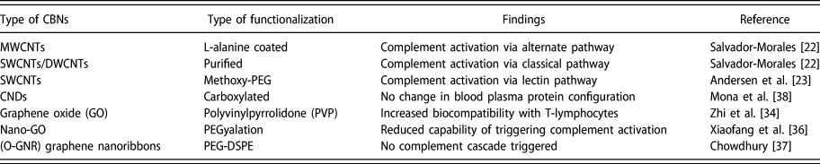
Several research groups also have focused on the role of surface modification in the immune response. For example, Zhi et al. proposed that apoptosis triggered in T-cells was much less prominent when GO coated with PVP was used [Reference Zhi, Fang, Bao, Shen, Zhang, Wang, Guo, Wan and Cui34]. Ni et al. further demonstrated that GO is capable of absorbing anti-IL10R receptor antibodies which are more effective as compared to free anti-IL10R antibodies [Reference Ni, Wang, Wu, Wang, Chen and Liu35]. Xiaofang et al. investigated the consequences of PEGylation of nano-GO sheets while exposed to serum proteins [Reference Tan, Feng, Zhang, Yang, Zhang, Liu and Peng36]. They found that when non-functionalized GO was compared with nGO-PEG, the latter demonstrated an intense reduction in both the capability to trigger complement C3 cleavage and protein binding. Therefore, it was assumed that PEGylation could selectively influence the interactions between multiple serum factors and nGO sheets. This was possibly due to the changes in surface chemistry that further leads to changes in the interactions patterns at the nano-biointerface. Researchers also accounted for the role of graphene nanoribbons in the activation of the complement system by functionalizing the graphene with an extensively used phospholipids-polymer conjugate [1,2-distearoyl-sn-glycero-3-phosphoethanolamine-polyethylene glycol (DSPE-PEG)] [Reference Chowdhury, Fang and Sitharaman37]. The study found under the given conditions that the interaction of complement proteins with O-GNR-PEG-DSPE did not trigger protein cleavage cascade. In a subsequent study, the same group proved that graphene nanoplatelets functionalized with dextran had no effect on the release of pro- and anti-inflammatory cytokines. The group concluded its research on the fact that graphene nanoribbons-PEG-DSPEs play no role in increasing the level of pro-inflammatory cytokine TNF-α in whole blood. However, a slight decrease in the release of the anti-inflammatory cytokine IL-10 (by 5–10%) was observed. Overall, it was anticipated that under the tested environment graphene nanoribbon-PEG-DSPEs had no effect on inflammatory cytokines release. The overall role of surface functionalization in order to reduce the chances of complement activation is summarized in Table I.
Effect of functionalization of various CBNs on complement system, showing the possibility of the variation in the biological responses.

Carbon NDs
Carbon nanodiamonds (NDs) which are produced by impact events like an explosion have been fascinating researcher owing to its various rare properties like high biocompatibility, low cytotoxicity and chemical inertness. These tiny carbon-based nanoscale objects (3–100 nm) are able to permeate the cell plasma membrane and even infiltrate through the nuclear membrane, hence it’s considered as a promising drug delivery candidate. Importantly, its ability to cross the blood–brain barrier might help in the delivering chemotherapy drugs to the brain. These also offer the scope of delivery of a broad range of therapeutics in addition to hydrophilic and hydrophobic molecules, fluorophores, proteins and nucleic acids. With the rapid increase in the application of engineered NDs, there is a need to completely understand their interactions with the haemostatic system. Mona et al. conducted a study on the adsorption of blood plasma protein components to different sizes of carboxylated NDs with the help of FTIR and UV-visible spectroscopy [Reference Mona, Kuo, Perevedentseva, Priezzhev and Cheng38]. The study revealed that 5–100 nm sized carboxylated NDs adsorbed human plasma proteins, however, protein absorption increased with the decrease in the size of NDs. While more blood plasma components were adsorbed by 5 nm NDs in comparison to 100 nm. This behavior was accredited to the fact that the surface area and structure of NDs decides the amount of adsorption of the plasma protein.
There are also reports which suggested that NDs may affect various blood parameters, because of its high adsorption potential that causes firm interactions of several plasma components with NDs. It may have several impacts on properties and functions of the blood cells like it may modify cell aggregation, may alter oxygen-carrying capacity, rheological properties of RBC and influence osmotic properties of blood plasma. There are reports which established that a range of plasma proteins, such as insulin, albumin, histone, fibrinogen and immunoglobulins may also adsorb on the surface of NDs [Reference Perevedentseva, Su, Su, Lin, Cheng, Karmenyan, Priezzhev and Lugovtsov39, Reference Lück, Paulke, Schröder, Blunk and Müller40, Reference Green, Davies, Roberts and Tendler41]. Wasdo et al. investigated the interaction of detonated-NDs with albumin and γ-globulin which are two very important plasma proteins [Reference Wasdo, Barber, Denslow, Powers, Palazuelos, Stevens, Moudgil and Roberts42]. However, their in vitro study revealed that NDs offered low affinity for both albumin and γ-globulin. Tsai et al. conducted a study using rat serum album, they found that adsorb serum albumin remained stable on the carboxylated-NDs (size 50 nm) for two days [Reference Tsai, Lin, Perevedentseva, Lugovtsov, Priezzhev and Cheng43]. Essentially, the oxygenation states of RBCs were also probed using Raman Spectroscopy, which showed that up to 100 µg/mL concentration of NDs did not affect the oxygenation state. The results supported the fact that NDs are comparatively safe nanoscale materials as compared to other nanoscale materials suitable for various biomedical applications.
Effects of CNBs on RBCs and its functions
CNTs
Although general hemocompatibility test is more important for biomedical applications of any CNTs, however, several groups have been doing research to address the effects of CNTs on RBCs lysis. As this is a very crucial aspect since RBC lysis may cause immediate health-issues to host body due to the scarcity of oxygen transportation. Donkor et al. reported that when human erythrocytes were put along with carboxylated SWCNTs in small concentration, it did not cause substantial RBC-lysis or internalization of CNTs by the RBCs [Reference Donkor, Su, Mandal, Jin and Tang44]. Conversely, in vivo results presented by Sachar and Saxena supported that in case of pristine and acid treated SWCNTs hemolysis occurred on mouse RBCs in case of carboxylated and not pristine SWCNT [Reference Sachar and Saxena45]. On the other hand, in another study carried out by Guo et al. it was seen that amine functionalization of MWCNTs showed reduced haemolysis [Reference Guo, Li, Zhao, Zhang, Deng, Geng, Li, Sun, Gu and Wan46]. Meng et al. studied the effect of various types of MWCNT’s (including long chain carboxylated and amine functionalized MWCNT as well as short chain carboxylated and amine functionalized MWCNT) on human RBC [Reference Meng, Cheng, Liu, Zhang, Li, Kong and Xu47]. TEM results confirmed that there is no sign of the internalization of any kind of CNTs by the RBC. However, a few MWCNTs were seen on the surface which was validated by SEM study. Interestingly, it was found that the interactions of RBC with MWCNTs led to the distortion in cell morphology, shrinkage, loss in cell membrane smoothness and in some cases disruption as well. In brief, the experiment proved that amine-functionalized MWCNTs are considered more harmful to erythrocytes than carboxylated ones, and short-MWCNTs showed more severe damage in comparison to long-MWCNTs.
Graphene
Overall interaction of graphene and its derivatives showed there were minimal adverse effects on RBCs. There was a report which showed the pharmacokinetic profiles of GO and its distribution in mice model along with the evaluation of its biocompatibility with RBCs [Reference Zhang, Yin, Peng, Hu, Zhu, Li, Fan and Huang48]. GO was tagged with Rhenium and used to examine the clearance and tissue distribution of (Re188) GO in mice. An efficient radiotracer method was used to determine its tissue distribution with subsequent evaluation of its biocompatibility with RBC via a number of bio-based assays. The results also suggested that Re188-GO provides excellent radiochemical stability and was suitable for in vivo study of ADMET characteristics of GO. The study proved that the comparatively higher half time in the blood circulation and its exceptional tissue compatibility with RBCs and target organs could be valuable in biomedical research. GO suspension displayed minimal effects on membrane integrity and morphology of erythrocytes at a dose of 10 µg/mL for 1–4 h. The haemolysis assay further proved the biocompatibility of GO with RBC. When comparisons were drawn with the previous study report by Crouzier et al., it was seen that biocompatibility of GO with RBC is more than SWCNT [Reference Crouzier, Nimmagadda, Nollert and McFetridge49]. The reports concluded that around 11% of erythrocytes were haemolysed within 15 min by unrefined SWCNT.
Another group explored effects of PEG-DSPE (1,2-distearoyl-sn-glycero-3-phosphoethanolamine-N-[amino(polyethylene glycol)]) functionalized graphene nanoribbons (O-GNR) on circulatory components of the vascular system. TEM images analysis exhibited prominent variations in RBC morphology. It was showing a definite loss of concavity in the shape of the RBCs due to the interactions with O-GNR-PEG-DSPE. It is worth mentioning that the loss of biconcave structure transformed these RBCs to spherocytes. Generally, these types of RBCs are considered abnormal by the vascular system and are hence removed from the circulation system. However, the group confirmed that very low concentrations (e.g., <80 µg/mL) of O-GNR-PEG-DSPE was not toxic to blood components and could be used in therapeutic and diagnostic applications [Reference Chowdhury, Fang and Sitharaman37].
Carbon NDs
The effect of ND on RBCs was also evaluated by various research groups. An in vitro study carried out by Lin et al. revealed that NDs did not cause hemolysis and had no effect on cell viability also. Moreover, the oxygenation and deoxygenation activities of the RBCs remained unaltered [Reference Lin, Tsai, Perevedentseva, Chang, Lin, Sun, Lugovtsov, Priezzhev, Mona and Cheng50]. However, it was observed that NDs induced structural deformability and aggregation based on the concentration of NDs. Overall, studies indicated that NDs can be used as efficient bio-labelling agents without compromising the physio-chemical properties of blood. Another study performed in this line of interest was to probe the comparative haemolytic properties of both NDs and GO where the researchers incubated GO and NDs of different size (35, 100, 250, and 500 nm) along with RBCs at room temperature [Reference Li, Hsieh, Chen, Chang, Hsieh, Chen, Hung, Wu and Chang51]. Interestingly, irrespective of the size, none of these NDs showed any sign of damage to RBCs or change in the morphology of the cells. On the other hand, while the membranes of RBCs incubated with GO showed considerable damage, resulting in a free release of haemoglobin too.
Another report suggested that carboxylated NDs shielded RBCs from gamma irradiation which is commonly performed during radiation therapy [Reference Santacruz-Gomez, Silva-Campa, Melendrez-Amavizca, Teran Arce, Mata-Haro, Landon, Zhang, Pedroza-Montero and Lal52]. Generally, gamma-irradiation leads to deoxygenation of RBCs and stimulate oxidative damage. It is seen that when NDs were functionalized with carboxylated (cNDs) groups these become reducing agents capable of regulating oxidative damage to cells or cell membranes. This was validated by AFM and Raman spectroscopy. The study concluded that possibly cNDs are neutralizing the free radicals formed due to oxidative stress during gamma-irradiation and as a result, it conserved the quality of stored blood after gamma-irradiation. The overall role of surface functionalization on RBC lysis is summarized in Table II.
Different type of surface modification of CBNs and its effects on RBCs lysis.

Interactions of CBNs with WBCs and inflammations
CNTs
Several functionalized CNTs have been found to be served as immunostimulatory agents that induce immune responses by affecting the WBCs, especially monocytes [Reference Pescatori, Bedognetti, Venturelli, Ménard-Moyon, Bernardini, Muresu, Piana, Maida, Manetti, Sgarrella, Bianco and Delogu53]. CNTs provoked activation of inflammasome (i.e., a multiprotein intracellular complex) which causes the release of interleukin (IL)-1β in case of primary human monocyte-derived macrophages [Reference Palomäki, Välimäki, Sund, Vippola, Clausen, Jensen, Savolainen, Matikainen and Alenius54]. In a similar study, it was noted that surface functionalization of CNTs mitigated the inflammatory responses. There was a drastic reduction in the aggregation of monocytes and reduced the production of IL-1β [Reference Yang, Flavin, Kopf, Radics, Hearnden, McManus, Moran, Villalta-Cerdas, Echegoyen, Giordani and Lavelle55].
Carbon NDs
Studies suggested that NDs were also interacted with WBCs by activating the active oxygen species (AOSs) [Reference Puzyr, Tarskikh, Makarskaya, Chiganova, Larionova, Detkov and Bondar56]. The results suggested that ND induced damage to WBCs by following two different mechanisms such as irreversible inhibition of cell membrane proteins due to direct binding with NDs and imbalance in osmotic equilibrium due to adsorption of NDs to plasma protein components in blood.
Another type of WBCs are the dendritic cells (DCs) which are considered as antigen presenting cells (APCs) that activate the native T-cells and are capable of processing endogenous as well as exogenous antigens. This plays an important role in the development of an immune response against pathogens. Also, it’s responsible for the secretion of cytokines and T-cell differentiation which are essential components of the host immune system. A study conducted recently showed that a composite nanoscale material (e.g., DPV576) which comprised of NDs and nano platinum could activate human DCs [Reference Ghoneum, Ghoneum and Gimzewski57]. Interestingly, these activated DCs could effectively employ in order to improve immune response in cancer treatment. It was proposed that possibly the composite nanoscale materials helped in the maturation of DCs by cytokine production, although, the process was not fully understood. The interaction between NDs and cells of the immune system was of prime importance as it was responsible for cellular internalization of foreign invasion followed by subsequent elimination of it. It was also observed that low concentrations of NDs could alter the activity of mouse cells extracted from the site of inflammation in such a manner that it enhanced the action of bactericidal agents (bacterial formylpeptide or OZ) [Reference Karpukhin, Avkhacheva, Yakovlev, Kulakova, Yashin, Lisichkin and Safronova58]. However, moderate amounts of ND can prevent additional cell activation during OZ phagocytosis. This may be effective in controlling treatment at the inflammation site.
Graphene
A recent report suggested that graphene nanoplatelets (i.e., short stacks of platelet-shaped graphene sheets) functionalized with dextran (i.e., a branched polymer of anhydroglucose), decreased the rate of histamine release from rat mast cells but higher doses increased the rate of complement activation by 12–20% [Reference Chowdhury, Kanakia, Toussaint, Frame, Dewar, Shroyer, Moore and Sitharaman59]. However, in comparison to SWCNTs, these could not cause any endothelial dysfunction. Another study confirmed that cells exposed to O-GNR-PEG-DSPE or only DSPE-PEG (1,2-distearoyl-sn-glycero-3-phosphoethanolamine-N-[amino(polyethylene glycol)-2000] (ammonium salt)) didn’t cause any significant rise in the release of histamine [Reference Chowdhury, Fang and Sitharaman37]. However, their effects on TNF-α (i.e., proinflammatory cytokine), as well as IL-10 (i.e., anti-inflammatory cytokine), were found to be significant. Besides, Chowdhury et al. studied the interaction of dextran-coated GO nanoplatelets with mast cells and concluded that it caused a decrease in the release of histamine in contrast to unexposed experimental controls [Reference Chowdhury, Fang and Sitharaman37]. Furthermore, they noticed that a concentration of upto 80 μg/mL of O-GNR-PEG-DSPE could not stimulate release of histamine from the mast cells. Hence it can be said that different morphological forms of graphene are capable of stimulating different allergic response when exposed to the same cells and tissues.
Role of CBNs on activation of platelets and blood coagulations
CNTs
Platelets or thrombocytes play a very essential role in blood coagulation at the location of the vascular injury. These are anuclear cells, derived from megakaryocytes in the bone marrow. Generally, a normal adult has 150,000 to 450,000 platelets per microliter of blood. An initial study conducted by Movat et al. was the first attempt to find out the link between phagocytosis and nanoparticles induced thrombosis [Reference Movat, Weiser, Glynn and Mustard60]. Since then many studies were made to understand the mechanism of thrombogenicity due to CBNs. MWCNT, SWCNT, mixed carbon nanoparticles (MCNs) and urban particulate matter (SRM1648) were examined with respect to their ability to stimulate platelet aggregation and was found to follow the order MCN > SWCNT > MWCNT > SRM1648. SRM1648 resulted in the weakest whereas MCN the strongest platelet aggregation ability [Reference Frame, Dewar, Mullick Chowdhury and Sitharaman61].
It was anticipated that exposure of MCN caused degranulation, ATP release and expression of P-selectin on the surface of platelet that ultimately induced the platelet aggregation. Interaction of platelets with MCN resulted in cell surface activation of glycoprotein IIb/IIIa which eventually correlated strongly with glycoprotein IIb/IIIa up-regulation. Considering that platelet aggregation by MCN was equal to that evoked by collagen, the authors further tried to elucidate the mechanism involved in signalling events that lead to platelet aggregation and compared it with that of collagen. They revealed that platelet activation by collagen depended on protein kinase C pathway. However, activation of the protein kinase C by SWCNT, MWCNT and MCN was different for each case [Reference Radomski, Jurasz, Alonso-Escolano, Drews, Morandi, Malinski and Radomski62]. Interestingly, unlike the other stimulants of platelet aggregation carbon-based NPs aggregate platelets through a pathway which is matrix metalloproteinase-dependent. Activation of platelets by CNTs occurs as they tend to imitate cell–cell bridges. Bihari et al. further established the activation of platelets by CNTs [Reference Bihari, Holzer, Praetner, Fent, Lerchenberger, Reichel, Rehberg, Lakatos and Krombach63]. Similar studies were carried out by various other research groups including Simak et al. [Reference Semberova, De Paoli Lacerda, Simakova, Holada, Gelderman and Simak64, Reference Lacerda, Semberova, Holada, Simakova, Hudson and Simak65] This group has further shown that water-insoluble derivatives of fullerene-like C60(OH)24 and nC60, as well as MWCNTs, stimulated platelet aggregation. Along with platelet activation, there was a noticeable increase in extracellular Ca2+ ions which could not be owed to membrane damage as it was suppressed by Ca2+ ions entry blockers like 2-aminoethoxy-diphenyl borate. Electron microscopy studies confirmed that cell membranes perforated by MWCNTs led to an increase in the intracellular Ca2+ ions. The study highlighted an important structural aspect of the platelets by suggesting that plasma membranes differ from intracellular membranes, whereas the former was a closely-packed tubular system that was possibly more susceptible to CNT perforation. The decrease in intracellular Ca2+ ions concentration caused an influx of extracellular Ca2+ ions. An important fact to note here is that although these water-soluble fullerenes were present within the platelets, but could not stimulate the release of intracellular Ca2+ ions. Along with this huge change in intracellular levels of Ca2+ ions, the MWCNTs led to the production of microparticles from the platelet membrane, which are further capable of stimulating other platelets and cells [Reference Khandoga, Stoeger, Khandoga, Bihari, Karg, Ettehadieh, Lakatos, Fent, Schulz and Krombach66, Reference Khandoga, Stampfl, Takenaka, Schulz, Radykewicz, Kreyling and Krombach67]. Furthermore, it was found that CNTS elicits intrinsic pathway of blood coagulation which dependents on coagulation factor IX. It was established that MWCNTs could bind with platelet cells significantly that ultimately enhanced the enzymatic activity of IXa (i.e., an activated form of IX). However, amidated MWCNTs induced the extrinsic pathway of blood coagulation [Reference Burke, Singh, Carroll, Owen, Kock, D’Agostino, Torti and Torti68].
Few studies have tried to address the issues of platelet activation and its aggregation by using graphene. Recently, platelet activation was demonstrated by probing the expression of granular membrane protein CD62P and resting platelet surface membrane glycoprotein CD42b (Glycoprotein Ib) in case of pristine graphene (p-G) and functionalized graphene (f-G). They used SEM and FACS analysis to validate the hypothesis. High level of platelet activation was reported in case of hydrophobic functionalized graphene (f-G) as compared to pristine graphene. There are also reports on the functionalization of the graphene with PEG. This PEGylated nanographene study was performed in order to find out the biological behaviors in xenograft tumor models in mice. The functionalized graphene was showing higher accumulation near the tumor site which was seen by in vivo fluorescence imaging. They also demonstrated that photothermal therapy could be possible by using this PEGylated nano-graphene using NIR optical absorption. Importantly, no significant toxicity was observed in mice which were confirmed by histology examination, blood chemistry, and whole blood panel analysis.
Although the mechanism through which CNBs induce platelet adhesion is still a matter of concern which needs to be understood properly as there have been several on-going studies directed to explore the same minutely. One such study was carried out on five types of CBNs including C60CS, SWCNT, MWCNTs, urban particulate matter and MCNs. These CNBs triggered platelet activation via glycoprotein (GP) integrin receptor GP IIb/IIIa. Studies showed that the size of the particles plays a critical role in affecting this receptor mediated the pathways. For example, micro-sized particles need protein kinase C (PKC) whereas nano-sized particles stimulated integrin receptor without PKC [Reference Radomski, Jurasz, Alonso-Escolano, Drews, Morandi, Malinski and Radomski62]. Besides, the importance of surface modification of SWCNTs was demonstrated by Vakhrusheva et al. [Reference Vakhrusheva, Gusev, Gusev and Vlasova69] The study demonstrated that thrombogenic effects were exerted by both PEGylated and carboxylated SWCNTs. On the other hand, while CNTs were modified with albumin that reduced the possibilities of the platelet agglomeration.
Carbon NDs
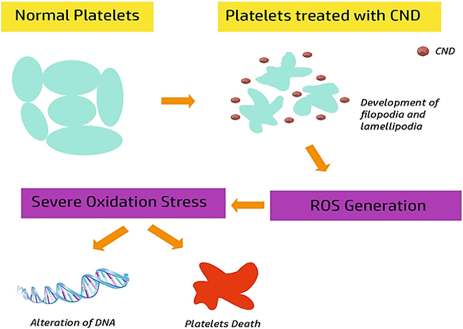
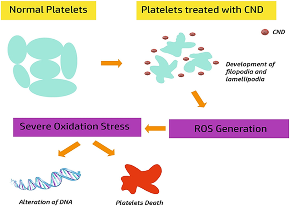
Since platelet cells are one of the vital components in thrombosis or blood clotting, thus, it is very much essential to address the same in case of nano-diamonds (NDs) too. According to the recent study carried out by Kumari et al. it was observed that carboxylated NDs (cNDs) had the significant potential to induce activation of platelet cells. It was seen that cNDs ranging in size from 4 to 10 nm stimulated platelet activation at a very low concentration like 1 μg/mL. Moreover, while it was injected in mice intravenously, these NDs caused extensive pulmonary thromboembolism that gave further evidence of its thrombus inducing potential [Reference Kumari, Singh, Singh, Grácio and Dash70]. Additionally, the experimental evidence also illuminated several other aspects of NDs interactions with platelets. For example, the group demonstrated that NDs increased free intracellular calcium levels in human platelets. That also adversely affected the viability and morphology of platelet. Cell viability assay (i.e., MTT assay) confirmed the reduced platelet viability whereas ultrastructural morphological changes were examined by both TEM and SEM analysis. It was found that normal platelets appeared spherical in shape whereas ND-treated platelet cells were seen to have developed filopodia or lamellipodia. Along with these morphological changes of platelet cells, a significance level of elevation of ROS was measured due to the treatment of NDs treated platelets (Fig. 3).
Change in shape of platelet after treatment with CND. Interactions of the CNDs with platelets lead to generation of ROS which causes DNA damages and cell death.
Graphene
Research carried out by Singh et al. [Reference Singh, Singh, Kulkarni, Sonkar, Grácio and Dash11, Reference Singh, Singh, Nayak, Kumari, Shrivastava, Grácio and Dash71] suggested that GO was extremely thrombogenic in mice and elicited an integrin RIIbβ3-mediated clumping response in human platelets. Since platelets play an indispensable role in thrombogenesis, hence use of GOs triggered the issues of pulmonary thromboembolism. Thus, possibilities of inter thrombo toxicity of GO possibly restrict its widespread in vivo biomedical applications. The research conducted by the above group also stated that amine-modified graphene (GO–NH2) was comparatively more biocompatible as it did not evoke any pulmonary thromboembolism or cause any adverse effect on human platelets. They also discovered that there was no notable change in ROS level in the platelets exposed to GO–NH2. The findings above revealed an unambiguous difference between GO and GO–NH2, suggesting that GO–NH2 is potentially a safer option for in vivo biomedical applications. This implies that carbon nanomaterials having different structures or chemical composition may illicit specific hematotoxic reactions. In view of this, another research group recently examined the effect of graphene nanoribbons (O-GNR) on platelets [Reference Chowdhury, Fang and Sitharaman37]. To find out the effect of surface modification, O-GNR was also coated with amphiphilic polymer PEG-DSPE. These two different types of GNRs were considered to study the platelet Factor 4 (PF4) which is a heparin-binding protein is specifically found in platelets when they are activated. The enzyme-linked immunosorbent assay (ELISA) test was carried out by using anti-PF4 antibodies in order to determine the extent of PF4 in blood. Eventually, no significant changes in levels of PF4 were seen in these two samples indicating that O-GNR-PEG-DSPE did not alter platelet activation in human blood. The overall role of surface functionalization on platelet activation and blood coagulation are summarized in Table III.
Effect of functionalization of CBNs on platelets aggregations and blood coagulation.

Fullerenes and carbon quantum dots
Fullerenes
Fullerenes are allotropic forms of carbon which have a highly symmetrical structure. Harold Kroto and Richard Smalley in 1985 have received a Nobel Prize for the discovery of fullerenes. The use of these compounds for biomedical applications has been explored due to their stable geometrical structure and unique properties like chemical inertness, ability to cross biological membranes without affecting the cell viability [Reference Pacor, Grillo, Đorđević, Zorzet, Lucafò, Da Ros, Prato and Sava72]. However, in some cases, it may cause toxicity due to localization in the liver. It may cause lipid peroxidation due to the presence of aπ bond in the sp 2 carbon [Reference Vesnina, Mamontova, Mykytiuk, Kutsenko, Bobrova, Kutsenko and Kaĭdashev73]. Moreover, low solubility in polar solvents is another disadvantage of fullerene. To overcome these challenges, several surface modifications have been made to make the fullerenes more hydrophilic, thus increasing its hemocompatibility. These molecules have mainly been used in the design of nanocarriers [Reference Lee, Mahendra and Alvarez74], enzyme inhibitors [Reference Lee, Nimmanpipug, Aruksakunwong, Promsri, Sompornpisut and Hannongbua75] and free radical scavengers [Reference Magoulas, Garnelis, Athanassopoulos, Papaioannou, Mattheolabakis, Avgoustakis and Hadjipavlou-Litina76].
Recently, fullerenes have been proved to be very effective as anti-inflammatory agents. C60 fullerenes preincubated with peripheral blood basophils and human mast cells were shown to suppress type I hypersensitivity [Reference Ryan, Bateman, Stover, Gomez, Norton, Zhao, Schwartz, Lenk and Kepley77]. Very little research on the interaction of fullerenes with blood proteins was done till date. This might be due to the rapid clearance of C60 from the blood (1% of the administered dose remains after 1 min in the circulation). However, the clearances of hydrophilic fullerene derivatives like quaternary ammonium salt-derivatized C60 have a slower clearance (9% of the administered dose remains after 1 min in the circulation) [Reference Monti, Moretti, Salvioli, Straface, Malorni, Pellicciari, Schettini, Bisaglia, Pincelli, Fumelli, Bonafè and Franceschi78]. It was then observed that this rapid clearance from the blood circulation may be the reason for endothelial damage. Monique et al., have shown the same in their article stating that the oxidative product of C60 fullerenes caused inflammation, inhibit cell proliferation and causes apoptosis in the endothelial cells [Reference Gelderman, Simakova, Clogston, Patri, Siddiqui, Vostal and Simak79].
Fullerenes protect the human peripheral blood mononuclear cells (PBMCs) from apoptosis by retaining the membrane potential of mitochondria [Reference Bunz, Plankenhorn and Klein80]. Neither polyhydroxy-C60 (poly-C60) nor N-ethyl-polyamino-C60 (nepo-C60) fullerene was able to activate T cells (adaptive immunity). But, it was observed that some parts of PBMCs which are related to the innate immune system have been activated. This was confirmed by the increased production of interleukin-6 and CD56 (natural killer cells) upon treatment of PBMCs with these fullerenes [Reference Liu, Jiao, Qiu, Li, Qu, Tian, Li, Bai, Lao, Zhao, Chai and Chen81]. The fullerene derivate like fullerenol [C60(OH)20] which was capable of activating the T cells also showing that these properties may vary based on the type of modification [Reference Liu, Jiao, Qiu, Li, Qu, Tian, Li, Bai, Lao, Zhao, Chai and Chen81].
Based on the above discussion, we can say that the biomedical applications of fullerenes are having lots of ambiguities. To summarize it may be said that desired results cannot be promised unless there is an in-depth understanding of the behavior of these materials.
Carbon quantum dots (CQDs)
CQDs are a class of fluorescent carbon nanomaterials with size less than 10 nm. They were accidently discovered during the production of SWCNTs in 2004 [Reference Xu, Ray, Gu, Ploehn, Gearheart, Raker and Scrivens82]. These materials have several applications in the field of biomedical research (e.g., bioimaging, catalysis, drug delivery and theranostics) [Reference Lategan, Fowler, Bayati, Fidalgo de Cortalezzi and Pool83] due to the properties like small size, solubility in water, excellent photostability and biocompatibility. They can form both crystalline and amorphous structure. The carbon atoms are predominantly sp 2 hybridized but sp 3 hybridized carbon atoms have also been reported [Reference Zheng, Ananthanarayanan, Luo and Chen84]. The fluorescence property of CQDs is due to the conjugated π-domains or surface defects i.e., sites which do not have perfect sp 2 carbon [Reference Cao, Meziani, Sahu and Sun85]. The CQDs have a high rate of clearance from the blood (6 h for complete clearance) with very low toxicity. Owing to all these characters, these materials have been highly utilized in the cancer bioimaging and blood glucose monitoring. Efforts have also been made to increase the circulation time of these particles for proper of targeting the tumor site. This was achieved with the use of hydrophilic polymer coating on the surface of CQDs [Reference Wang, Meng, Wang, Li, Shi, Chen, Wang and Huang86]. Drug delivery through the blood–brain barrier is an important application of the CQDs. These particles easily cross the blood–brain barrier however the detailed mechanism of the above mentioned applications is not yet fully understood.
Although this is an emerging research field, however many crucial aspects of the CQDs remain unexplored. Especially the interaction of these particles with the blood and its proteins is not yet understood. A recent study using proteome analysis concluded that high concentrations of CQDs cause inflammation. Signalling molecules like MIP-2, MIP-1α, MIP-1β, IL-8, TNF-α, and SDF-1 were up-regulated upon CQD exposure indicating that they are responsible for hypersensitivity and autoimmunity [Reference Lategan, Fowler, Bayati, Fidalgo de Cortalezzi and Pool83].
Effect of protein corona on biological functions of CBNs
Once the nanoparticles are administered into blood, they start interacting with the macromolecules (e.g., mainly proteins and ions) present in the body depending on their physicochemical properties. While protein molecules get adsorbed on the surface of the nanoparticles forming layer(s) is commonly known as ‘protein-corona’. Based on the stability of this adsorbed layer, the corona is classified as ‘Hard corona’ (stable) and ‘Soft corona’ (unstable) [Reference Lundqvist, Stigler, Cedervall, Berggård, Flanagan, Lynch, Elia and Dawson87]. The protein-corona plays several important roles in altering the biological responses of any nanoscale materials explored by various recent studies. The fate of the nanomaterial greatly relies on the characteristic of the protein corona which modulates its toxicity, time of circulation, specificity and targeting abilities.
Actually, the proteins present in larger concentrations and/or opposite charge of the nanoscale materials are first absorbed. However, these are quickly replaced selectively with proteins having higher affinity which helps in the formation of a stable hard corona. Sometimes, the proteins in the hard corona may have weak interactions with other proteins in the surrounding which is then called the soft corona. This is a dynamic process whereas the absorbed molecules are constantly replaced with the molecules of higher affinities. Thus, the features of the protein corona constantly change while the nanoparticles circulating within the blood and move from one organ to another [Reference Lundqvist, Stigler, Cedervall, Berggård, Flanagan, Lynch, Elia and Dawson87]. And depending on the type of molecules absorbed, the properties of the nanomaterials keep changing with the time that ultimately decides the biological response of the nanomaterials.
Several research groups have been working in this area to explore the effects of protein corona in presence of CBNs like GO and CNTs. Most of the recent studies found that in some cases, the formation of a protein-corona actually reducing the cytotoxicity of the nanoparticles in the cell. For example, it was observed that the formation of the corona has reduced the ability of the GO nanomaterials to penetrate the cells, thus reducing its cytotoxicity [Reference Duan, Kang, Tian, Garate, Zhao, Ge and Zhou88]. On the other hand plenty of studies demonstrated that GO might be a good option for drug delivery as it showed better cellular uptake. Another study on immune perturbations caused by MWCNTs showed that are chemically modified have switched from a mannose receptor to a scavenger receptor [Reference Gao, Zhang, Mu, Bai, Li, Zhou, Butch, Powell, Snyder, Jiang and Yan89]. However, the exact mechanism in which this phenomenon takes place was not fully understood yet. Although, recent studies conducted using microarray revealed that the serum proteins help in the macrophage recognition of SWCNTs. This occurred due to the activation of Toll-like receptors by the proteins in the protein-corona [Reference Mukherjee, Bondarenko, Kohonen, Andón, Brzicová, Gessner, Mathur, Bottini, Calligari, Stella, Kisin, Shvedova, Autio, Salminen-Mankonen, Lahesmaa and Fadeel90]. It is worth mentioning that characteristic of protein corona is unique to each nanoscale materials. Previous studies carried out in this area are not able to fully explore several important aspects about the dynamics of protein corona formation by considering similar physicochemical properties of the CBNs, hence, plenty of ambiguities and contradictions are there among the outcomes of various studies. Therefore, in-depth understanding is essential to get conclusive ideas about the detailed outcome of the protein corona. It would be mentioned here that the effect of protein-corona on fullerenes and CQDs is very less explored yet. However, the studies on protein adsorption on the surface of fullerenes explored that the spherical surface of the fullerenes helps in adsorption of more amount of proteins as compared to flat surfaces [Reference Asuri, Karajanagi, Vertegel, Dordick and Kane91].
Conclusion
In brief, carbon-based nanostructures are showing incredible prospects that involve the development of smart, programmable advanced material suitable for many application including drug and gene delivery. Further, it is obvious that rapid progress in this line of interest may introduce a large number of novel CBNs in the field of biomedicine. However, as discussed, one must evaluate the key hemocompatibility parameters before willing to employ these nanomaterials for practical purposes. It appears that one of the main factors that control hemocompatibility of these nanomaterials depends on surface properties like availability of surface area, surface charge and functional groups. Functionalization and surface modifications have indeed proven to considerably change the reactivity of the nanomaterials. Step-wise and methodical engineering of such nanomaterials is needed to reduce their toxicity without compromising their inherent properties. In fact, it is prudent to say that more experimental proof is needed to decide whether the data available in vitro is enough to translate theirs in vivo usage. Therefore, future studies must aim to develop new methods and/or techniques to avoid thrombogenicity, complement activation, haemolysis, cytotoxicity and inflammation of these materials which are crucial for their real-life biomedical applications.
Acknowledgment
Authors are thankful to DBT, Gov. of India for financial support (BT/IN/Indo-US/Foldscope/39/2015). This work also received financial support from Seed grant of IIIT Allahabad.