Introduction
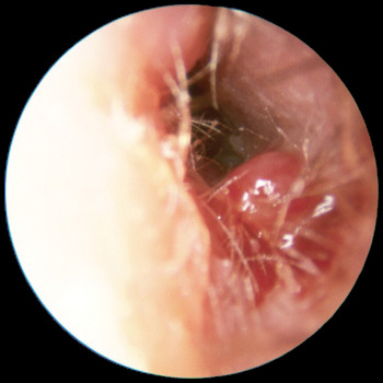
Skull base osteomyelitis typically presents in immunocompromised individuals, often elderly diabetic patients, with severe otalgia and unilateral otorrhoea.Reference Chandler1 There may be invasion of local structures and necrosis of skin, cartilage and bone. The presence of ear canal granulations (such as in Figure 1) and excessive pain in the setting of chronic otitis externa are highly suggestive of skull base osteomyelitis. Cranial nerve palsies may ensue, typically affecting the facial nerve, but spread to the petrous apex may involve the abducens, trigeminal and optic nerves.Reference Holder, Gurucharri, Bartels and Colman2 Spread to the jugular foramen may affect the lower cranial nerves.
Auriscopic view showing ear canal granulation suggestive of skull base osteomyelitis.

Cross-sectional imaging is required to exclude malignancy, and the diagnosis is confirmed by radionuclide scanning to identify skull base inflammation.
Pseudomonas aeruginosa is the commonest pathogenic micro-organism,Reference Rubin and Yu3 although others have been identified, including fungal species.Reference Gordon and Giddings4Pseudomonas aeruginosa is a Gram-negative, motile rod of low virulence which is an uncommon commensal of the external auditory canal. It causes opportunistic otitis externa following minor trauma or water exposure, but in an immunocompromised host can cause coagulative necrosis of soft tissues by production of endotoxin, collagenase and elastase.Reference Chandler5, Reference Slattery and Brackmann6 The infection spreads from the external auditory canal along the tympano-mastoid suture to the retromandibular fossa, or via the fissures of Santorini to the parotid space. Infection of the soft tissues around the jugular and stylomastoid foramina predisposes to lower cranial nerve palsies and venous thrombosis.Reference Mani, Sudhoff, Rajagopal, Moffat and Axon7 Further spread of infection can occur along venous channels (e.g. the lateral venous sinus and petrosal sinuses) and fascial planes, to involve the petrous apex, with secondary involvement of bone. Pseudomonas preferentially spreads along vascular channels, and therefore infection may not be in bony continuity, unlike typical acute mastoiditis, which spreads along pneumatised tracts.
Worldwide reports of pseudomonas resistance to fluoroquinolones are now emerging.Reference Bernstein, Holland, Porter and Maw8, Reference Berenholz, Katzenell and Harell9 We therefore sought to review our experience of skull base osteomyelitis, especially the causative organisms and their antibiotic sensitivities. We compared this with contemporary literature, and reviewed our treatment algorithm based on our findings.
Methods
A retrospective chart review from January 2004 to August 2011 was performed. Patients were identified by their admission diagnostic code, and their computerised records examined. Demographic data and details of investigations, microbiology, management and follow up were collected.
Results
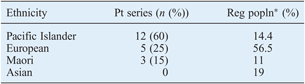
Twenty patients (13 men and 7 women) were identified, with a mean age of 66 years (range, 42–90 years). Twelve (60 per cent) patients identified their ethnic origin as Pacific Islander, compared with a regional proportion of 14.4 per cent. An overview of patients’ ethnic origin is shown in Table I. Seven (35 per cent) patients were not New Zealand residents at presentation, but had been referred from, or had recently arrived from, South Pacific Islands.
Ethnicity: patient series vs regional population
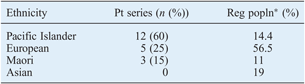
* Auckland, from 2006 census data.10 Pt = patient; Reg popln = regional population
Nineteen (95 per cent) patients were immunocompromised, 18 (90 per cent) had diabetes mellitus and 1 had acute myeloblastic leukaemia. None had a history of previous ear surgery. The mean patient total hospital stay was 31 days (range, 7–194 days), with a median of 15 days. On admission, a facial palsy was noted in 12 (60 per cent) patients, and lower cranial nerve palsies (glossopharyngeus and vagus) were also present in 3 (15 per cent).
Investigation
Eighteen blood cultures were performed for six patients; none yielded any positive growth.
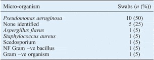
The results of ear canal swab analysis are shown in Table II. Ten (50 per cent) swabs grew P aeruginosa; one of these strains was resistant to ciprofloxacin, but all were sensitive to ceftazidime. Two fungal infections were identified, with scedosporium and Aspergillus flavus. Ear canal granulations were biopsied in seven patients (35 per cent), but only two yielded any growth.
Dominant micro-organism identified in ear canal swabs

NF = non-fermenting; –ve = negative; +ve = positive
Cross-sectional imaging was performed in 19 patients (computed tomography (CT) in 18, and magnetic resonance imaging (MRI) performed additionally in 6 and alone in 1). This was followed by diagnostic radionuclide imaging: technetium-99m scintigraphy in 12 (60 per cent) patients and gallium-67 scintigraphy in 6 (30 per cent). The median timing of radionuclide scanning was day four after admission.
Management and outcome
A wide variety of antimicrobials was prescribed in our series. However, 50 per cent of patients were prescribed a single intravenous agent, ceftazidime; 60 per cent also received ciprofloxacin orally.
A mastoidectomy was performed for four patients (primarily to obtain tissue for microbiological culture and to exclude malignancy), with facial nerve decompression for complete facial palsy in two.
Three patients died in the treatment period, giving a mortality rate of 15 per cent. Two died of intracranial causes: subarachnoid haemorrhage and cerebral infarction. One was considered unfit for surgical debridement of fungal osteomyelitis and was treated palliatively.
Discussion
There are a number of interesting findings within this cohort of patients with skull base osteomyelitis.
There was a high rate of facial palsy on admission – 60 per cent – compared with 0–40 per cent in contemporary case series.Reference Mani, Sudhoff, Rajagopal, Moffat and Axon7, Reference Djalilian, Shamloo, Thakkar and Najme-Rahim11, Reference Ali, Meade, Anari, ElBadawey and Zammit-Maempel12 No pathological organism was identified in 25 per cent of cases. Both these findings probably reflect late presentation and prolonged community-based management for ‘simple’ otitis externa.
Pseudomonas aeruginosa was identified in only half of the patients admitted to our unit, while in five (25 per cent) no organism was identified. Historically, P aeruginosa was the pathogen found in over 90 per cent of skull base osteomyelitis cases,Reference Rubin and Yu3 with other organisms (such as staphylococcus species, Proteus mirabilis, Klebsiella pneumonia and aspergillus species) rarely isolated.Reference Babiatzki and Sade13–Reference Cunningham, Yu, Turner and Curtin16
The management of culture-negative skull base osteomyelitis, now a common clinical entity, has been described, with good outcomes using a combination of intravenous cephalosporin and oral ciprofloxacin.Reference Djalilian, Shamloo, Thakkar and Najme-Rahim11 Prolonged and targeted systemic antimicrobial therapy is required: a broad spectrum cephalosporin combined with ciprofloxacin is the treatment of choice, with a high cure rate.Reference Gehanno17 Hyperbaric oxygen has been used with some benefit as an adjunct to antimicrobials;Reference Shupak, Greenburg, Hardoff, Gordon, Melamed and Meyer18 however, a Cochrane review found that no randomised, controlled trials had been performed to support this treatment.Reference Phillips and Jones19
Surgery has a limited role in skull base osteomyelitis, being performed primarily to obtain specimens for culture and to exclude malignancy.Reference Grandis, Branstetter and Yu20 Facial nerve decompression has been used for cases with complete facial palsy,Reference Mani, Sudhoff, Rajagopal, Moffat and Axon7, Reference Soudry, Joshua, Sulkes and Nageris21 but without a significant improvement in function, as in our series; numbers are small in all series, so we conclude that it has unproven therapeutic benefit.
Unfortunately, microbial resistance to ciprofloxacin is emerging,Reference Bernstein, Holland, Porter and Maw8, Reference Berenholz, Katzenell and Harell9 and must be suspected in patients with deteriorating clinical features (e.g. increasing otalgia and new cranial nerve palsies) or rising levels of inflammatory markers. We emphasise the need for early collection of specimens for culture and prolonged antimicrobial treatment to prevent further selection of resistant strains.
Cross-sectional imaging is necessary to exclude other pathology. Magnetic resonance imaging has some advantages as it can detect changes in bone marrow, rather than the mineral cortex changes detectable on CT.Reference Grandis, Curton and Yu22 However, we favour CT as it is more accessible, and we reserve MRI scanning for cases of diagnostic difficulty, or in order to identify soft tissue complications such as meningeal involvement.
Radionuclide imaging is the diagnostic modality of choice. Technetium-99m localises to areas of osteoblastic activity, and is thus a sensitive indicator of osteomyelitis; furthermore, scintigraphy becomes positive earlier in the course of infection as bone demineralisation is not required (unlike CT scanning, for example).Reference Grandis, Curton and Yu22 However, this osteoblastic activity persists for months to years after infection has resolved, so this scan is not useful in gauging response to treatment.Reference Ali, Meade, Anari, ElBadawey and Zammit-Maempel12 Gallium-67 scintigraphy is used to monitor progress, as this scan normalises early after the resolution of infection.Reference Okapala, Siraj, Nilssen and Pringle23 We perform gallium-67 scintigraphy after six weeks of antimicrobial treatment; if it is still positive, antimicrobials must be continued and the scan repeated at six-weekly intervals until resolution. There are no high-level evidence-based guidelines to suggest the optimum treatment duration or scanning interval, so this strategy is based on case series and our own experience.
Antimicrobial treatment should be continued as long as the gallium-67 scan remains positive.Reference Soudry, Joshua, Sulkes and Nageris21 Once the appropriate treatment has been initiated, symptoms generally settle early and patients may feel inclined to discontinue treatment prematurely; the importance of treatment completion must be emphasised. Once an organism and its antibiotic sensitivities are available, antimicrobials should be rationalised. Ideally, treatment would continue in the community with peripheral intravenous catheters or oral ciprofloxacin in appropriately selected cases. In our series, the median in-patient stay was 15 days, although in the most recent six cases this had reduced to 8 days.
• Skull base osteomyelitis typically occurs in immunocompromised patients, due to Pseudomonas aeruginosa
• Pseudomonas resistance to ciprofloxacin has now been reported, with one such case in this series
• Two cases is this series were due to fungal species
• Prolonged antimicrobial treatment is required, typically a cephalosporin and a fluoroquinolone, dependent on culture and sensitivities
• Early culture specimen collection, close clinical monitoring and follow-up imaging are essential to ensure resolution
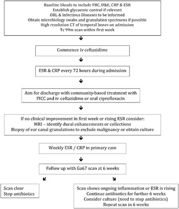
The purpose of our treatment algorithm (Figure 2) is to standardise and rationalise treatment for the local population and local pathogenic organisms. The algorithm has been designed to emphasise the need for: obtaining culture material early in the infection; multidisciplinary input; close monitoring of the infection with regular review; and awareness-raising among otolaryngologists and primary care physicians regarding the condition. Imaging plays a crucial role in diagnosis and follow up, but the various modalities discussed above may not be universally available; thus, a local management algorithm should be developed based on local resources.
Algorithm for management of a patient admitted with suspected skull base osteomyelitis. FBC = full blood count; U&E = urea and electrolytes; CRP = C-reactive protein; ESR = erythrocyte sedimentation rate; ORL = otorhinolaryngology; CT = computed tomography; iv = intravenous; PICC = peripherally inserted central cannula; MRI = magnetic resonance imaging

Conclusion
Skull base osteomyelitis remains a challenging condition to manage as the diagnosis is often delayed. Culture-negative skull base osteomyelitis is not uncommon, and must be managed empirically with close follow up. Pseudomonas resistance to fluoroquinolones is emerging in many countries and will make this condition even more challenging, as prolonged intravenous antimicrobial treatment may represent the only possibility of cure. The imaging modalities used are dependent on local availability, but ideally should combine cross-sectional imaging with radionuclide scintigraphy.
Acknowledgements
The authors would like to acknowledge the work of H Herbert and D Bowyer in data collection.