Extracorporeal life support has become a widely accepted short-term mechanical circulatory support in the setting of profound circulatory collapse, both succeeding cardiac surgery and during acute illnesses such as myocarditis, decompensated cardiomyopathy, and intractable arrhythmia. A major setback frequently encountered in these patients is left heart hypertension, which may result in pulmonary oedema, ventricular distension, and subendocardial ischaemia.Reference Aiyagari, Rocchini, Remenapp and Graziano 1 Hence, it is crucial to ensure an adequate left heart decompression. In this article, we reviewed our experience in left heart decompression during central extracorporeal life support through direct placement of a left ventricular vent. The pathophysiological considerations and significant aspects in post-operative management are highlighted.
Material and methods
This study was a retrospective, single-centre case series. Between August, 2011 and August, 2012, we identified eight patients who were placed on paediatric extracorporeal life support with additional left ventricular venting. In these cases, left ventricular venting was indicated to ensure an adequate left ventricular drainage. We reviewed patient demographics, cardiac catheterisation reports, surgical reports, extracorporeal flow data, inpatient charts, and the routinely performed echocardiograms and chest X-rays.
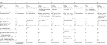
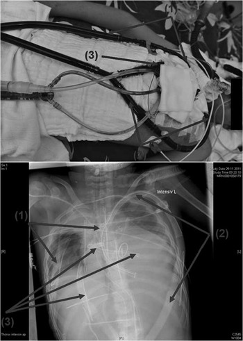
Of the patients, seven underwent the procedure in the operating room and only one (patient 5, see Table 1) underwent the procedure in the paediatric cardiac intensive care unit. Among the eight patients, four underwent primarily surgical extracorporeal life support implantation for an acute illness and four were implanted after surgical repair of a congenital cardiac defect. All patients were approached through median sternotomy. An arterial cannula was placed in the ascending aorta, and the right atrium was cannulated for venous return. An appropriately sized venting cannula was inserted through the right superior pulmonary vein and was connected to the venous line of the circuit with a Y-connector such that active left heart decompression was achieved (Fig 1). The targeted extracorporeal support flow is calculated as follows: 2.8 L/minute/m2 body surface area for patients weighing up to 20 kg and 2.6 L/minute/m2 body surface area for patients weighing above 20 kg. Direct flow measurement of the left ventricular venting cannula was performed intermittently.
Patient demographic and procedural data and outcomes.

AV = atrioventricular valve; BiVAD = biventricular assist device; cAVSD = complete atrioventricular septal defect; ECLS = extracorporeal life support; ICU = intensive care unit; IGF = insulin-like growth factor; LV = left ventricle; MAPCAs = major aortopulmonary collateral arteries; VSD = ventricular septal defect
Venting and cannulation for extracorporeal life support. An arterial cannula (1) was placed in the ascending aorta. The right atrium was cannulated for venous return (2). A left ventricular vent (3) was inserted through the right superior pulmonary vein and was connected to the venous line on the circuit such that complete left heart decompression was achieved.

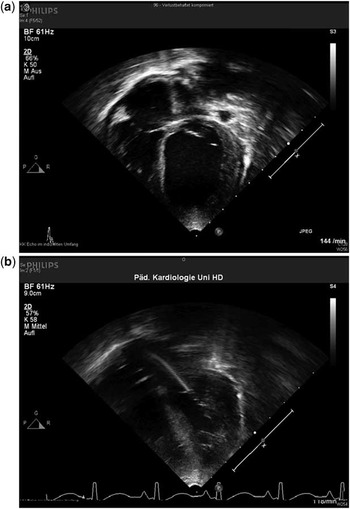
Transthoracic echocardiography while on extracorporeal life support. Left ventricular distension before venting ( a ) and left ventricular decompression after insertion of a left ventricular vent ( b ).

In patient 1, a left ventricular vent was inserted after 9 hours on support and in patient 2 after 13.5 hours on support. These patients required venting because of left ventricular dilatation (see Fig 2) and beginning pulmonary oedema. With adequate left heart decompression, marked clinical improvements were observed in both patients. On the basis of this brief experience, a left ventricular vent was inserted at the time of extracorporeal support initiation in the subsequent six patients. Intraoperative transoesophageal echocardiography was performed to assess the placement of all cannulas and to ensure that the tip of the venting cannula remains in the left ventricle. In the setting of pulmonary dysfunction, a three-way tap was inserted in the proximal venous line and the left ventricular vent line. This allowed separate blood gas analyses from each line.
We used a Levitronix CentriMag blood pump (Levitronix LLC, Waltham, Massachusetts, United States of America) and a D902 ECMO (Dideco, Sorin Group, Milan, Italy) apparatus as a long-term oxygenator with phosphorylcholine P.h.i.s.i.o-coated circuit tubing (Sorin Group, Milan, Italy).
The intraoperative management of anticoagulation to institute extracorporeal life support was as follows: if heparin had not been initiated or if it had been reversed with protamine, then support was instituted with a 300 IU bolus of unfractioned heparin per kilogram body weight. If the patient failed to be weaned off cardiopulmonary bypass, then support was instituted in the fully heparinised patient. Unfractioned heparin was stopped, but no protamine sulphate was administered unless the bleeding could not be controlled surgically. Post-operatively, if bleeding was not an issue, a continuous intravenous infusion of unfractioned heparin was initiated immediately. We aimed for an activated clotting time of 130–160 seconds, which was measured routinely. For a patient with significant haemorrhage, in whom a surgical cause had been excluded, heparin was discontinued and not restarted until the bleeding had stopped.
Delayed sternal closure was an essential part of the management of central extracorporeal life support. The chest was left open and the sternal wound was covered with a polyvinyl chloride membrane, which was individually trimmed from a regular blood bag. This facilitated a second look procedure for direct control of haemorrhage.
Total circuit flow was recorded routinely until decannulation. The readiness to be weaned from support was evaluated by clinical assessment and echocardiography at a reduced pump flow; it was first considered after ∼48–72 hours on support. All patients underwent a series of follow-up echocardiographies, and procedural success was defined as a reduction in left ventricular dilatation and an improvement in left ventricular contractility.
In patients with a bodyweight under 6 kg, delayed sternal closure was performed 3–7 days after extracorporeal circuit decannulation depending on their haemodynamic stability (see Table 1). Older children with a bodyweight above 6 kg tolerated sternal closure better; their chests were closed at the same time as circuit decannulation.
Results
Patient demographics, procedural data, and outcomes are summarised in Table 1.
All the patients included in our study have a biventricular heart. None of the study patients died while on support. Of the patients, seven (87.5%) were successfully weaned from support. Patient 2 was transitioned to a Berlin Heart Excor Paediatrics biventricular assist device (Berlin Heart, Berlin, Germany) after 10 days of support. A transthoracic echocardiography showed a dilated and hypokinetic left ventricle without improved contractility. He was stable on the assist device and his blood pump was replaced at the 177th day of support. During this intervention, the inflow cannula of the left-sided blood pump was seen to be severely damaged, leading to a fatal air embolism and eventually death. Thus, the late mortality rate was 12.5% (n = 1).
All patients who underwent surgical repair of a congenital cardiac defect were placed on support in the operating room, except for patient 5. This patient was first placed on extracorporeal support in the paediatric cardiac intensive care unit, 6.5 hours after her primary surgery. Changing the venous and the left ventricular vent cannulas was deemed necessary after 3 days of initiation of support because of in-line thrombus development. She was successfully weaned at the eighth day on support. After 4.5 hours of stable weaning, her haemodynamics started to deteriorate acutely. Both parents decided to terminate all life-prolonging procedures. This corresponded to an early mortality rate of 12.5% (n = 1).
Patient 1 was stable after successful weaning from support and was discharged home. Owing to the development of chronic low output syndrome after parvovirus B19-associated myocarditis, he was placed on a Berlin Heart Excor Paediatrics biventricular assist device, 4 months after successful weaning from extracorporeal life support. He remained stable on the assist device and orthotopic cardiac transplantation was successfully carried out at the 71st day of assist support.
Thus, in total, six study patients (75%) survived, and all were alive and discharged from the hospital (see Table 1). Major mechanical and circuit problems that required circuit changes occurred only in patient 5, as described above. Sternal wound infection and limb ischaemia occurred in none of the study patients. None of the patients suffered an embolic stroke or developed relevant intracranial bleeding resulting in neurological dysfunction during and after extracorporeal life support. Patient 1 developed cerebral microbleeding, as shown on magnetic resonance imaging, while on support, without any sustained neurological deficit. Three patients had a culture-proven bacterial infection that developed during support. The causative organisms were Staphylococcus aureus (n = 2) and Staphylococcus epidermidis (n = 1). All these infections were controlled with antibiotics on the basis of their culture and sensitivity testing; none of them led to a prior weaning. No systemic mycosis was observed; airway fungal colonisations were treated with appropriate antimycotics.
Comment
Potential benefits in left ventricular venting during extracorporeal life support
Central extracorporeal life support represents a feasible and effective method to support critically ill children with a low cardiac output. It raises the aortic blood flow and substantially increases both mean and diastolic arterial pressures. Thus, in case of cardiogenic shock, initiation of support increases the systemic afterload.Reference Bavaria, Ratcliffe, Gupta, Wenger, Bogen and Edmunds 2 In the presence of profoundly impaired left ventricular contractility and the added afterload from the arterial cannula, the aortic valve may fail to open, resulting in an impaired left ventricular ejection. In these cases, inadequate right atrial drainage, bronchial circulation, aortic regurgitant flow, as well as an extracardiac left-to-right shunt can lead to left ventricular distension. In such cases, the left ventricular end-diastolic volume progressively rises and increases myocardial wall stress and oxygen consumption and decreases coronary perfusion. All the above-mentioned conditions lead to subendocardial ischaemia, which endangers myocardial recovery.Reference Aiyagari, Rocchini, Remenapp and Graziano 1 , Reference Kato, Seo, Ando, Takagi and Ito 3 An impaired left ventricular ejection while on support, as described above, causes intraventricular blood stagnation and also predisposes to thrombus formation.Reference Moubarak, Weiss, Leprince and Luyt 4 Accordingly, an adequately sized left ventricular vent for satisfactory decompression should constantly be attempted. Intracardiac thrombus occurred in none of our study patients, and routinely preformed echocardiography showed adequate left ventricular drainage via the venting cannula. No left heart distension was observed. In our series, intraoperative transoesophageal echocardiography showed that a better left ventricular decompression was achieved when the tip of the venting cannula was in the left ventricle instead of the left atrium, in children both with and without systemic atrioventricular valve regurgitation. Thus, all venting cannulas were placed in the left ventricle.
Incomplete left ventricular decompression can remain clinically silent for several hours. It increases left atrial pressure, which occurs along with potential severe consequences such as haemoptysis and pulmonary oedema,Reference Aiyagari, Rocchini, Remenapp and Graziano 1 especially in children with significant systemic atrioventricular valve regurgitation. In patient 1, the left ventricular vent was inserted after 9 hours on support because of echocardiographic left ventricular dilatation and beginning pulmonary oedema on chest X-rays. The development of pulmonary oedema was more progressive in patient 2, in whom significant mitral valve regurgitation was eminent. After insertion of a left ventricular vent, chest X-rays improved and, more importantly, the oxygenation improved significantly, suggesting a clinically relevant reduction in pulmonary oedema. With adequate left ventricular venting, no pulmonary oedema or haemorrhage were observed. The marked clinical improvement in both patients changed our management of extracorporeal life support. We believed that all children had profound myocardial dysfunction with a nearly absent left ventricular ejection when they were placed on extracorporeal support, and as a result all children with a biventricular heart and an insufficient interatrial shunt underwent left ventricular venting at the time of extracorporeal support initiation. During the study period, we encountered three additional cases of children with biventricular hearts who did not undergo left ventricular venting. Among them, two had an atrial septal defect and one had an intact atrial septum. Only one child with an atrial septal defect survived; the other two children died of left ventricular failure. A series of echocardiographies performed on the two children while on extracorporeal support showed inadequate left ventricular decompression, and both patients may have benefited from additional venting.
Left heart decompression in children and young adult patients with extracorporeal life support can be accomplished through a variety of techniques including surgical or balloon atrial septostomy or combined blade and balloon atrial septostomy.Reference Seib, Faulkner and Erickson 5 With this method, a residual atrial septal defect may persist, requiring interventional or surgical closure once the patient has been weaned. Another potential benefit of our technique, and in contrast to atrial septostomy, would be that the presence of an indwelling venting cannula allows separate blood gas analyses from the venous cannula and the left ventricular vent. In case of pre-existing pulmonary oedema or other pulmonary dysfunctions, this method allows the monitoring of pulmonary recovery and helps detect possible coronary hypoxia when the left ventricle begins to recover. When the left ventricle starts to eject the lower saturated blood from the dysfunctional lungs, the coronary arteries may not obtain the well-oxygenated blood from the arterial cannula, which is located in a more distal part of the ascending aorta. If this occurs, it is our first choice to increase the ventilator support. Delaying weaning and increasing the circuit flow to limit left ventricular ejection were rarely required. Secker–Walker et al demonstrated that cannulation of the ascending aorta in sheep did not result in adequate oxygenation of the coronary until the bypass flows were very high (85%).Reference Secker-Walker, Edmonds, Spratt and Conn 6 This phenomenon causes sustained myocardial dysfunction, which has been documented to occur in neonates while on extracorporeal life support for primary respiratory failure.Reference Hirschl, Heiss and Bartlett 7 For this reason, performing blood gas analysis via a left ventricular vent is superior to arterial gas analysis at the right upper extremity. Further, in infants and toddlers, it is our institutional preference to avoid keeping an indwelling upper extremity arterial cannula for several days because of a high risk of vascular injury that can cause upper limb ischaemia.
Critical pitfalls in extracorporeal life support and additional left ventricular venting
Our institution uses a centrifugal blood pump that provides an active venous drainage during support. As the venting cannula becomes a part of the venous return to the circuit, it provides active left heart decompression. Assuming there is no venting cannula distortion, obstruction, or end-hole occlusion, the flow rate on the venting line depends mainly on cannula diameter and length. Patient 5, who performed poorly and died after extracorporeal support explantation, had the smallest left ventricular vent, that is, 10 French, and a very low flow rate on the venting cannula, which caused progressive in-line thrombus formation refractory to the increased anticoagulation. This required circuit exchange on the third day of support. Since this encounter, we placed a minimum vent size of 12 French and have never observed in-line thrombus formation again, even when the venous cannula was bigger than the venting cannula. Aiyagari et al reported in their study passive left atrial decompression during support.Reference Aiyagari, Rocchini, Remenapp and Graziano 1 They pointed out that left ventricular readiness could be tested by clamping the venting cannula as a “weaning trial” before discontinuing left drainage or extracorporeal support. We did not perform partial or complete clamping of the venting cannula to avoid in-line thrombus formation. As part of the extracorporeal life support procedure, transiently reducing the circuit flow was preferred. This not only increased the left ventricular preload, but also decreased its afterload, allowing the left ventricle to eject. Left ventricular ejection should be tested at a low afterload, allowing exclusion of a completely akinetic left ventricle.
The timing of extracorporeal life support initiation in a controlled fashion before circulatory collapse is critically important in avoiding an irreversible organ injury. Chaturvedi et al reported their experience in post-operative extracorporeal life support and pointed out the higher survival rates among patients cannulated in the operating room (64%) than in the cardiac intensive care unit (29%).Reference Chaturvedi, Macrae and Brown 8 They believed that this is due to early effective support preventing prolonged hypoperfusion. Similarly, our study showed a case of lethality in a patient who was put on support because of a prolonged low cardiac output 6.5 hours after the primary cardiac surgery, that is, the case of patient 5. This delayed support initiation together with an in-line thrombus formation complicated her post-operative course. She died 4.5 hours after successful weaning because of recurrent haemodynamic deterioration refractory to medical management. This was the only early mortality reported in our series.
Continuous monitoring of cerebral oxygenation via near-infrared spectroscopy is also critical to monitor the safe cerebral flow rate while on central extracorporeal life support. An increase in cerebral blood flow velocity was shown to be associated with an increased risk of intracranial haemorrhage in neonatal lambs on an extracorporeal life support.Reference Rosenberg and Kinsella 9 Unfavourable cannula positioning in the ascending aorta, wherein the arterial flow selectively enters the brachiocephalic trunk, can cause cerebral hyperperfusion. This altered cerebral blood flow and the required anticoagulation on support raise the concern of an intracranial haemorrhage. Thus, cranial ultrasound examination was performed routinely in babies with an open fontanel. As the risk of haemorrhage is greatest during the first few days of support, daily ultrasounds have been recommended.Reference Bulas and Glass 10 In the presence of neurological abnormalities after successful weaning, cranial magnetic resonance imaging was carried out in older children. The presence of a left ventricular vent also raises a concern of a thromboembolic stroke; therefore, serial echocardiograms should be performed while on extracorporeal support. In our series, no patients developed relevant intracranial bleeding resulting in permanent neurological dysfunction during and after extracorporeal support, and no intracardiac thrombus or embolic stroke was observed.
In peripheral extracorporeal life support, the risk of cerebral hypoxia may be higher in patients with pulmonary dysfunction, given the fact that well-oxygenated blood needs to travel retrogradely and blood mixing may occur in the distal aortic arch or descending aorta. Another concern that arises with peripheral support is the risk of thrombus formation in the aortic root.Reference Leontiadis, Koertke, Bairaktaris and Koerfer 11 Peripheral support also requires peripheral arterial access with a possible vascular injury that can cause limb ischaemia by dissection, thrombosis, or an inadequate flow around the cannula. Neck cannulation can damage the jugular vein or carotid artery, necessitating carotid ligation. Foley et al reported a 21% incidence of limb ischaemia during femoral cannulation for cardiopulmonary support.Reference Foley, Morris and Woo 12 Their patients required fasciotomy, and limb amputation was also performed in one case. Although left ventricular decompression could also be achieved via a percutaneously placed transaortic catheterReference Fumagalli, Bombino and Borelli 13 , Reference Barbone, Malvindi, Ferrara and Tarelli 14 in peripheral extracorporeal life support, based on the above aspects and given the size of the required cannulas relative to the paediatric vascular system, central extracorporeal life support is preferred at our institution. Limb ischaemia occurred in none of our patients.
Conclusion
On the basis of our study, we identified that in the setting of a low cardiac output and insufficient interatrial shunt additional left ventricular decompression via a venting cannula could help avoid left heart distension and might promote myocardial recovery. It prevents pulmonary congestion and its associated pulmonary haemorrhage. Separate blood gas analyses from the venous cannula and the left ventricular vent allow monitoring of pulmonary recovery and help detect possible coronary hypoxia when the left ventricle begins to recover. Therefore, we recommend the use of central extracorporeal life support with an integrated left ventricular vent in children with a low cardiac output.
Acknowledgements
The authors thank all the dedicated medical staff and perfusionists at the Department of Cardiac Surgery and Paediatric Cardiology, University of Heidelberg.
Financial Support
This research received no specific grant from any funding agency, commercial or not-for-profit sectors.
Conflicts of Interest
None.