Introduction
Human onchocerciasis is caused by the filarial nematode Onchocerca volvulus (O. volvulus), mainly affecting skin and eyes. Humans are the only known host of the adult worms, but to complete their development, the microfilariae (mf) need to be taken up by blackflies (Simulium species). Simulium damnosum is the main vector transmitting O. volvulus in our study area (Mahenge district, southern Tanzania; Raybould and White, Reference Raybould and White1979). The mf then re-enter the human host during a bloodmeal and develop into adult worms. These mate and produce mf, which migrate through skin and underlying tissue, and may be found occasionally in sputum, urine, blood, and cerebrospinal fluid (CSF) (Fuglsang and Anderson, Reference Fuglsang and Anderson1974; Duke et al. Reference Duke, Vincelette and Moore1976). Range and intensity of pathological changes depend on the host's immune response and the genotype of O. volvulus present in the area (Erttmann et al. Reference Erttmann, Unnasch, Greene, Albiez, Boateng, Denke, Ferraroni, Karam, Schulz-Key and Williams1987; Zimmerman et al. Reference Zimmerman, Dadzie, De Sole, Remme, Alley and Unnasch1992; Brattig, Reference Brattig2004).
Some studies suggest a connection between onchocerciasis and epilepsy in affected areas in Africa (Kipp et al. Reference Kipp, Kasoro and Burnham1994; Jilek-Aall, Reference Jilek-Aall and Rose1995; Kaiser et al. Reference Kaiser, Kipp, Asaba, Mugisa, Kabagambe, Rating and Leichsenring1996, Reference Kaiser, Asaba, Leichsenring and Kabagambe1998; Newell et al. Reference Newell, Vyungimana and Bradley1997; Boussinesq et al. Reference Boussinesq, Pion, Demanga-Ngangue and Kamgno2002), while others do not (Kabore et al. Reference Kabore, Cabore, Melaku, Druet-Cabanac and Preux1996; Druet-Cabanac et al. Reference Druet-Cabanac, Preux, Bouteille, Bernet-Bernady, Dunand, Hopkins, Yaya, Tabo, Sartoris, Macharia and Dumas1999; Farnarier et al. Reference Farnarier, Diop, Coulibaly, Arborio, Dabo, Diakite, Traore, Banou, Nimaga, Vaz and Doumbo2000). Overall a link between the two diseases seems probable (Druet-Cabanac et al. Reference Druet-Cabanac, Boussinesq, Dongmo, Farnarier, Bouteille and Preux2004; Pion et al. Reference Pion, Kaiser, Boutros-Toni, Cournil, Taylor, Meredith, Stufe, Bertocchi, Kipp, Preux and Boussinesq2009). One hypothesis is that mf may cause inflammation or direct destruction of brain tissue similar to the mechanism known from the analysis of affected skin and eyes (Marin et al. Reference Marin, Boussinesq, Druet-Cabanac, Kamgno, Bouteille and Preux2006). Mf of O. volvulus may act similar to larval stages of other non-filarial nematodes, which are able to cause a disease entity called larva migrans, which can affect the central nervous system (e.g. Toxocara spp., Angiostrongylus spp, Gnathostoma spinigerum; Marx et al. Reference Marx, Lin, Masruha, Rodrigues, da Rocha, Vilanova and Gabbai2007; Ramirez-Avila et al. Reference Ramirez-Avila, Slome, Schuster, Gavali, Schantz, Sejvar and Glaser2009).
The area around Vigoi division in Mahenge, Ulanga District, Morogoro Region, southern Tanzania, where this study was carried out, has a very high prevalence of epilepsy. This high prevalence of epilepsy had already been estimated by Jilek-Aall in the 1960s (Jilek and Jilek-Aall, Reference Jilek and Jilek-Aall1970) and has been confirmed by an epidemiological survey of the Ulanga District conducted in 1991 (Rwiza et al. Reference Rwiza, Kilonzo, Haule, Matuja, Mteza, Mbena, Kilima, Mwaluko, Mwang'ombola, Mwaijande, Rweyemamu, Matowo and Jilek-Aall1992). The mountainous area around Vigoi, division in Mahenge, is also known to be highly endemic for onchocerciasis. A possible association between onchocerciasis and the high prevalence of epilepsy in the Mahenge mountains was suspected by Jilek-Aall, and first published in 1995 (Jilek-Aall, Reference Jilek-Aall and Rose1995). Furthermore, in this area, Jilek-Aall and Rwiza (Reference Jilek-Aall and Rwiza1992) reported a mortality in PWE which doubled the rate observed in the rural Tanzanian population of similar age (Jilek-Aall and Rwiza, Reference Jilek-Aall and Rwiza1992). Despite identification of possible causes (such as malaria or perinatal hypoxia) in some of the PWE who have participated in previous and present studies in Mahenge, a large number of cases remained unexplained. All these observations suggested the need for further investigations into the topic of epilepsy in Mahenge and the possible role of O. volvulus. Therefore, the present study was carried out to assess whether in this particular area there is a relationship between epilepsy and O. volvulus.
MATERIALS AND METHODS
Study site and selection of participants
The study was conducted in The Mahenge Epilepsy Clinic, which was founded in 1960 by L. Jilek-Aall, today counting over 900 people with epilepsy (Aall-Jilek, Reference Aall-Jilek1965; Rwiza et al. Reference Rwiza, Kilonzo, Haule, Matuja, Mteza, Mbena, Kilima, Mwaluko, Mwang'ombola, Mwaijande, Rweyemamu, Matowo and Jilek-Aall1992; Jilek-Aall, Reference Jilek-Aall, Schachter and Andermann2003). The clinic is situated in the Government Hospital of Mahenge which cares for approximately 32 000 inhabitants of the Vigoi division, Ulanga district, southern Tanzania. The hospital of Mahenge is the only hospital for patients of the Vigoi division, which extends over mountainous terrain. Most people of the study area belong to the Pogoro tribe, a Bantu tribe. People in general are poor subsistence farmers, their staple food being corn, millet and manioc (Jilek and Jilek-Aall, Reference Jilek and Jilek-Aall1970). In the study area, there is an exceedingly high prevalence of epilepsy of up to 37·1/1000 depending on the location (Jilek-Aall and Rwiza, Reference Jilek-Aall and Rwiza1992). The reason for this is unknown. In addition, this region is highly endemic for onchocerciasis, which affected 58·6% of the population in 1990 (Mwaiko et al. Reference Mwaiko, Mtoi and Mkufya1990). Because of this, Mahenge was included in the African Programme for Onchocerciasis Control (APOC) in 1997, but this was poorly implemented.
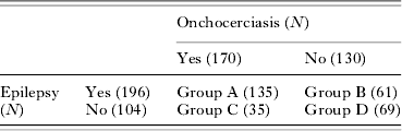
Between April and July 2005, people with epilepsy (PWE), who regularly attend The Mahenge Epilepsy Clinic to collect their medication, and their accompanying relatives were approached and asked whether they would like to participate in the study (convenience sampling). Pregnant women, young children (all children below the age of 8 years) and people who appeared medically unwell were excluded from the study, because lumbar puncture was part of it. People with inactive epilepsy (see below) were also actively excluded from the study. Participants were recruited into 4 main study groups depending on diagnosis of onchocerciasis, epilepsy or both: PWE and onchocerciasis (group A), PWE only (group B), individuals with onchocerciasis but without epilepsy (group C) and people with neither epilepsy nor onchocerciasis (group D, see Table 1).
Study groups and number of participants
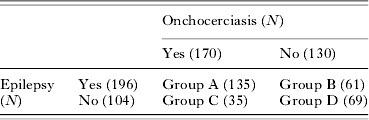
N, Number of participants in the respective groups.
An attempt was made to collect information about Ivermectin treatment in all participants. However, most participants did not know when they last got treatment or if they had ever received it. As the tablets are given to the head of a household for the whole family, no definite record of intake was available at the time of the study. Also, Ivermectin treatment per se would not have influenced the allocation of patients to study groups. The diagnosis of onchocerciasis can only be established on the basis of at least 1 mf on skin snip or a positive O. volvulus skin snip PCR or both. Because of the multitude and unreliability of dermatological presentations, clinical features were not used as a diagnostic criterion. Dermatological manifestation was present in virtually all of the patients, whereas ocular symptoms were not reported.
All PWE had active epilepsy with at least 1 seizure in the year preceding the survey. Patients with single epileptic seizures or epileptic seizures with an obvious precipitant were excluded. Epilepsy was defined as 2 or more afebrile seizures unrelated to acute metabolic disorders or withdrawal of drugs or alcohol (Senanayake and Roman, Reference Senanayake and Roman1993).
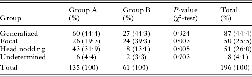
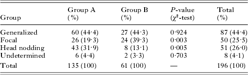
All participants were subject to further history taking and examination. PWE were evaluated by means of a standardized protocol presented by trained medical students. Thereafter PWE were examined by experienced neurologists (W. M., L. J. A., A. S. W., E. S.). The seizure type was determined according to an International League Against Epilepsy (ILAE) adjusted clinical classification for resource-poor countries suggested by Winkler et al. (ILAE, 1981; Winkler et al. Reference Winkler, Schaffert and Schmutzhard2007; see Table 2). The head nodding syndrome, described by Winkler et al. in 2008, was included in the classification (Winkler et al. Reference Winkler, Friedrich, König, Meindl, Helbok, Unterberger, Gotwald, Dharsee, Velicheti, Kidunda, Jilek-Aall, Matuja and Schmutzhard2008).
Seizure types in groups A and B
(According to ILAE (1981) and Winkler et al. (Reference Winkler, Schaffert and Schmutzhard2007, Reference Winkler, Friedrich, König, Meindl, Helbok, Unterberger, Gotwald, Dharsee, Velicheti, Kidunda, Jilek-Aall, Matuja and Schmutzhard2008). Group A, people with epilepsy and onchocerciasis; Group B, people with epilepsy only; ILAE, International League Against Epilepsy.)
Microfilarial load and density
Investigation of mf load and density was carried out on site in Tanzania. Altogether, mobile mf were found in the skin of 123 participants (41·0%). Two skin snips were taken from every participant, one from skin covering the right iliac crest and a second one from skin over the left iliac crest. In individuals showing changes compatible with onchocerciasis in other areas of skin, additional skin snips were taken from the respective region. The skin was cut in half and weighed. One half was given into a microtitre plate well containing normal saline, the other half was stored for PCR analysis. After 1 h and again after 4 h, the number of mf visible under the microscope was counted. If no mf were seen, the well was checked again after 12 h. The highest number/mg found in any skin snip of a patient was noted as mf load, and the total number/mg divided by the number of skin snips as mf density.
Skin PCR
The investigation was carried out in the Department of Infectious Diseases and Tropical Medicine, Ludwig-Maximilians-University, Munich, Germany. The DNA was extracted from the samples using the Puregene DNA Purification System by Gentra Systems (Minneapolis, USA). The genome of O. volvulus contains a tandemly repeated DNA sequence of 150 base pairs, which can serve as a template to produce specific short DNA fragments (primers; Meredith et al. Reference Meredith, Unnasch, Karam, Piessens and Wirth1989). These in turn can be used for selective amplification of O. volvulus DNA using PCR. The PCR was performed according to Zhang et al. (Reference Zhang, Li and Weil2000) using gel electrophoresis instead of paper chromatography to visualize the amplification product.
Blood
White blood cell counts were performed in the Neubauer chamber. Thin blood films were produced and stained with May-Grünwald Giemsa to evaluate differential blood counts. The presence of mf in the blood was determined by modified Knott concentration (WHO, 1997). Although O. volvulus mf are occasionally found in the blood of affected individuals (Duke et al. Reference Duke, Vincelette and Moore1976), this test was mainly used to evaluate the presence of blood-dwelling mf in the study population (e.g. Wuchereria bancrofti, Mansonella perstans). All of the above investigations were conducted on site in Tanzania.
IgG4 antibodies against O. volvulus were detected with enzyme-linked immunosorbent assay (ELISA; Lal and Ottesen, Reference Lal and Ottesen1988) performed at The Department of Infectious Diseases and Tropical Medicine, Ludwig-Maximilians-University, Munich, Germany. The antigen needed for the ELISA was obtained from adult O. volvulus found in 6 subcutaneous nodules excised from Tanzanian individuals with onchocerciasis. The optical densities measured were compared to a standard curve, using pooled sera with no, low, medium and high concentrations of O. volvulus specific antibodies. Results were given in factors calculated as multiples of normal activity (Funke et al. Reference Funke, Felgner and Geister1981). To detect false positive results, all positive samples were also tested for Wuchereria bancrofti antigen with an ELISA kit (Tropical Biotechnology Pty Ltd, Queensland, Australia).
Cerebrospinal fluid
Cell counts were performed on site in Tanzania, all other investigations were conducted in the CSF laboratory of the Department of Neurology, Medical University of Innsbruck, Austria. Out of ethical considerations, lumbar puncture was performed on participants of Group A, B and C only. In infections of the central nervous system, besides increased cell count, protein, albumin, glucose and immunoglobulins may lie outside the normal range. The presence of intrathecally produced specific antibodies against the suspected infectious agent may be of further diagnostic interest and the antibody index can be calculated.
Cells were counted in the Fuchs-Rosenthal chamber (cut-off 5 cells/μl), and CSF both native and stained with May-Grünwald Giemsa was checked for mf. Total protein was measured (reference value 15–45 mg/dl), and glucose, albumin, and unspecific IgG were determined and compared to their respective values in the blood. The reference range for the IgG index was 0·34–0·64 and that for the CSF/serum glucose ratio >0·5. The reference value for the albumin ratio changes with age and can be calculated (Zettl et al. Reference Zettl, Lehmitz and Mix2005). The O. volvulus IgG4-ELISA was performed according to the method described for blood, and the CSF/serum antibody index (AI) for O. volvulus was calculated (Reiber and Lange, Reference Reiber and Lange1991). The AI was considered positive above 4. The PCR for O. volvulus was performed according to the method used for skin snips.
Statistical analysis
The data were analysed using SPSS 14.0 (SPSS Inc., Chicago, USA). The frequency comparisons between groups on categorical data were performed with the χ2-test or the Fisher's exact test. For continuous data the mean with the standard deviation (s.d.) or the median with the range was given, depending on the distribution pattern of the data. For comparison of groups on one variable the Mann-Whitney U test or the Kruskal-Wallis test was used. Correlations were performed with Spearman's rank correlation.
Ethical considerations
The study was cleared by the Ethics Committee of the Muhimbili University College of Health Sciences, Dar es Salaam. Informed consent was obtained from all participants. All patients of the Mahenge Epilepsy Clinic received free treatment irrespective of whether they participated in the study or not.
RESULTS
Demographic data
Three hundred individuals were included in the study. Of these, 174 (58·0%) were female and 126 (42·0%) were male. Of all participants, 196 (65·3%) had epilepsy. After identification of individuals with onchocerciasis (n=170), the study population consisted of 135 PWE with onchocerciasis (group A, 45·0%), 61 PWE only (group B, 20·3%), 35 participants with onchocerciasis only (group C, 11·7%), and 69 people with neither epilepsy nor onchocerciasis (group D, 23·0%; see Table 1). The median age of PWE was 19·0 years (range 8–65) in group A and 20·0 years (range 10–36) in group B, and of participants without epilepsy 43·0 years (range 22–72) in group C and 42·0 years (range 14–70) in group D. The two latter were mainly composed of older relatives accompanying PWE to the clinic, which explains the older median age.
Epilepsy
In 87 PWE (44·4%) seizures were generalized, in 50 PWE (25·5%) focal, 51 PWE (26·0%) had head nodding syndrome alone or in combination with other types of seizures, and in 8 PWE (4·1%) the type of seizure could not be determined because of insufficient data (see Table 2). More individuals with head nodding syndrome were found in PWE who suffered from onchocerciasis (group A; χ2-test, P=0·005), whereas patients with focal seizures were more common in PWE only (group B; χ2-test, P=0·003; see Table 2).
In total, 102 PWE (52·0% of 196 PWE) had a positive family history (FH) of epilepsy outside the study population. No significant difference was seen between PWE with and without onchocerciasis (groups A and B; χ2-test, P=0·369). Twenty-eight PWE (14·3%) had learning disabilities. Possible causes in their past medical history included cerebral malaria, hypoxic conditions or measles encephalitis (no difference between groups; χ2-test, P=0·592). In addition, 17 of them had a positive FH for epilepsy. Thirty-four PWE (17·3%) had experienced febrile fits during their childhood without obvious neurological sequelae (more in PWE only (group B); χ2-test, P=0·010). Of these, 18 had a positive FH for epilepsy. In 44 PWE (22·4%), no personal or FH that could explain the origin of their seizures was found (no difference between groups; χ2-test, P=0·943), and in 8 PWE (4·1%) we could not obtain sufficient data to exclude possible causes for their seizures.
Onchocerciasis
Microfilariae in skin and skin PCR: definition of study groups
Six of the 123 participants with mobile mf had a negative PCR result of the skin. All 6 were PWE and were included in the onchocerciasis group (group A), as they had IgG4 antibodies against O. volvulus in serum, no mf on Knott concentration and a negative W. bancrofti ELISA. Forty-seven individuals (15·7%) with a negative microscopic result had a positive skin PCR for O. volvulus, raising the total number of individuals affected by onchocerciasis to 170 (56·7%). Skin snips of 7 participants with mobile mf were not tested with PCR, because they were either lost in transfer or not big enough to be used for direct microscopy and PCR. In none of these participants were mf found on Knott concentration and all were negative in the W. bancrofti ELISA. Six of them were positive on O. volvulus ELISA in serum, 1 was borderline. The person with a borderline result had skin changes compatible with onchodermatitis. All 7 were included into the respective onchocerciasis groups (6 PWE with onchocerciasis, 1 participant with onchocerciasis only).
The median load was 3·0 mf/mg (range 0·33–80·0) and the median density was 1·5 mf/mg (range 0·2–51·5). PWE with onchocerciasis (group A) and participants without epilepsy but onchocerciasis (group C) did not differ significantly in their load or density. Age, in which PWE and participants without differed significantly (Mann–Whitney U test, P<0·001), did not correlate with mf load (Spearman's rank correlation, P=0·550) or mf density (Spearman's rank correlation, P=0·377). Comparing mf load and density between group A and group C within different age groups, the numbers of participants falling into any of these groups were found too small for reliable statistical interpretation. There was no significant difference in mf load or density between participants suffering from different types of seizure.
Microfilariae in blood: exclusion of blood-dwelling microfilariae
In 7 of 163 blood samples tested by the Knott concentration method, mf were seen (6 in PWE with onchocerciasis, 1 in a person with onchocerciasis only). Only 1 of these samples, which was taken from a participant with epilepsy and onchocerciasis, was positive for W. bancrofti. All these 7 participants had a positive result on skin snip PCR, therefore all of them were included in the onchocerciasis groups.
Analysis of cerebrospinal fluid
CSF was obtained from 197 participants. PCR and cell count was performed on these 197 samples. In total, 191 samples were analysed regarding other CSF parameters (see Table 3), as 6 samples went missing en route to the CSF laboratory. Participants who did have neither epilepsy nor onchocerciasis (group D) did not undergo lumbar puncture, 34 participants from the other groups refused CSF to be taken.
Results of CSF parameters distributed by patient group
(N, Number of participants in the respective groups. Group A, people with epilepsy and onchocerciasis; Group B, people with epilepsy only; Group C, participants with onchocerciasis without epilepsy, Group D, participants with neither onchocerciasis nor epilepsy. KW test, Kruskal-Wallis one-way analysis of variance.)

No mf were seen in any of the 197 CSF samples, all of which were also negative on PCR. Twenty individuals (10·5%) had more than 5 cells/μl CSF (19 PWE and one person without epilepsy; range 5·3–58·7 cells/μl), 10 of them had onchocerciasis. The mean values are shown in Table 3, the difference between PWE with and without onchocerciasis (groups A and B) was significant (Mann–Whitney U test, P=0·054), the average amount of cells being higher in PWE only (see Table 3). In participants with more than 5 cells, there was no association with the level of serum antibodies (Mann–Whitney U test, P=0·409), mf load (Mann–Whitney U test, P=0·413), mf density (Mann–Whitney U test, P=0·475) or number of eosinophils (Mann–Whitney U test, P=0·898).
Only PWE had elevated protein, albumin ratio or unspecific IgG Index (for mean values see Table 3). The glucose ratio was above 0·45 in all participants, no difference was seen between the groups (Kruskal-Wallis test, P=0·517, see Table 3). Likewise, there was no difference in total protein concentration (Kruskal-Wallis test, P=0·169).
Comparison of the absolute results of albumin ratio between groups showed higher values in participants with onchocerciasis only (group C, Kruskal-Wallis test, P=0·001), most likely due to the fact that these participants were considerably older. However, when taking age into consideration, the albumin ratio was above the age adjusted limit in 2 PWE with onchocerciasis (group C) and 2 PWE alone only (group B; 2·1%; range 0·006–0·008).
The 4 study groups did not differ significantly in the unspecific IgG Index (Kruskal-Wallis test, P=0·724).
O. volvulus IgG4 ELISA in blood and cerebrospinal fluid and antibody index
Of the 298 blood samples tested in the O. volvulus IgG4 ELISA, 244 (81·3%) contained antibodies. The amount of IgG4 increased with age of the participants (Spearman's rank correlation, correlation coefficient 0·242, P<0·001). After matching the groups for age, a tendency was seen for higher values in individuals with onchocerciasis, which was significant when comparing the 2 groups of PWE (groups A and B; Mann–Whitney U test, P=0·002), whereas in the 2 groups without epilepsy it was not (groups C and D; Mann–Whitney U test, P=0·444).
The factors found in the CSF ELISA (n=194) showed the same tendency with higher median factors in PWE and onchocerciasis (group A; median factor 3, range 0–26) than in PWE only (group B; median factor 1, range 0–8; Mann–Whitney U test, P=0·013), but no difference between PWE and onchocerciasis (group A as above) and participants with onchocerciasis only (group C; median factor 7, range 0–14; Mann–Whitney U test, P=0·408).
The CSF/blood AI (n=63, including all those with a positive unspecific IgG Index) based on the above O. volvulus IgG4-ELISA results was positive (>4) in 2 participants (1·0%) and borderline (2–4) in 2 other participants (1·0%). Results were distributed equally in the 2 groups with onchocerciasis (groups A and C). One participant with epilepsy and onchococerciasis with a positive AI had a slightly elevated CSF cell count (5·6 cells/μl). The other CSF values were within normal range in all 4 individuals.
Other blood values
White blood cell count was done in all 300 participants. The median number of eosinophils was 333/μl (range 0–3740) and the median percentage of eosinophils in the differential blood count was 6·0% (range 0–44). PWE and onchocerciasis (group A) had significantly higher values of eosinophils than PWE only (group B; Mann–Whitney U test, P=0·004 for total number and P=0·019 for percentage). After matching the groups for age, PWE had significantly higher percentages of eosinophils compared to all other participants without epilepsy (Mann–Whitney U test, P=0·026), while participants with onchocerciasis did not have significantly higher values than those without onchocerciasis (Mann–Whitney U test, P=0·115).
DISCUSSION
It has been hypothesized that adult worms or the larval stages of O. volvulus may be able to enter CSF or to penetrate brain tissue (Marin et al. Reference Marin, Boussinesq, Druet-Cabanac, Kamgno, Bouteille and Preux2006). This theory was supported by the finding of mf in the CSF of individuals with onchocerciasis in Mexico after treatment and in Cameroon before and after treatment with filaricidal drugs (Mazzotti, Reference Mazzotti1959; Duke et al. Reference Duke, Vincelette and Moore1976). Interestingly, the finding by Duke et al. was not accompanied by an inflammatory CSF syndrome, which is to be expected if mf of O. volvulus were to lodge in the CSF space (Duke et al. Reference Duke, Vincelette and Moore1976).
Several study groups examined the skin mf density/load in individuals both with and without epilepsy, to check whether higher mf numbers could be found in PWE, making them more prone to microfilariaemia and therefore to migration of mf into the central nervous system (CNS). Studies from Burundi (Newell et al. Reference Newell, Vyungimana and Bradley1997) and Cameroon (Boussinesq et al. Reference Boussinesq, Pion, Demanga-Ngangue and Kamgno2002) were able to show a significant relationship between skin mf density/load and epilepsy in PWE compared to those without epilepsy. Results from our study support findings from Mali (Farnarier et al. Reference Farnarier, Diop, Coulibaly, Arborio, Dabo, Diakite, Traore, Banou, Nimaga, Vaz and Doumbo2000), Burkina Faso (Kabore et al. Reference Kabore, Cabore, Melaku, Druet-Cabanac and Preux1996) and the Central African Republic (Druet-Cabanac et al. Reference Druet-Cabanac, Preux, Bouteille, Bernet-Bernady, Dunand, Hopkins, Yaya, Tabo, Sartoris, Macharia and Dumas1999), showing no significant difference in mf density or load between PWE and onchocerciasis and those with onchocerciasis only. Other studies examined the relationship of the prevalence of onchocerciasis and the prevalence/incidence of epilepsy; again showing conflicting results (Ovuga et al. Reference Ovuga, Kipp, Mungherera and Kasoro1992; Kipp et al. Reference Kipp, Kasoro and Burnham1994; Kaiser et al. Reference Kaiser, Kipp, Asaba, Mugisa, Kabagambe, Rating and Leichsenring1996, Reference Kaiser, Asaba, Leichsenring and Kabagambe1998; Farnarier et al. Reference Farnarier, Diop, Coulibaly, Arborio, Dabo, Diakite, Traore, Banou, Nimaga, Vaz and Doumbo2000). A meta-analysis on these epidemiological studies in 2004 failed to demonstrate a clear causal relationship of onchocerciasis and epilepsy (Druet-Cabanac et al. Reference Druet-Cabanac, Boussinesq, Dongmo, Farnarier, Bouteille and Preux2004). A recent meta-analysis by Pion et al. (Reference Pion, Kaiser, Boutros-Toni, Cournil, Taylor, Meredith, Stufe, Bertocchi, Kipp, Preux and Boussinesq2009) was, however, able to show an association between onchocerciasis and epilepsy, unfortunately, because of timing, data from our study could not be included.
Whereas the mf count in skin of healthy individuals in comparison to that of PWE has been reported by some of the above research groups (Newell et al. Reference Newell, Vyungimana and Bradley1997; Druet-Cabanac et al. Reference Druet-Cabanac, Preux, Bouteille, Bernet-Bernady, Dunand, Hopkins, Yaya, Tabo, Sartoris, Macharia and Dumas1999; Farnarier et al. Reference Farnarier, Diop, Coulibaly, Arborio, Dabo, Diakite, Traore, Banou, Nimaga, Vaz and Doumbo2000; Boussinesq et al. Reference Boussinesq, Pion, Demanga-Ngangue and Kamgno2002), further CSF analyses in PWE and onchocerciasis have not been undertaken so far. No other filarial nematode infesting humans is known to migrate to the CNS apart from Loa loa, of which mainly the adult worms have been found to be able to enter the brain causing acute and severe neurological symptoms mainly when an individual co-infected with O. volvulus is treated with filaricidal drugs. Occasional cases were reported, but are rare in comparison to the number of individuals affected by L. loa (Lukiana et al. Reference Lukiana, Mandina, Situakibanza, Mbula, Lepira, Odio, Kamgno and Boussinesq2006). However, the geographical distribution of L. loa mainly includes West and Central Africa and therefore seems to be absent from our study area (Simonsen, Reference Simonsen, Cook and Zumla2003). The larval forms of other non-filarial nematodes are known to affect areas of the nervous system, causing symptoms as radiculomyelitis or meningitis (Walker and Zunt, Reference Walker and Zunt2005). As with L. loa, none of these nematodes is frequently associated with development of epilepsy and, despite their regional high prevalence, neurological signs and symptoms do not seem to appear in clusters. Nematodes known to affect the CNS also frequently leave some trace in the CSF, leading to findings on CSF analysis such as pleocytosis, increased protein content, high amount of antibodies or rarely, detection of larvae (Walker and Zunt, Reference Walker and Zunt2005; Ramirez-Avila et al. Reference Ramirez-Avila, Slome, Schuster, Gavali, Schantz, Sejvar and Glaser2009). If O. volvulus or its mf were able to enter the brain, we should have observed some mf or DNA of O. volvulus in the CSF or at least more pronounced changes of the CSF parameters in PWE and onchocerciasis. If the contact of the parasite or its mf with CSF had happened some time ago, the CSF PCR may be negative as was the case in our patients, but one would expect more individuals with an intrathecal antibody production and therefore positive AI. It is unlikely that migration of mf to the CNS is a singular, clear cut event, not leaving any trace of inflammatory or immunological response. We found slight CSF pleocytosis in some PWE, but only half of them had onchocerciasis. The average amount of cells in CSF was actually higher in PWE without onchocerciasis. CSF pleocytosis in some of the patients may be explained by recent seizure activity. In general, pathological CSF parameters were seen in a very small number of participants tested.
One should not conclude this paper without mentioning that the findings in CSF in PWE and onchocerciasis and the different results of prevalence and case-control studies in different parts of sub-Saharan Africa may underlie possible geographical variation of the genotype of O. volvulus. It is a well-known phenomenon that O. volvulus of the savannah-type causes river blindness, whereas ocular involvement in people with onchocerciasis from forests is less prevalent (Erttmann et al. Reference Erttmann, Unnasch, Greene, Albiez, Boateng, Denke, Ferraroni, Karam, Schulz-Key and Williams1987; Zimmerman et al. Reference Zimmerman, Dadzie, De Sole, Remme, Alley and Unnasch1992). Blindness in our study area of eastern Africa is almost unheard of. If migration of O. volvulus to the eye is possible, penetration of the parasite into the brain seems possible too. A report of the presence of O. volvulus mf in the CSF of people with onchocerciasis comes from Cameroon and relates to 8 patients with heavy infestation of O. volvulus (5 from the forest and 3 from the savannah), all of them with ocular involvement (Duke et al. Reference Duke, Vincelette and Moore1976). Before treatment mf were seen in the CSF of all savannah patients, but only of 2 of the 5 forest patients, supporting the idea that, although there was no genotyping performed in this study from Cameroon, the assumed forest and savannah-type of O. volvulus may behave differently when it comes to CSF involvement. We do not know whether the recent meta-analysis by Pion et al. (Reference Pion, Kaiser, Boutros-Toni, Cournil, Taylor, Meredith, Stufe, Bertocchi, Kipp, Preux and Boussinesq2009) would have shown a different result, if the 2 main genotypes had been considered separately, but we assume that this information was not at hand.
Another possibility for a connection between O. volvulus and epilepsy is that an immunological response against mf or adult filariae may result in the production of cross-reacting antibodies causing an autoimmune affection of cerebral tissue (Marin et al. Reference Marin, Boussinesq, Druet-Cabanac, Kamgno, Bouteille and Preux2006). Sowda, a localized, chronic, hyper-reactive onchodermatitis, seems to be associated with the presence of autoantibodies to proteins found in neutrophils in affected individuals (Gallin et al. Reference Gallin, Jacobi, Buttner, Schonberger, Marti and Erttmann1995). This may be similar to the way autoimmunity has been postulated to play a part in the development of ocular onchocerciasis (McKechnie et al. 1993). Different autoantibodies could be identified in patients suffering from ocular onchocerciasis and a shared antigen (OV39) between ocular tissue and O. volvulus parasite has been found, its implication, however, is unclear (Ottesen, Reference Ottesen1995). As part of the retina represents nervous tissue and the OV39 antigen has also been identified in the neural retina and the optic nerve, a shared antigen within the CNS may be present as well. Although there are no data on autoantibodies cross-reacting with an antigen in the CNS and the significance of the known autoantibodies in the development of chorioretinitis has been questioned, it seems possible that tissue destruction through inflammatory response to mf in a site with limited or no ability to regenerate is later complicated by autoantibodies leading to perpetuation of the immune response even after mf have been destroyed by chemotherapy (Cooper et al. Reference Cooper, Guderian, Proaño and Taylor1996). The human immunobiological response to filarial infestation is complicated and certainly not clarified yet, but hypereosinophilia, resulting from host hypersensitivity to the microfilarial stage of the parasite, could be involved in the pathogenesis of epilepsy (Ottesen et al. Reference Ottesen, Neva, Paranjape, Tripathy, Thiruvengadam and Beaven1979). PWE in our study certainly had higher levels of eosinophils than participants without epilepsy. However, this may also be due to infestation with intestinal worms that PWE may be especially prone to due to poor living conditions. An interesting finding which may point into the direction of autoimmunity is a positive although weak association between positivity of O. volvulus skin PCR and pathological lesions such as glioses on MRI in children and young adults with head nodding, an epilepsy syndrome endemic in the area the study took place (Winkler et al. Reference Winkler, Friedrich, König, Meindl, Helbok, Unterberger, Gotwald, Dharsee, Velicheti, Kidunda, Jilek-Aall, Matuja and Schmutzhard2008). This seems to be supported by the fact that people with head nodding in our cohort of patients were mainly found in group A, i.e. in people with epilepsy and onchocerciasis, this association was significant.
Head nodding, which was present in 26% of the PWE, consists of a repetitive short loss of neck muscle tone in children resulting in a nodding of the head and may represent atypical absence seizures as has been demonstrated recently by our group using electroencephalogram (unpublished results). In these children, milestones prior to onset of epileptic seizures are mainly normal, but impairment of cognitive function may become obvious in 40% of them, mainly after seizure onset (Winkler et al. Reference Winkler, Friedrich, Meindl, Kitunda, Jilek-Aall, Matuja and Schmutzhard2010). In the majority of these children head nodding is associated with or will later develop into generalized epileptic seizures; some individuals may suffer from a more widespread neurological involvement, although in our cohort this applies only to a minority (Winkler et al. Reference Winkler, Friedrich, König, Meindl, Helbok, Unterberger, Gotwald, Dharsee, Velicheti, Kidunda, Jilek-Aall, Matuja and Schmutzhard2008). A subset of 62 patients with head nodding was described previously and a weak association of positivity of O. volvulus skin PCR and pathological lesions such as gliosis on MRI was found in a cohort of 12 patients who underwent MRI investigations (Winkler et al. Reference Winkler, Friedrich, König, Meindl, Helbok, Unterberger, Gotwald, Dharsee, Velicheti, Kidunda, Jilek-Aall, Matuja and Schmutzhard2008). In these children, gliotic lesions in the brain may be part of an autoimmune process possibly associated with O. volvulus infection. Cerebral MRI investigations of a larger number of PWE and onchocerciasis may shed some more light as to the presence of changes compatible with an autoimmune process and is currently underway.
Although Mahenge, our study area, has been part of the African Programme for Onchocerciasis Control (APOC) since 1997, distribution of Ivermectin seems to have been erratic, with most people not remembering whether they had ever received treatment. The majority of our study population still showed mf on microscopy and/or had a positive skin PCR, which demonstrates that onchocerciasis is endemic, although people may have been treated with filaricidal drugs. Ivermectin, however, is not macrofilaricidal, therefore mf of O. volvulus may still be present. As reported above, findings from CSF were overwhelmingly negative in people with and without onchocerciais, rendering treatment with Ivermectin as a possible confounder less important. Also, the O. volvulus AI index, which may be positive in infections that happened some time ago (this has to be postulated in our patients as the possible infection would have to pre-date the onset of epilepsy), was only positive in 2 people, one with epilepsy and onchocerciasis and one with onchocerciasis alone. Even if treatment with Ivermectin had been effective in our study population, at least some PWE would have been expected to show a positive AI index, if infection with O. volvulus was the cause for their epileptic seizures.
Beside the extensively discussed role of O. volvulus in the pathogenesis of epilepsy in the Mahenge area, non-infectious causes for the high prevalence of epilepsy also need to be considered. Many inhabitants of the Mahenge mountains originate from endogamic marriage unions and a genetic component is believed to be involved in the clustering of epilepsy in certain families (Neuman et al. Reference Neuman, Kwon, Jilek-Aall, Rwiza, Rice and Goodfellow1995). Therefore, the high number of PWE in this area may also be explained by an inherited disorder or a higher susceptibility of them to changes caused during infestation with O. volvulus, which to date may not be known.
In conclusion, according to our results, migration of O. volvulus or its mf into the CNS, which may have explained the high prevalence of epilepsy in the Mahenge area, seems unlikely. Nevertheless, based on the knowledge of human reactions to dying mf with the involvement of a possible autoimmune process, and the difference in symptoms of onchocerciasis in different geographical regions, the research of onchocerciasis may take a new direction.
ACKNOWLEDGMENTS
We are very grateful to the staff of The Mahenge Epilepsy Clinic as well as to the staff of the Mahenge District Hospital who were extremely helpful. We especially thank the laboratory staff of the Department of Infectious Diseases and Tropical Medicine, University of Munich, Germany, for helping us with the ELISA and guiding us through the PCR analyses. We are also grateful to the team of the CSF laboratory of the Department of Neurology, Medical University of Innsbruck, Austria, and the team of the laboratory of the Mahenge District Hospital for their contribution. Last but not least we would like to thank our co-author Professor Louise Jilek-Aall, who founded the Mahenge Epilepsy Clinic. Her tireless efforts have helped to raise awareness for the individuals in Mahenge suffering from epilepsy over the past 4 decades.
FINANCIAL SUPPORT
This work was financially supported by the Savoy Epilepsy Foundation, Canada, and private donations.