INTRODUCTION
Endoparasites such as Fasciola hepatica live in hostile environments, being exposed to the host's immune system, host enzymes and various foreign substances excreted in bile, for example. The ability to detoxify xenobiotic compounds is an important defence mechanism that contributes to their overall survival strategy. Helminth parasites are controlled largely by use of anthelmintic drugs and their capacity to inactivate them will have an influence on their natural susceptibility to a particular drug. This ability also has a bearing on whether the parasite can develop resistance to that drug; resistance can be achieved, for example, by an increase in the parasite's metabolizing activity. For F. hepatica, it is known that the fluke can metabolize both triclabendazole (TCBZ) and albendazole (ABZ) to their sulphoxide metabolites (Solana et al. Reference Solana, Rodriguez and Lanusse2001, Reference Solana, Scarcella, Virkel, Ceriani, Rodríguez and Lanusse2009; Mottier et al. Reference Mottier, Virkel, Solana, Alvarez, Salles and Lanusse2004; Alvarez et al. Reference Alvarez, Solana, Mottier, Virkel, Fairweather and Lanusse2005); also, triclabendazole sulphoxide (TCBZ.SO) to triclabendazole sulphone (TCBZ.SO2) (Robinson et al. Reference Robinson, Lawson, Trudgett, Hoey and Fairweather2004). Furthermore, an increased ability to metabolize TCBZ and TCBZ.SO has been linked to the development of resistance to this drug by the fluke (Robinson et al. Reference Robinson, Lawson, Trudgett, Hoey and Fairweather2004; Alvarez et al. Reference Alvarez, Solana, Mottier, Virkel, Fairweather and Lanusse2005).
The initial phase of detoxification of xenobiotics (Phase I) is carried out by enzyme systems such as the flavin mono-oxygenase (FMO) and cytochrome P450 (CYP P450) systems. The FMO system probably represents the main pathway for the metabolism of TCBZ to TCBZ.SO by F. hepatica (Alvarez et al. Reference Alvarez, Solana, Mottier, Virkel, Fairweather and Lanusse2005). Inhibition of the FMO system by methimazole (MTZ) led to severe morphological changes in a TCBZ-resistant fluke isolate; the changes were greater than with TCBZ or TCBZ.SO on their own (Devine et al. Reference Devine, Brennan, Lanusse, Alvarez, Trudgett, Hoey and Fairweather2009). Evidence has been advanced for the existence of CYP P450-mediated enzyme systems in various helminths: the trematodes Dicrocoelium dendriticum, F. hepatica and Schistosoma mansoni; the cestode, Moniezia expansa; and a number of nematodes, including Haemonchus contortus and Caenorhabditis elegans (Kerboeuf et al. Reference Kerboeuf, Soubieux, Guilluy, Brazier and Riviere1995; Kotze, Reference Kotze1997, Reference Kotze1999, Reference Kotze2000; Gotoh, Reference Gotoh1998; Alvinerie et al. Reference Alvinerie, Dupuy, Eeckhoutte, Sutra and Kerboeuf2001; Solana et al. Reference Solana, Rodriguez and Lanusse2001, Reference Solana, Scarcella, Virkel, Ceriani, Rodríguez and Lanusse2009; Saeed et al. Reference Saeed, Mostafa, O'Connor, Rafferty and Doenhoff2002; Kotze et al. Reference Kotze, Dobson and Chandler2006; Cvilink et al. Reference Cvilink, Skálová, Szotáková, Lamka, Kostiainen and Ketola2008, Reference Cvilink, Szotáková, Krížová, Lamka and Skálová2009). A CYP P450-mediated pathway has been implicated in the metabolism of TCBZ by microsomal fractions of F. hepatica (Alvarez et al. Reference Alvarez, Solana, Mottier, Virkel, Fairweather and Lanusse2005). It probably plays a greater role in the conversion of TCBZ.SO to TCBZ.SO2 than the initial conversion of TCBZ to TCBZ.SO (Alvarez et al. Reference Alvarez, Solana, Mottier, Virkel, Fairweather and Lanusse2005). The present study was carried out to assess the effect of inhibition of CYP P450 activity on the ability of TCBZ-susceptible (TCBZ-S) and TCBZ-resistant (TCBZ–R) fluke isolates to respond to treatment ex vivo with TCBZ.SO. Piperonyl butoxide (PB) was used in this investigation: it is a potent inhibitor of CYP P450 oxidation (Hodgson and Levi, Reference Hodgson, Levi and Jones1998) and is frequently used as a pesticide synergist (Jones, Reference Jones1998). Surface changes to the flukes were assessed by means of scanning electron microscopy. Attention was focused on the tegument because it is the main route of entry of TCBZ compounds into the fluke (Mottier et al. Reference Mottier, Alvarez, Ceballos and Lanusse2006a; Toner et al. Reference Toner, McConvery, Brennan, Meaney and Fairweather2009) and because it carries out a number of important functions for the fluke (Fairweather et al. Reference Fairweather, Threadgold, Hanna and Dalton1999). The study complements the previous morphological investigation with MTZ (Devine et al. Reference Devine, Brennan, Lanusse, Alvarez, Trudgett, Hoey and Fairweather2009) and the biochemical pharmacology investigation involving MTZ and PB (Alvarez et al. Reference Alvarez, Solana, Mottier, Virkel, Fairweather and Lanusse2005). Since biochemical data pertaining to PB action against fluke isolates has already been published, the current study was confined to obtaining morphological data on the consequences of enzyme inhibition. It is part of a series of studies to determine the role of altered drug metabolism in TCBZ-resistant flukes.
MATERIALS AND METHODS
The protocol used in this investigation parallels that used in a recent study involving the FMO inhibitor, MTZ (Devine et al. Reference Devine, Brennan, Lanusse, Alvarez, Trudgett, Hoey and Fairweather2009); the reader is referred to that study for full details. Briefly, adult flukes of the Cullompton TCBZ-susceptible and Oberon TCBZ-resistant fluke isolates were pre-incubated in PB at a concentration of 1×10⁻⁴ m for 2 h at 37°C, before transfer to fresh NCTC 135 culture medium for 22 h at 37°C. The fresh medium contained either PB; PB+NADPH (1 nm); PB+NADPH+TCBZ (15 μg/ml); or PB+NADPH+TCBZ.SO (15 μg/ml). A stock solution of PB was initially prepared at a concentration of 1×10⁻¹ M in methanol. NADPH was added to the incubation medium to promote the oxidative metabolism of TCBZ and TCBZ.SO. Controls at 0 h and 24 h were also prepared. After incubation, flukes were fixed and prepared for scanning electron microscopy as described by Devine et al. (Reference Devine, Brennan, Lanusse, Alvarez, Trudgett, Hoey and Fairweather2009). A minimum of 4 flukes were prepared for each treatment.
RESULTS
A large number (16) of experiments was carried out in this study and this precludes a detailed description of surface changes for each one. The text will focus on the main changes observed and will be supported by a selection of appropriate micrographs and summary Tables of results.
Controls
The surface architecture of the control specimens appeared normal. For images of normal morphology, the reader is referred to the paper by McConville et al. (Reference McConville, Brennan, Flanagan, Hanna, Edgar, Castillo, Hernández-Campos and Fairweather2009: Figs 1–6).
Scanning electron micrographs (SEMs) of the tegumental surface of the liver fluke, Fasciola hepatica (Cullompton and Oberon isolates) following in vitro treatment for 24 h with piperonyl butoxide (A–B) and PB+NADPH (C–D). (A) Ventral surface of the oral cone region, Cullompton isolate, showing the surface morphology of the tegument and spines (S) which appear normal. (B) Dorsal surface of the apical cone region, Oberon isolate, showing slight swelling (arrow) of the tegument between the spines (S). (C) Ventral surface of the anterior midbody region, Cullompton isolate, showing slight swelling of the inter-spinal tegument (arrow). S, spine. (D) Ventral surface of the posterior midbody region, Oberon isolate, showing swelling (arrow) and furrowing (F) of the inter-spinal tegument and swelling of the tegument covering the spines (double arrow). S, spine.

Cullompton and Oberon isolates treated with PB and PB+NADPH
The Cullompton isolate showed a normal surface morphology following incubation in PB for 24 h (Fig. 1A). The only change seen in the Oberon isolate was a slight swelling of the inter-spinal tegument (Fig. 1B). After treatment with PB+NADPH, there was a slight swelling of the inter-spinal tegument in the Cullompton isolate (Fig. 1C). In addition to this, furrowing of the tegument and swelling of the tegument covering the spines were observed in the Oberon isolate, but the changes were mild (Fig. 1D).
Cullompton isolate treated with TCBZ and TCBZ.SO
Descriptions of surface changes brought about by treatment with the two drugs have been published elsewhere and will not be repeated here. The surface changes observed in the present study match these descriptions. Therefore, the reader is referred to the paper by Halferty et al. (Reference Halferty, Brennan, Trudgett, Hoey and Fairweather2009) for changes brought about by TCBZ and TCBZ.SO (Figs 1B, 2A, D and 3B and Figs 1C, 2B, E and 3C, respectively) and the paper by Toner et al. (Reference Toner, McConvery, Brennan, Meaney and Fairweather2009: Figs 1 and 2) for changes induced by TCBZ.SO.
Scanning electron micrographs (SEMs) of the tegumental surface of the liver fluke, Fasciola hepatica (Cullompton isolate) following in vitro treatment for 24 h with PB+NADPH+TCBZ (A–C) or PB+NADPH+TCBZ.SO (D–F). (A) Dorsal surface of the oral cone region showing swelling of the inter-spinal tegument (arrow) and disruption to the spine tips (double arrow). S, spine. Inset shows a high magnification image of disruption to the tegument covering a spine (arrow). S, spine. (B) Ventral surface of the lateral margin in the anterior midbody region showing swelling of the inter-spinal tegument (arrow). The swollen tegument between the spines (S) has become furrowed (F). (C) A high-power image of the ventral surface of the posterior midbody region showing swelling (arrow), furrowing (F) and blebbing (B) of the tegument between the spines (S). (D) A high-power image of the ventral surface along the lateral margin in the anterior midbody region showing swelling of the inter-spinal tegument (arrow), furrowing (F) of the tegument, swelling of the tegument covering the spines (double arrow) and quite extensive blebbing (B) on the surface of the fluke. S, spine. (E) Ventral surface of the posterior midbody region showing swelling of the inter-spinal tegument (arrow) and of the tegument covering the spines (double arrow). S, spine. Inset shows a high magnification image of the swelling to the tegument between the spines (arrow) and blebbing (B) associated with the spine tips. S, spine. (F) Ventral surface of the tail region showing swelling (arrow) and furrowing (F) of the tegument. Patches of blebs (B) and microvillus-like projections (MV) are present on the tegumental surface.

Cullompton isolate treated with PB+NADPH+TCBZ
Following 24 h incubation in vitro, a predominantly normal morphology was observed on the apical cone region. There was some slight swelling of the inter-spinal tegument (Fig. 2A), but the spines on the ventral surface were unaffected, whilst some disruption to the spines was visible on the dorsal surface (Fig. 2A, inset). Along the lateral margins in the ventral anterior midbody region, the tegument was swollen and furrowed (Fig. 2B). In the ventral posterior midbody region, the tegument was swollen both between and covering the spines, and blebbing and furrowing were associated with the tegumental surface (Fig. 2C). On the dorsal surface of this region only slight swelling of the tegument was observed, especially around the base of the spines. Both the dorsal and ventral surfaces of the tail region showed swelling and furrowing of the tegument.
Cullompton isolate treated with PB+NADPH+TCBZ.SO
Swelling of the inter-spinal tegument was observed on the ventral surface of the apical cone region, while on the dorsal surface the tegument retained a normal appearance. Along the lateral margin in the anterior midbody region, there was extensive swelling, furrowing and blebbing of the tegumental surface (Fig. 2D). In contrast, general swelling of the inter-spinal tegument was observed on the dorsal surface in this region. On the ventral surface in the posterior midbody region, the tegument between and covering the spines was swollen (Fig. 2E), with some blebbing associated with the spine tips (Fig. 2E inset). The inter-spinal tegument on the dorsal surface in this region was swollen and furrowed. The ventral surface of the tail region showed swelling and furrowing of the tegument and had a ‘roughened’ appearance, due to the presence of microvillus-like projections (Fig. 2F). The dorsal tail surface showed slight swelling of the inter-spinal tegument.
Oberon isolate treated with TCBZ and TCBZ.SO
Descriptions of surface changes brought about by treatment with the two drugs have been published elsewhere and will not be repeated here. The surface changes observed in the present study are similar to those described by Devine et al. (Reference Devine, Brennan, Lanusse, Alvarez, Trudgett, Hoey and Fairweather2009: Fig. 4) and Walker et al. (Reference Walker, McKinstry, Boray, Brennan, Trudgett, Hoey, Fletcher and Fairweather2004: Figs 17–24) for changes following treatment with TCBZ and TCBZ.SO, respectively.
Oberon isolate treated with PB+NADPH+TCBZ
Swelling and blebbing of the inter-spinal tegument was observed on both surfaces of the apical cone region (Fig. 3A). On the ventral surface of the lateral margins in the anterior midbody region, extensive swelling and blebbing of the tegument was observed, with the spines appearing to be sunken due to the severe swelling of the tegument surrounding them. On the dorsal surface of this same area, swelling and blebbing of the tegument was also observed, but was not as severe (Fig. 3B). On the ventral surface of the posterior midbody region, there was swelling of both the inter-spinal tegument and of the tegument covering the spines; some disruption to the spine tips was also observed (Fig. 3C). On the dorsal surface in this region, swelling, blebbing and furrowing of the tegument between the spines was observed, with the tegumental surface displaying a ‘roughened’ appearance due to the presence of microvillus-like projections (Fig. 3D). The tegument on both surfaces of the tail region was swollen and furrowed. In addition, there was a carpet of fine blebs on the dorsal surface.
Scanning electron micrographs (SEMs) of the tegumental surface of the liver fluke, Fasciola hepatica (Oberon isolate) following in vitro treatment for 24 h with PB+NADPH+TCBZ. (A) Ventral surface of the oral cone region showing swelling (arrow) and blebbing (B) of the tegument between the spines (S). Inset shows a high-power image of the swelling of the tegument covering a spine (S) and associated blebbing (B). (B) A high-power image of the dorsal surface of the lateral margin in the anterior midbody region showing swelling (arrow), furrowing (F) and blebbing (B) of the inter-spinal tegument. S, spine. (C) A high-magnification image of the ventral surface of the posterior midbody region showing swelling (arrow) and furrowing (F) of the inter-spinal tegument and the tegument covering the spines (*). Some disruption is also associated with the spine tips (double arrow). S, spine. (D) Dorsal surface of the posterior midbody region showing swelling (arrow) and furrowing (F) of the tegument between the spines (S). The surface appears roughened due to the presence of microvillus-like projections (MV).

Oberon isolate treated with PB+NADPH+TCBZ.SO
Swelling of the inter-spinal tegument was observed on both surfaces of the oral cone region, together with extensive blebbing (Fig. 4A). Along the lateral margins in the anterior midbody region, there was swelling, blebbing and furrowing of the tegument (Fig. 4B). The ventral tegument in the posterior midbody region was swollen and the surface assumed a ‘roughened’ appearance, due to the presence of microvillus-like projections (Fig. 4C). Swelling of the inter-spinal tegument and the tegument covering the spines was observed on the dorsal surface in this region (Fig. 4D), with disruption to the spine tips visible at a higher magnification (Fig. 4D inset). There was general swelling of the inter-spinal tegument on both surfaces in the tail region; furrowing and blebbing also occurred on the ventral surface.
Scanning electron microscope micrographs (SEMs) of the tegumental surface of the liver fluke, Fasciola hepatica (Oberon isolate) following in vitro treatment for 24 h with PB+NADPH+TCBZ.SO. (A) Dorsal surface of the oral cone region showing swelling (arrow) and blebbing (B) of the tegument between the spines (S). Inset is a high-power image of the tegumental surface highlighting the extensive blebbing (B). (B) Ventral surface of the lateral margin in the anterior midbody region showing swelling (arrow), blebbing (B) and furrowing (F) of the inter-spinal tegument. Double arrow, sunken spine; S, spine. (C) Ventral surface of the posterior midbody region showing swelling (arrow) and furrowing (F) of the tegument between the spines (S). The surface appears roughened due to the presence of microvillus-like projections (MV). Numerous small blebs (B) are pressent on the tegumental surface. Inset shows a high-power image of the microvillus-like projections (MV) on the tegument covering the spines (S). B, blebs. (D) Dorsal surface of the posterior midbody region showing swelling of the inter-spinal tegument (arrow) and of the tegument covering the spines (double arrow). The tegument also has a furrowed appearance (F). S, spine. Inset shows a high-power image of the swelling of the tegument (arrow) between and covering the spines (S); also, disruption associated with the spine tips (double arrow).

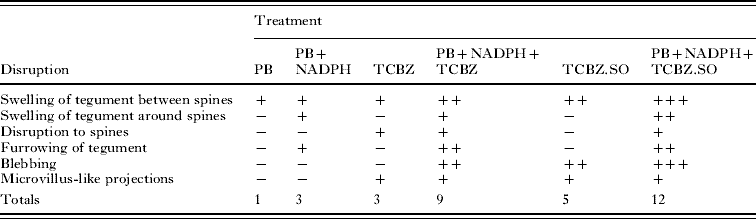
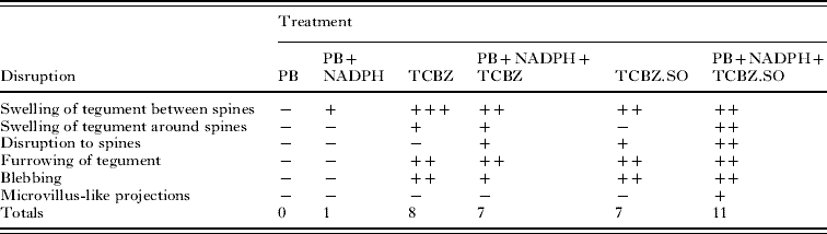
The main changes brought about by drug action and the relative severity of these changes are summarized in Tables 1 and 2.
Oberon isolate of Fasciola hepatica. Summary of surface changes following different drug treatments
(−, No noticeable disruption; +, general/mild disruption; ++, severe disruption; +++, very severe disruption; PB, piperonyl butoxide; NADPH, nicotinamide adenine dinucleotide phosphate; TCBZ, triclabendazole; TCBZ.SO, triclabendazole sulphoxide.)

Cullompton isolate of Fasciola hepatica. Summary of surface changes following different drug treatments
(−, No noticeable disruption; +, general/mild disruption; ++, severe disruption; +++, very severe disruption; PB, piperonyl butoxide; NADPH, nicotinamide adenine dinucleotide phosphate; TCBZ, triclabendazole; TCBZ.SO, triclabendazole sulphoxide.)

DISCUSSION
A number of strategies have been advanced to try to deal with the growing threat of drug resistance in parasite control. One of them is to improve the pharmacokinetics of existing anthelmintics, by enhancing their bioavailability, with the aim of increasing their efficacy. This may serve to extend the useful lifespan of the drugs as few new drugs are appearing on the market. Modulation of drug pharmacokinetics can be achieved by co-administration of inhibitors of drug metabolizing enzymes. Cytochrome P450 enzymes are known to be involved in the metabolism of benzimidazole drugs in mammals (Gottschall et al. Reference Gottschall, Theodorides and Wang1990; Velík et al. Reference Velík, Baliharová, Fink-Gremmels, Bull, Lamka and Skálová2004). Co-administration of fenbendazole or oxfendazole with PB has been shown to improve the pharmacokinetic parameters of each benzimidazole (BZ) (McKellar et al. Reference McKellar, Gokbulut, Muzandu and Benchaoui2002; Sanchez et al. Reference Sanchez, Small, Jones and McKellar2002). In separate studies involving the same BZs, the improvement lead to greater efficacy against nematode parasites; more significantly, in one of the studies, the combination showed increased efficacy against BZ-resistant nematodes (Benchaoui and McKellar, Reference Benchaoui and McKellar1996; Sanchez-Bruni et al. Reference Sanchez-Bruni, Fusé, Moreno, Saumell, Álvarez, Fiel, McKellar and Lanusse2005). Of more direct relevance to the present study, the pharmacokinetics of TCBZ in sheep have been shown to be enhanced by co-treatment with PB (Virkel et al. Reference Virkel, Lifschitz, Sallovitz, Ballent, Scarcella and Lanusse2009). PB is widely used as a synergist in pesticide preparations to make them more potent (Jones, Reference Jones1998).
Few ex vivo and in vitro studies have been carried out to examine the impact of metabolic inhibitors on drug pharmacokinetics within the parasite, as distinct from the host. In one study, synergism was demonstrated between PB and rotenone in vitro against the nematode, H. contortus, producing enhanced activity of rotenone (Kotze et al. Reference Kotze, Dobson and Chandler2006). The results of the present study have demonstrated that PB, a CYP P450 inhibitor, can potentiate the action of TCBZ.SO and (to a lesser degree) TCBZ against the TCBZ-R Oberon isolate of F. hepatica. Although TCBZ and TCBZ.SO alone induced greater disruption of the Cullompton isolate than the Oberon isolate, PB only enhanced the action of TCBZ.SO in this TCBZ-S isolate.
For the Oberon isolate, with TCBZ alone there was limited tegumental swelling, disruption to the spine tips and roughening of the surface by microvillus-like projections. Addition of PB to the incubation medium resulted in greater swelling and furrowing of the tegument, along with blebbing of the surface membrane. Treatment with TCBZ.SO on its own led to more severe changes than those observed with TCBZ alone, in that there was greater swelling and blebbing. When flukes were incubated in TCBZ.SO in the presence of PB, there was more severe disruption than with TCBZ.SO treatment alone: that is, a higher level of swelling, furrowing and blebbing was observed, together with disruption to the spines. So, for this isolate, there was greater disruption with TCBZ.SO than with TCBZ; PB co-incubation led to greater disruption with both drugs; and the combination of PB+TCBZ.SO was the most damaging of all treatments.
With the Cullompton isolate, following incubation with TCBZ, there was severe and widespread swelling and furrowing of the tegument, together with blebbing and, when it was combined with PB, the overall level of disruption was similar. This TCBZ-S isolate does not have enhanced sulphoxidative metabolic activity, so the addition of PB does not change the fluke's metabolic pattern; this may help to explain why PB did not enhance the TCBZ-induced damage in the Cullompton isolate.
TCBZ.SO alone induced widespread swelling of the tegument and severe blebbing of the surface membrane of the Cullompton isolate, changes that were increased by co-incubation with PB. With the latter combination, there was greater disruption to the spines and roughening of the surface membrane as well. So, for this TCBZ-S isolate, there was slightly greater surface disruption with TCBZ than TCBZ.SO; PB co-incubation lead to greater disruption only with TCBZ.SO; and the most disruptive treatment was with PB+TCBZ.SO. The CYP P450-mediated system seems to be involved primarily in the conversion of TCBZ.SO into the arguably less active sulphone metabolite. Thus, even in the TCBZ-S flukes, a reduced formation of the less active sulphone metabolite induced by a PB-mediated inhibition may account for the increased morphological disruption observed for TCBZ.SO co-incubated with PB.
The results show that inhibition of a drug metabolism pathway has a comparatively greater effect in TCBZ-R than-S flukes. It is known that the TCBZ-R Sligo isolate has a greater ability than the Cullompton isolate to metabolize TCBZ to TCBZ.SO (Alvarez et al. Reference Alvarez, Solana, Mottier, Virkel, Fairweather and Lanusse2005) and TCBZ.SO to TCBZ.SO2 (Robinson et al. Reference Robinson, Lawson, Trudgett, Hoey and Fairweather2004). This may be due to the over-expression of the CYP P450 enzyme pathway in TCBZ-R flukes, rendering them more sensitive to enzyme inhibition. Interestingly, PB had a similar (though limited) impact on TCBZ sulphoxidation in the two isolates (Alvarez et al. Reference Alvarez, Solana, Mottier, Virkel, Fairweather and Lanusse2005). It had less impact than MTZ, so the CYP P450 system is more likely to be involved in the conversion of TCBZ.SO to TCBZ.SO2 than the initial conversion of TCBZ to TCBZ.SO. Enhancement of the FMO-mediated metabolic activity responsible for the conversion of TCBZ to TCBZ.SO may also contribute to the mechanism of resistance to TCBZ in the Sligo and Oberon isolates (Alvarez et al. Reference Alvarez, Solana, Mottier, Virkel, Fairweather and Lanusse2005; Devine et al. Reference Devine, Brennan, Lanusse, Alvarez, Trudgett, Hoey and Fairweather2009). To complicate matters further, the accumulation of TCBZ and TCBZ.SO is greatly reduced in the Sligo isolate (Alvarez et al. Reference Alvarez, Solana, Mottier, Virkel, Fairweather and Lanusse2005; Mottier et al. Reference Mottier, Alvarez, Fairweather and Lanusse2006b), so the mechanism of resistance may be multi-factorial. Altered uptake and metabolism would significantly limit the amount of drug reaching the tissue target molecule(s) of TCBZ.
In conclusion, this study has shown that it is possible to alter the susceptibility to TCBZ of a TCBZ-R fluke isolate and convert it to a more TCBZ-S state by co-treatment with the CYP P450 inhibitor, PB. The result is compatible with the biochemical pharmacology data presented by Alvarez et al. (Reference Alvarez, Solana, Mottier, Virkel, Fairweather and Lanusse2005). The latter experiment was performed with microsomal fractions of flukes, whereas the present study was done on whole flukes and this perhaps gives a better overall impression of what metabolic inhibition can do to the intact fluke. The combined results provide a growing body of evidence that altered drug metabolism in F. hepatica is involved in TCBZ resistance, although, as mentioned previously, it may not be the sole mechanism (Fairweather, Reference Fairweather2005, Reference Fairweather2009). To put the result into some kind of context, a number of strategies have been proposed to deal with anthelmintic resistance, including development of new drugs, use of drug combinations and better use of existing drugs. No new drugs are being developed for Fasciola and the use of drug combinations may promote rather than delay drug resistance. So, gaining a better understanding of drug pharmacokinetics in both host and parasite could be important in helping to develop ways to optimize drug use and maintain the activity of TCBZ in the face of drug resistance. The results of this and other studies suggest that the concept of modulation of drug pharmacokinetics appears to be a valid one that could be developed further.
This work was supported by a DARDNI Post-graduate Studentship to C. D. It was also partly supported by a grant from the European Union (DELIVER grant, no. FOOD-CT-200X-023025) and by a BBSRC/Defra grant (C00082X/1).