Introduction
Conotruncal anomalies represent a heterogeneous group of CHDs. We report an exceptionally rare, complex, and previously undescribed conotruncal lesion in a newborn.
Case summary
A full-term male infant weighing 3.5 kg was born via spontaneous vaginal delivery following an uncomplicated pregnancy, with normal Apgar scores at birth. At 25 minutes of age, he developed respiratory distress and cyanosis, requiring endotracheal intubation and mechanical ventilation. On physical examination, no dysmorphic features were noted. The chest was hyperinflated, but breath sounds were equal bilaterally. Cardiovascular examination revealed a grade 3/6 systolic murmur over the upper sternal border.
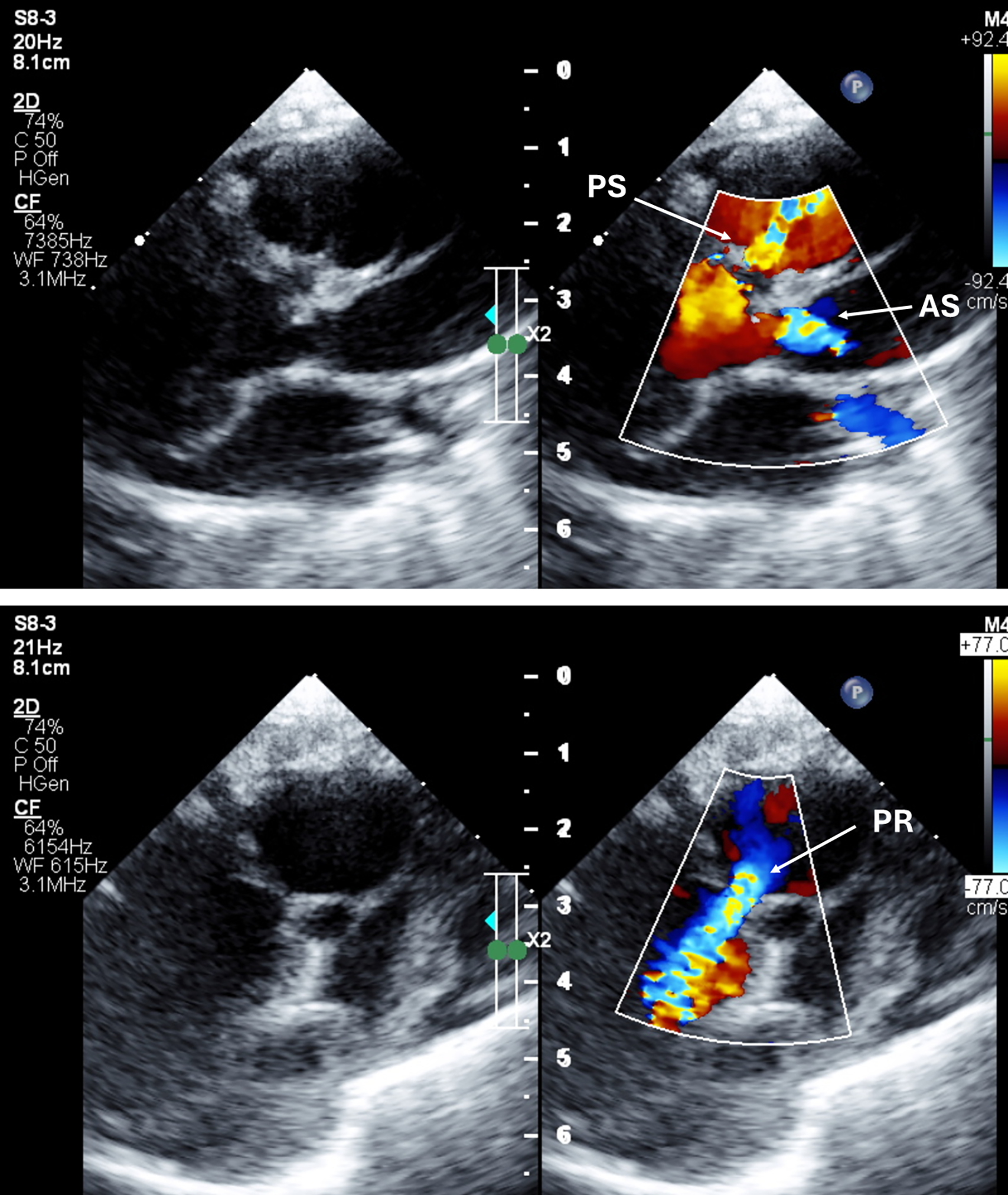
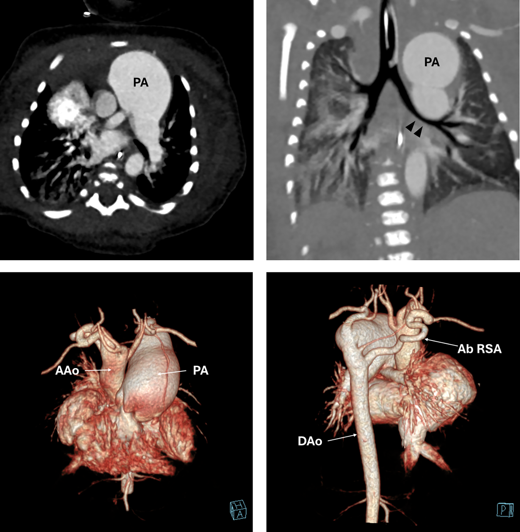
Echocardiography demonstrated features of absent pulmonary valve syndrome alongside a large outlet ventricular septal defect and severe aortic valve stenosis. CT angiography demonstrated a Type B interrupted aortic arch and an aberrant right subclavian artery (Figures 1, 2). Furthermore, the pulmonary arteries were massively dilated causing severe extrinsic airway compression at the level of the mid-trachea and left bronchus, confirmed via bronchoscopy. Given the complex nature of the cardiac lesions and the severity of the airway compromise, the overall prognosis was poor. Following a multidisciplinary team discussion and counselling, the parents elected for palliative care.
Echocardiogram images. Upper panel—long axis view demonstrating severe aortic stenosis with a narrow left ventricular outflow tract. The pulmonary stenosis jet is also evident on colour flow doppler. Lower panel—long axis view of the right ventricular outflow tract demonstrating significant pulmonaryregurgitation from a dilated pulmonary artery. (AS, aortic stenosis; PS, pulmonary stenosis; PR, pulmonary regurgitation).

CT angiography. Upper panel—the massively dilated pulmonary artery causing significant compression on the left main bronchus (double arrowheads). Lower panel—3D reconstruction images showing type B interrupted aortic arch and aberrant right subclavian artery arising from the descending aorta. (AAo, ascending aorta; Ab RSA, aberrant right subclavian artery; DAo, descending aorta; PA,pulmonary artery).

Discussion
Conotruncal anomalies encompass a diverse and heterogeneous group of CHDs, accounting for approximately 10–12% of all CHDs. Reference Hoffman and Kaplan1 The coexistence of absent pulmonary valve syndrome, type B interrupted aortic arch, and severe aortic stenosis represents a novel and exceptionally complex phenotype involving both the right and left ventricular outflow tracts. Multimodality imaging was instrumental in accurately delineating the anatomy and establishing the diagnosis.
To better understand the embryological basis of this constellation of abnormalities, a review of conotruncal development is warranted. The conotruncus, comprising the muscular conus and truncus arteriosus, arises from the primitive right ventricle. Reference Allen, Moss and Adams2 During normal cardiac development, it undergoes translocation to align with the interventricular septum, septation into the aorta and pulmonary artery, and rotation to achieve the mature configuration of the great arteries. The morphology of the conotruncal septum in our patient is particularly noteworthy. Its involvement in both the right and left ventricular outflow tracts appears to be balanced, resulting in the absence of aortic overriding despite significant conotruncal maldevelopment.
The associated aortic arch abnormalities strongly suggest involution of both embryonic fourth aortic arches. Involution of the left fourth aortic arch results in a type B interrupted aortic arch, whereas involution of the right fourth aortic arch gives rise to an aberrant right subclavian artery. The coexistence of a conotruncal anomaly and aortic arch abnormality is highly suggestive of a 22q11.2 deletion syndrome. Unfortunately, genetic testing was not performed because the parents declined further investigation.
Severe airway obstruction caused by massively dilated pulmonary arteries mandated urgent surgical consideration. Contemporary management of absent pulmonary valve syndrome favours a single-stage primary repair. This strategy typically includes reduction pulmonary arterioplasty or plication, relocation of the pulmonary artery bifurcation away from the tracheobronchial tree via the Lecompte manoeuvre, and, when indicated, tracheobronchial stenting. These procedures are performed alongside right ventricular outflow tract reconstruction (with or without a valved conduit) and concomitant repair of associated lesions, which include ventricular septal defect, interrupted aortic arch, and aortic stenosis.
However, a single-stage complete repair in this patient would have required prolonged cardiopulmonary bypass and cross-clamp times, substantially elevating perioperative morbidity and mortality risks. Furthermore, given the strong association with underlying tracheobronchomalacia, prolonged postoperative ventilatory support was anticipated. Although a staged surgical strategy could represent a theoretical alternative, the combination of complex intracardiac malformations and severe airway compression ultimately conferred a grave prognosis.
Consequently, given the anticipated procedural morbidity and low likelihood of long-term success, palliative care was chosen as the most appropriate and compassionate management strategy.
Supplementary material
The supplementary material for this article can be found at https://doi.org/10.1017/S1047951126124172.
Acknowledgements
None.
Financial support
This research received no specific grant from any funding agency, commercial, or not-for-profit sectors.
Competing interests
The authors have no relationships relevant to the contents of this paper to disclose.