Introduction
Since the first description of superior semi-circular canal dehiscence syndrome by Minor et al. in 1998, further evidence has emerged and improved our understanding of this condition.Reference Minor, Solomon, Zinreich and Zee1 The diagnosis is based on primarily clinical grounds, including findings such as sound- and/or pressure-induced vertigo, oscillopsia, and autophony and conductive hyperacusis. However, detailed audiovestibular assessment, particularly vestibular-evoked myogenic potentials testing, as well as radiological confirmation of the bony dehiscence of the superior semi-circular canal, determine the final diagnosis.Reference Minor, Solomon, Zinreich and Zee1–Reference Lookabaugh, Kelly, Carter, Niesten, McKenna and Curtin3
With respect to imaging studies, computed tomography (CT) of the temporal bones and cone beam tomography are the most popular imaging modalities of choice for identifying the osseous dehiscence in patients with a clinical suspicion of superior semi-circular canal dehiscence syndrome.Reference Minor, Solomon, Zinreich and Zee1–Reference Lookabaugh, Kelly, Carter, Niesten, McKenna and Curtin3 However, since the first description of superior semi-circular canal dehiscence syndrome in 1998, magnetic resonance imaging (MRI) has advanced significantly, and scanners of increased field strength and are now widely available. Recent studies have focused on the ability of MRI to identify the bony defect in patients with superior semi-circular canal dehiscence syndrome, with promising results.Reference Browaeys, Larson, Wong and Patel2,Reference Beyazal, Özgür, Çeliker, Beyazal, Turan and Terzi4–Reference Krombach, Di, Martiny, Prescher, Haage and Buecker6 However, most of these studies either focused solely on the radiological presence of a bony defect, without clinical correlation and a clinical suspicion of superior semi-circular canal dehiscence syndrome, or utilised 3 Tesla MRI scanners, which are still not widely available in all hospitals.Reference Browaeys, Larson, Wong and Patel2,Reference Beyazal, Özgür, Çeliker, Beyazal, Turan and Terzi4–Reference Krombach, Di, Martiny, Prescher, Haage and Buecker6
We aimed to determine the value of 1.5T MRI for radiological confirmation of a bony defect in patients diagnosed with superior semi-circular canal dehiscence syndrome based on clinical, audiovestibular and radiological (CT scan of temporal bones) criteria, in a retrospective case–control setting.
Materials and methods
Basic settings and patient selection
We performed a retrospective, blinded analysis of patients referred to our tertiary university centre for investigations of superior semi-circular canal dehiscence syndrome as approved by the Caldicott Guardian.
We identified symptomatic patients with superior semi-circular canal dehiscence syndrome who were referred to our service for tertiary input through the senior author's prospectively collected database. Out of 30 symptomatic patients with superior semi-circular canal dehiscence syndrome seen within a three-year period, we included 12 patients (12 ears) with unilateral superior semi-circular canal dehiscence syndrome evident on the CT scan who had also undergone MRI scans of the internal auditory meatus as part of their investigations. These patients underwent detailed clinical assessment and had typical presenting features. Specifically, all patients had: pressure- or sound-induced vertigo, oscillopsia, autophony and bone conduction hyperacusis, high-resolution CT temporal bone scans indicating superior semi-circular canal dehiscence, and low-threshold cervical vestibular-evoked myogenic potentials.
The control group consisted of 12 patients, assessed in our department for otological or neurotological symptoms other than superior semi-circular canal dehiscence syndrome symptoms. Specifically, all patients from the control group were investigated for unexplained hearing loss or atypical dizziness, and they all had had CT scans of the temporal bone and MRI scans of the internal auditory meatus. Clinically (including detailed vestibular testing) and radiologically, these patients did not meet the criteria for superior semi-circular canal dehiscence syndrome.
We excluded patients with a previous history of ear or lateral skull base surgery, a history of meningitis or skull base fracture, suboptimal imaging because of artefacts, and those with incomplete datasets or incomplete imaging studies.
We followed the Strengthening the Reporting of Observational Studies in Epidemiology (‘STROBE’) case–control reporting guidelines.Reference von, Altman, Egger, Pocock, Gøtzsche, Vandenbroucke and ;7
Imaging settings and assessment
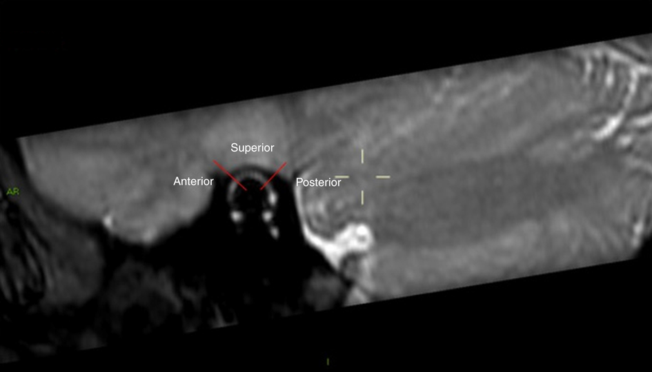
All patients underwent MRI of the internal auditory meatus with volumetric high-resolution T2-weighted sequences, either in a 1.5T Siemens Aera (‘TrueFISP’ (true fast imaging with steady-state free precession) sequences, 0.6 mm thickness) or Philips Achieva (balanced fast field echo, 0.6 mm thickness) MRI scanner. The scans were anonymised, and reconstructed in the sagittal oblique plane parallel and vertical to the superior semi-circular canal to facilitate accurate reporting (Figure 1).
The superior semi-circular canal on sagittal oblique magnetic resonance imaging (MRI) with marking indicating the site of a potential bony defect; this was a guide for the radiologists with respect to the site of the dehiscence. It is worth commenting that while the image is of adequate resolution, it does highlight the challenge of assessing the bony eminence on MRI. AR = anterior/right; I = inferior

The MRI and CT images were kept completely anonymous throughout the study.
Three experienced head and neck radiologists (with 5, 10 and 20 years’ experience at consultant level, respectively) blindly reviewed the scans of the superior semi-circular canal dehiscence syndrome and control groups. The scans were prepared in one file and presented to the radiologists in a random order.
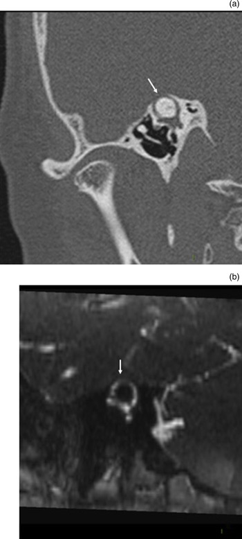
The radiologists were asked to identify whether a superior semi-circular canal bony defect was present based on the MRI scans. For those scans where the radiologist thought superior semi-circular canal dehiscence was present, the radiologist also had to identify the site of the dehiscence, grading it as posterior, anterior or superior (arcuate eminence) (Figure 2).
Superior semi-circular canal dehiscence shown on sagittal oblique computed tomography (a) and the corresponding magnetic resonance imaging (b); the arrows highlight the bony dehiscence.

Statistical analysis
We calculated the average sensitivity, specificity, positive predictive value and negative predictive value of the internal auditory meatus MRI scan in diagnosing superior semi-circular canal dehiscence syndrome, with Cohen's kappa to measure inter-rater reliability.
Results
There were 12 patients in each group. The superior semi-circular canal dehiscence syndrome group comprised five male and seven female patients, with an average age of 46.2 years (range, 26–68 years). The control group consisted of six male and six female patients, with an average age of 45.25 years (range, 26–64 years). There was no statistical difference between the two groups in terms of age (p = 0.71).
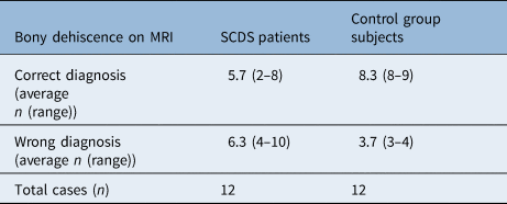
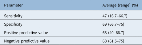
The average sensitivity and specificity of 1.5T MRI to identify the bony defect were 47 per cent (range, 16.7–66.7 per cent) and 69 per cent (range, 66.7–75 per cent) respectively (Table 1). Interestingly, the site of the bony defect was reported correctly by all radiologists in all true positive cases (100 per cent). However, there were cases of overdiagnosis and underdiagnosis of the bony defect (Table 2). Careful review of the radiological assessment showed that for defects within the mastoid cavity, all radiologists scored a falsely negative result.
Correct radiological diagnosis on 1.5T MRI

MRI = magnetic resonance imaging; SCDS = superior semi-circular canal dehiscence syndrome
Synopsis of results

Cohen's kappa = 0.75
Figure 2 shows a true positive result for dehiscence on CT (a) and on MRI (b); this was reported correctly by all radiologists.
Cohen's kappa was 0.75, indicating substantial inter-rater reliability.
Discussion
Main findings
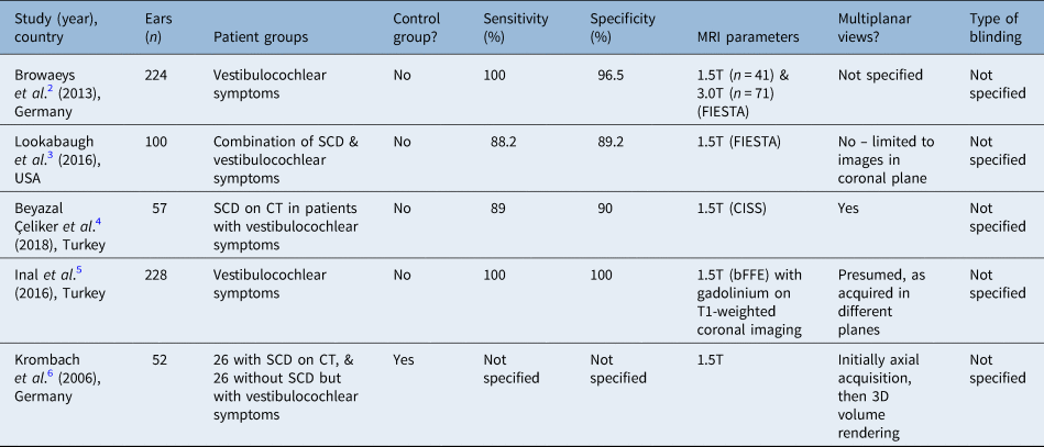
Utilising 1.5T MRI and a blinded setting involving three experienced radiologists, we found lower sensitivity and specificity of MRI in identifying the bony defect in patients with superior semi-circular canal dehiscence syndrome compared to reported data in the literature (Table 3). While for the true positive cases MRI can detect the dehiscent site accurately, our results do not indicate that 1.5T MRI could replace CT in our practice. Previous studies in the literature – based on a mixture of 1.5T and 3T MRI scans – showed much higher specificity and sensitivity, indicating that MRI could potentially replace CT, as described in Table 3.Reference Browaeys, Larson, Wong and Patel2–Reference Krombach, Di, Martiny, Prescher, Haage and Buecker6
Summary of studies in literature: sensitivity, groups, blinding and MRI parameters

MRI = magnetic resonance imaging; FIESTA = fast imaging employing steady-state acquisition; SCD = superior semi-circular canal dehiscence; CT = computed tomography; CISS = constructive interference in steady state; bFFE = balanced fast field echo; 3D = three-dimensional
Our study included only patients with definite superior semi-circular canal dehiscence syndrome, and confirmed no superior semi-circular canal dehiscence syndrome cases based on full diagnostics. In contrast, most previous studies focused on identifying superior semi-circular canal dehiscence from a pooled list of scans or patients. A diagnosis of superior semi-circular canal dehiscence syndrome should be based on clinical grounds and be supported by the diagnostic test results.
Comparison with other studies
If MRI was equivalent to CT in terms of radiological confirmation of superior semi-circular canal dehiscence syndrome, MRI could suffice as a single imaging modality of choice, leading to cost and time savings, and to reduced population radiation exposure. Two previous studiesReference Browaeys, Larson, Wong and Patel2,Reference Lookabaugh, Kelly, Carter, Niesten, McKenna and Curtin3 found higher sensitivity (88.2–100 per cent) and specificity (89.2–96.5 per cent) for MRI compared to CT for radiological confirmation of canal dehiscence. Even with such high sensitivity and specificity, the positive predictive values (62.5–100 per cent) and negative predictive values (94.7–61 per cent) varied to the extent that one study could confirm the diagnosis using MRIReference Lookabaugh, Kelly, Carter, Niesten, McKenna and Curtin3 and the other could exclude it with MRI.Reference Browaeys, Larson, Wong and Patel2
As mentioned, radiological identification of bony dehiscence in isolation is not diagnostic of superior semi-circular canal dehiscence syndrome – a combination of otological symptoms, and audiometric and vestibular testing, in conjunction with radiology, is used to make the diagnosis.Reference Ward, Carey and Minor9 However, this is not fully taken into account in some of studies reported, as these often refer to a ‘confirmation of diagnosis’ when what is actually meant is lack of bone covering seen on imaging. All patients with CT appearances of superior semi-circular canal dehiscence in the literature confirmed by radiologyReference Browaeys, Larson, Wong and Patel2–Reference Krombach, Di, Martiny, Prescher, Haage and Buecker6,Reference Spear, Jackson, Mehta, Morel, Miller and Anderson8 were unselected otology patients – no further audiological or vestibular testing was given for the greatest likelihood of superior semi-circular canal dehiscence. There is no absolute diagnostic ‘gold standard’ that shows a ‘presence’ or ‘absence’ with which to compare either CT or MRI radiological confirmation of superior semi-circular canal dehiscence, and any imaging has to be considered in the context of the overall clinical picture (Table 3). Finally, it is worth mentioning that the bony anatomy as visualised through CT is also important in pre-operative planning.Reference Tikka and Kontorinis10
Strengths and limitations
All studies to date, including ours, have been retrospective, and have relied on dual imaging being performed for clinical reasons, which may skew the data. Only one other studyReference Krombach, Di, Martiny, Prescher, Haage and Buecker6 had a control group (n = 26), and only one other studyReference Browaeys, Larson, Wong and Patel2 described how blinding occurred with two to three experienced neuroradiologists. The relatively small number of enrolled subjects corresponds to the rarity of the condition and is similar to what has been published so far.
• Previous studies showed high specificity and sensitivity of magnetic resonance imaging (MRI) for detecting superior semi-circular canal dehiscence
• However, most used pooled patient images rather than definite superior semi-circular canal dehiscence syndrome diagnosis images
• This novel retrospective case–control study compared MRI (1.5T) with computed tomography (CT) in superior semi-circular canal dehiscence syndrome and control groups
• The imaging scans were reviewed blindly and independently by three experienced radiologists
• Average sensitivity and specificity of 1.5T MRI to identify bony defect were 47 per cent (range, 16.7–66.7) and 69 per cent (range, 66.7–75) respectively
• Magnetic resonance imaging (1.5T) is important for inner-ear imaging, but cannot replace CT when superior semi-circular canal dehiscence syndrome is suspected
Most of the images were acquired using 1.5T MRI in all available studies, with one studyReference Browaeys, Larson, Wong and Patel2 using a mix of 1.5T and 3T. Our study used images reconstructed on the sagittal oblique plane, parallel and vertical to the superior semi-circular canal images; in addition, patient data were removed and presented to the radiologists in a blinded fashion. This ensured the similar blind and independent assessment of all images. In the literature, the images presented varied from full multiplanar reconstructions on a radiology workstationReference Beyazal, Özgür, Çeliker, Beyazal, Turan and Terzi4 to MRI images only in the coronal plane.Reference Spear, Jackson, Mehta, Morel, Miller and Anderson8
Despite the above limitations, which are common for most studies, we used patient groups with clinical, audiological or vestibular evidence of superior semi-circular canal dehiscence syndrome (or no superior semi-circular canal dehiscence syndrome) with true blinding of this information from the three radiologists.
Conclusion
Despite the technological advances of MRI, we found low specificity and sensitivity for 1.5T MRI in identifying the bony defect in patients with superior semi-circular canal dehiscence syndrome in a retrospective, blinded, case–control-matched, review study. Computed tomography should still be used, not only to support the clinical suspicion, but also to facilitate with surgical planning whenever this is considered.
Competing interests
None declared