Introduction
Cisplatin (cis-diaminedichloroplatinum(II)) is a complex composed of platinum, ammonium and chloride ions. It is an important chemotherapeutic agent frequently used in the treatment of solid organ tumours, notably head and neck cancers,Reference Jordan, Schwade and Truelson1 urogenital system cancers, central nervous system tumours, neuroblastoma, osteosarcoma and oesophageal cancers.Reference Fram2 Dose limitation is necessary due to ototoxicity, nephrotoxicity, neurotoxicity, bone marrow suppression and gastrointestinal side effects.Reference Guneri, Serbetçioglu, Ikiz, Guneri and Ceryan3, Reference Feghali, Liu and Water4
There have been several studies on cisplatin toxicity. Studies on rats have established that cisplatin use results in ototoxicity, due to loss of inner hair cells and outer hair cells in the organ of Corti,Reference Cardinaal, De Groot, Huizing, Veldman and Smoorenburg5, Reference Blakley, Cohen, Doolittle, Muldoon, Campbell and Dickey6 degeneration in the stria vascularis, and a notable decrease in spiral ganglion cells as a result of apoptosis.Reference Cardinaal, De Groot, Huizing, Veldman and Smoorenburg5, Reference Lynch, Gu, Pierce and Kil7, Reference Rybak, Whitworth and Somani8 Hearing loss due to cisplatin use is of the sensorineural type and is generally bilateral, progressive, irreversible, high frequency sparing and accompanied by tinnitus.Reference Ekborn, Anderssen, Ehrsson, Eksborg, Laurell and Wallin9, Reference Schweitzer10 Hearing loss is related to the cisplatin dose and dosing frequency;Reference Sakamoto, Kaga and Kamio11 in adults, bolus doses cause more severe damage.Reference Reddel, Kefford, Grant, Coats, Fox and Tattersall12 The reported incidence of hearing loss due to adult cisplatin usage ranges from 9 to 91 per cent.Reference Sakamoto, Kaga and Kamio11–Reference Rybak, Husain, Evenson, Morris, Whitworth and Somani13
In rodent studies, several different cisplatin doses and methods of administration have been used to attain ototoxicity. The intraperitoneal route is most frequently used. It is well known that chronic cisplatin treatment causes less damage than acute cisplatin treatment.Reference Saito and Aran14
Distortion product otoacoustic emission (DPOAE) and auditory brainstem response (ABR) tests have been used experimentally to determine ototoxicity, and are reported to be the ‘gold standards’ for such assessment.Reference Giordano, Lorito, Ciorba, Martini and Hatzopoulos15
This study examined whether cisplatin-induced ototoxicity, assessed via DPOAE and ABR testing, could be decreased or prevented using vitamins E, B and C and l-carnitine; it also investigated the relative effects of these compounds.
Materials and methods
Animals
Experimental animal study approval (decree number 2010-57-286) was received from the Ankara University local ethical board for animal experiments. In our study, a total of 25 (50 ears) healthy, adult, male, Albino–Wistar rats were used, taken from the health laboratory and weighing between 250 and 270 g. After transportation, the rats were left to rest for 48 hours under an appropriate temperature and humidity environment, to acclimatise them to their new environment. Animals were housed in the Ankara University Veterinary School surgery clinic in a controlled environment at a temperature of 20–22°C and 65–70 per cent relative humidity, with a 12 hour light–dark cycle, and were given pellets and water ad libitum except at drug administration times. All the provisions of the Helsinki Final Act on experimental studies were followed.
Anaesthesia and drug administration
All rats were anaesthetised with 60 mg/kg intraperitoneal ketamine hydrochloride (Ketalar, Eczacıbaşı Parke-Davis, İstanbul, Turkey) and 10 mg/kg intraperitoneal xylazine hydrochloride (Alfazyne, Alfasan International BV, Woerden, Holland).
The rats' external ear canals and tympanic membranes were inspected otoscopically. Ears with plugs were cleaned. We excluded from the study any rats with an infection in the external ear canal, opacification and/or perforation of the tympanic membrane, or infection in the tympanum.
Rats were placed on warm blankets which kept their body temperature at approximately 35°C; their rectal temperatures were measured regularly while under anaesthesia. The rats were divided into five groups and their ABR and DPOAE results were recorded, prior to drug administration.
Group one (the control group; n = 5), received 15 mg/kg intraperitoneal cisplatin (Cisplatin DBL, Hospira Australia, Melbourne, Victoria, Australia) via slow infusion.
Group two (n = 5) received 2 ml/kg intraperitoneal vitamin B complex (Bemiks Kompoze, Eczacıbaşı İlaç Sanayi ve Ticaret AŞ, İstanbul, Turkey); 2 ml contained 25 mg vitamin B1, 2 mg vitamin B2, 5 mg vitamin B6, 50 mg nicotinamide and 8.6 mg d-pantenol. Thirty minutes later, 15 mg/kg intraperitoneal cisplatin was administered.
Group three (n = 5) received 50 mg/kg intraperitoneal vitamin E (Evigen 2 ml, Aksu Farma Tıbbi Ürünler İlaç Sanayi, İstanbul, Turkey), followed 30 minutes later by 15 mg/kg intraperitoneal cisplatin.
Group four (n = 5) received 50 mg/kg intraperitoneal vitamin C (Vitabiol C 5 ml, İE Ulugay İlaç Sanayi, İstanbul, Turkey), followed 30 minutes later by 15 mg/kg intraperitoneal cisplatin.
Group five (n = 5) received 50 mg/kg intraperitoneal l-carnitine (Carnitine 1 g, Santa Farma İlaç Sanayi, İstanbul, Turkey), followed 30 minutes later by 15 mg/kg intraperitoneal cisplatin.
Seventy-two hours after cisplatin administration, ABR and DPOAE results were recorded in all groups.
Distortion product otoacoustic emissions
Distortion product otoacoustic emissions were recorded and analysed. We measured 2f1-f2 cubic distortion product components using a GSI Audio screener (MEDers) device (Grason-Stadler Viasys Healthcare, Wisconsin, USA). The ratio between f2 and f1 frequencies (f2/f1) was kept at 1.22. Stimulus intensity was taken as L1 for f1 frequency and as L2 for f2 frequency, and the difference between L1 and L2 levels was maintained at a level of 10 dB SPL (L1 = 65 dB SPL and L2 = 55 dB SPL). Distortion product otoacoustic emissions were measured at a frequency of 2f1-f2 with a microphone in the external auditory canal, using frequencies of 2000, 3000 and 4000 Hz at geometrical averages of f1 and f2. The duration of testing differed; the mean duration was 60 seconds. Emission amplitudes 3 dB over the noise threshold were considered significant. Measurements were made in a chamber where the noise level did not exceed 50 dB.
A signal-to-noise ratio generated at a geometrical average of 2f1-f2 cubic distortion products was used when evaluating DPOAE results; frequency bands of 2000, 3000 and 4000 Hz were used. The signal-to-noise ratio was determined to be more reliable than DPOAE amplitudes when evaluating DPOAE responses.
Auditory brainstem response
Auditory brainstem response was measured with a Medelec Synergy five-channel electromyography and Evoked Potentials (EP) device (Oxford Instruments, Abingdon, UK). Neonatal external ear canal probes were fitted in the ear on the measurement side. Subdermal stainless steel monopolar pin electrodes (017K103; Oxford Instruments) were fitted at the vertex (positive), mastoid region (negative) and dorsum (earth).
Electrode impedances were controlled to ensure that they were less than 2 kg Ω. In order to evaluate each ear individually, a white noise mask was applied to the other ear while one ear was being stimulated.
Stimulations were produced in the first 10 milliseconds. Each click was filtered (0.1–3 kHz). The stimulation level was decreased in steps of 10 dB, starting from 100 dB HL at 11 pulses per second. The threshold of hearing was defined as the lowest stimulation intensity at which a visible and reproducible ABR response was seen. A mean of 1500 clicks/stimulus was applied for all levels.
Data analysis
Data were analysed using the Statistical Package for the Social Sciences for Windows version 11.5 software program. The distribution of continuous variables (both normal and non-normal) was assessed using the Shapiro–Wilk test. The homogeneity of variations was assessed using the Levene test. Auditory brainstem response descriptive statistics are shown as means ± standard deviations, for wave I–IV intervals at 95 dB. Auditory brainstem response threshold values and DPOAE signal-to-noise ratio are shown as medians (minimum–maximum).
The significance of differences between group mean values was evaluated using one-way analysis of variance (ANOVA). The significance of differences in median values was evaluated using the Kruskal–Wallis test. The conditions causing the observed differences were determined using the post hoc Tukey test, where the results of one-way ANOVA were considered to be significant, or using the non-parametric multiple comparisons test, where the results of the Kruskal–Wallis test were considered to be significant.
The significance of variation between the five groups' initial versus final wave I–IV interval difference was evaluated using the Bonferroni corrective dependent t-test. The significance of variation in threshold values and signal-to-noise ratios was evaluated using the Bonferroni corrective Wilcoxon sign test.
Results were considered statistically significant at p values of less than 0.05. The Bonferroni correction was used to control type I errors in all possible multiple comparisons.
Results and analysis
During the study, one rat from the vitamin E group died on the second day after cisplatin administration, and one rat from the vitamin B group died on the third day after cisplatin administration; both were excluded from the study.
When pre-treatment ABR and DPOAE results were evaluated, no significant differences were observed between the groups (p > 0.05).
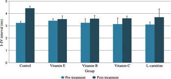
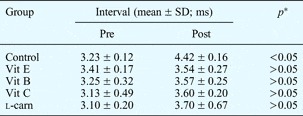
The pre- and post-treatment mean wave I–IV interval values are presented in Table I. In the control group, statistically significant wave I–IV interval differences were observed, comparing pre- and post-treatment values (p < 0.05). Statistically significant variation in wave I–IV interval differences was observed between the control group and the vitamin E and B groups (p < 0.05 for both comparisons, Figure 1). However, no statistically significant variation in wave I–IV interval differences was seen when the vitamin E and B groups were compared with the vitamin C and l-carnitine groups (p > 0.05).
Auditory brainstem response wave I–IV intervals in the different treatment groups, before and after treatment.

Pre- and post-treatment ABR wave I–IV intervals

*Analysis of variance. ABR = auditory brainstem response; SD = standard deviation; pre = pre-treatment; post = post-treatment; vit = vitamin; l-carn = l-carnitine
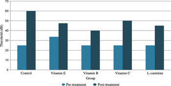
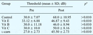
Cisplatin caused a very wide range of ABR threshold changes. However, no statistically significant difference was observed between pre- and post-treatment ABR threshold values, as shown in Table II (p > 0.05). A statistically significant difference was observed between the control group and other groups in ABR hearing threshold value changes (p < 0.05, Figure 2). A statistically significant difference (p < 0.05) between threshold value variations was observed when the vitamin E group was compared with the vitamin B, vitamin C and l-carnitine groups, and also when the vitamin B group was compared with the vitamin C and l-carnitine groups.
Auditory brainstem response thresholds in the different treatment groups, before and after treatment.

Pre- and post-treatment ABR thresholds

*Wilcoxon signed-rank test. ABR = auditory brainstem response; SD = standard deviation; pre = pre-treatment; post = post-treatment; vit = vitamin; l-carn = l-carnitine
The mean pre- and post-treatment signal-to-noise ratios are given in Table III. Comparison of the initial versus final signal-to-noise ratio indicated a significant decrease in the control, vitamin B and l-carnitine groups, for evaluations at 2000 and 3000 Hz (p < 0.01 for both). At 4000 Hz, comparison of the initial versus final signal-to-noise ratio indicated a significant decrease only in the control group (p < 0.01).
Pre- and post-treatment DPOAE signal-to-noise ratios

DPOAE = distortion product otoacoustic emission; SD = standard deviation; pre = pre-treatment; post = post-treatment; vit = vitamin; l-carn = l-carnitine
Discussion
It has been shown that chemotherapy using platinum components is a modern and effective treatment method for head and neck cancers.Reference Rybak, Whitworth and Somani8
In such patients treated with cisplatin, ototoxicity is known to be the chief side effect that decreases patients' quality of life and constrains the treatment protocol.Reference Blumenreich, Woodcock, Richman, Jones, Gentile and Kubota16 Unlike nephrotoxicity, which can be controlled by hydration and diuretics, ototoxicity develops cumulatively and irreversibly.Reference Li, Frenz, Brahmblatt, Feghali, Ruben and Berggren17
Therefore, research aiming to prevent cisplatin-associated ototoxicity is ongoing. Compounds which decrease the damage stimulated by oxidative stress have been used to prevent cisplatin-associated nephrotoxicity in rats.Reference Ajith, Usha and Nivitha18, Reference Ajith, Abhishek, Roshny and Sudheesh19 Likewise, several antioxidant agents have been used to prevent cisplatin-associated inner ear damage, with varying success. The agents most commonly used to help prevent cisplatin-associated ototoxicity are d-methionine,Reference Giordano, Lorito, Ciorba, Martini and Hatzopoulos15, Reference Wimmer, Mees, Stumpf, Welsch, Reichel and Suckfüll20l-methionine,Reference Li, Frenz, Brahmblatt, Feghali, Ruben and Berggren17 lipoic acid,Reference Lynch, Gu, Pierce and Kil7 N-acetylcysteine,Reference Feghali, Liu and Water4 glutathione ester,Reference Campbell, Larsen, Meech, Rybak and Hughes21 sodium salicylate,Reference Hyppolito, de Oliveira and Rossato22 sodium thiosulphate,Reference Wimmer, Mees, Stumpf, Welsch, Reichel and Suckfüll20 ebselenReference Lynch, Gu, Pierce and Kil7 and 4-methylthiobenzoic acid.Reference Kamimura, Whitworth and Rybak23
The ototoxic mechanism of cisplatin is not fully understood. However, it has been noted that free radicals, in particular superoxide anions,Reference Dehne, Lautermann, Petrat, Rauen and de Groot24 cause intracellular antioxidant depletion.Reference Rybak, Husain, Morris, Whitworth and Somani25 Glutathione is the major substrate in the antioxidant pathway. Rats given cisplatin have been observed to have significantly decreased glutathione and free radical production, together with decreased glutathione peroxidase levels and glutathione reductase activity.Reference Rybak, Husain, Morris, Whitworth and Somani25 Chemoprotective agents prevent ototoxicity by increasing glutathione levels and also by direct antioxidant activity.Reference Rybak, Husain, Morris, Whitworth and Somani25
Studies have shown that serum antioxidant levels decrease in cancer patients receiving cisplatin treatment. It has been noted that levels of vitamin E, vitamin C, uric acid and ceruloplasmin decrease after each cisplatin treatment and return to normal before subsequent treatments.Reference Weijl, Hopman, Wipkink-Bakker, Lentjes, Berger and Cleton26
Vitamins E, B and C and l-carnitine are known to be powerful antioxidants. Vitamin E is the most dominant lipid-soluble agent capable of preventing free radicals.Reference Teranishi, Nakashima and Wakabayashi27 It significantly reduces the chemical damage to DNA and lipid membranes which is mediated by free radicals.Reference Ajith, Abhishek, Roshny and Sudheesh19, Reference Hatano, Uramoto, Okabe, Furukawa and Ito28 In addition, the chemoprotective action of vitamins C and E facilitates the elimination of free radicals which develop due to cisplatin usage.Reference Ajith, Abhishek, Roshny and Sudheesh19
In previous studies of cisplatin-associated ototoxicity in rats, more expensive and advanced assessment methods were used, such as ABR, DPOAE and electrocochleography, rather than simple methods.Reference Freitas, Silva, Brito, Carvalho, Gomes and Ribeiro R de29 Today, assessment of ABR and DPOAE is frequently used in clinical studies.Reference Ekborn, Anderssen, Ehrsson, Eksborg, Laurell and Wallin9 Therefore, our study evaluation employed ABR threshold values and wave I–IV intervals and DPOAE signal-to-noise ratios.
The determination of electrophysiological ABR thresholds in rats is the subject of current debate. As in previous studies, we observed that ABR wave II had a large amplitude and disappeared as the stimulation intensity decreased. This wave has been used to determine the hearing threshold in animals.Reference Freitas, Silva, Brito, Carvalho, Gomes and Ribeiro R de29 Kamimura et al. Reference Kamimura, Whitworth and Rybak23 have suggested that the ABR wave IV–V complex generated in humans is similar to the wave IV complex observed in rats and cats.Reference Shaw30 However, there is no consensus on the late component of the ABR complex in small mammals.Reference Shaw30 Our study data indicated that wave III has a small amplitude and generally appears at the descendant leg of wave II. A definable positive peak was accepted as wave III.Reference Freitas, Silva, Brito, Carvalho, Gomes and Ribeiro R de29
Kalkanis et al. Reference Kalkanis, Whitworth and Rybak31 studied rats with cisplatin-induced ototoxicity and found a significant decrease in ototoxicity in animals treated with intraperitoneal vitamin E. Similarly, Teranishi et al. Reference Teranishi, Nakashima and Wakabayashi27 suppressed hearing loss at high frequencies, and also suppressed delayed cochlear outer hair cell loss, in guinea pigs treated with vitamin E. Leonetti et al. Reference Leonetti, Biroccio, Gabellini, Scarsella, Maresca and Flori32 studied cisplatin-administered rats and reported a significant decrease in side effects with vitamin E treatment; the protective effect of vitamin E treatment enabled the use of high concentrations of antitumour agents, increasing the therapeutic effect of cisplatin.
Our study found that ABR threshold variations and wave I–IV interval changes (at 95 dB) were significantly reduced in the vitamin E group compared with the control group. In addition, evaluation of DPOAE signal-to-noise ratios showed a protective effect (at all frequencies) in the vitamin E group. These findings indicate that cisplatin-induced ototoxicity was significantly decreased in animals treated with vitamin E.
Laevo-carnitine has antioxidant activity which is known to increase mitochondrial membrane sensitivity. Seidman et al. Reference Seidman, Khan, Bai, Shirwany and Quirk33 studied rats with hearing loss and found an increase in hearing thresholds with l-carnitine administration. Bielefeld et al. Reference Bielefeld, Coling, Chen and Henderson34 studied F344/NHsd rats treated with l-carnitine, and suggested that l-carnitine improves mitochondrial activity and thus decreases oxidative stress in these cells; however, no statistically significant differences in hearing thresholds were observed. Coleman et al. Reference Coleman, Kopke, Liu, Ge, Harper and Jones35 assessed the use of l-carnitine in a chinchilla model of noise-induced hearing loss, and found that l-carnitine had a protective effect by increasing mitochondrial activity and by decreasing noise-induced reactive oxygen type formation.Reference Coleman, Kopke, Liu, Ge, Harper and Jones35
In our study, a significant decrease in ABR threshold variation and in wave I–IV interval variation was observed, comparing the l-carnitine and control groups. In addition, there was a significant difference between pre- and post-treatment DPOAE signal-to-noise ratios at 2000 and 3000 Hz, but not at 4000 Hz. These results indicate that l-carnitine may be protective against cisplatin-induced ototoxicity at higher frequencies.
Guneri et al. Reference Guneri, Serbetçioglu, Ikiz, Guneri and Ceryan3 studied cisplatin-induced ototoxicity in guinea pigs, and found significantly increased transient evoked otoacoustic emissions in a group given vitamin B, compared with a group not given vitamin B; they concluded that combined vitamin B administration had a protective effect against cisplatin ototoxicity.Reference Guneri, Serbetçioglu, Ikiz, Guneri and Ceryan3
• Cisplatin chemotherapy can cause ototoxicity
• This study assessed the otoprotective effect of vitamins E, B and C and l-carnitine in rats given cisplatin
• These vitamins and l-carnitine significantly decreased cisplatin ototoxicity in rats
Our study included a group administered a combined vitamin B preparation consisting of vitamins B1, B2 and B6 plus nicotinamide and d-panthenol. This group was found to have significantly reduced ABR threshold value variations and wave I–IV interval variations, compared with the control group. Assessment of signal-to-noise ratios at 4000 Hz also indicated a protective effect of vitamin B, similar to l-carnitine.
A previous study assessing the use of vitamins C and E to treat idiopathic sudden hearing loss found a significant decrease in reactive oxygen species.Reference Hatano, Uramoto, Okabe, Furukawa and Ito28 Furthermore, Ajith et al. Reference Ajith, Usha and Nivitha18 found that vitamins C and E decreased nephrotoxicity in cisplatin-treated mice.
We found a significantly smaller difference in ABR threshold value variations and wave I–IV interval variations in our vitamin C group, and also in our vitamins E and B and l-carnitine groups. Signal-to-noise ratio data indicated that vitamin C had a protective effect at all frequencies.
Previous studies have evaluated many potentially otoprotective agents according to their mode of administration and dose, but have not compared the effects of two or more agents.Reference Guneri, Serbetçioglu, Ikiz, Guneri and Ceryan3, Reference Li, Frenz, Brahmblatt, Feghali, Ruben and Berggren17, Reference Hyppolito, de Oliveira and Rossato22, Reference Teranishi, Nakashima and Wakabayashi27, Reference Kalkanis, Whitworth and Rybak31 In contrast, our study compared four different antioxidant agents, both with a control group and with each other. In terms of ABR threshold variation and wave I–IV interval variation, the greatest differences were found in the vitamin E and B groups and the smallest differences in the vitamin C and l-carnitine groups, compared with the control group.
Conclusion
Using a rat model of cisplatin-induced ototoxicity, we evaluated the protective efficacy of vitamins E, B and C and l-carnitine. We evaluated ototoxicity using ABR results (measuring threshold variations and wave I–IV intervals) and DPOAE results (measuring signal-to-noise ratios). It should be remembered that comparisons can be made by determining amplitude with the DPOAE test. Vitamins and l-carnitine were found to make a significant contribution to decreasing or preventing cisplatin-induced ototoxicity in rats.
The use of additional treatments to decrease cisplatin ototoxic effects in humans is open to discussion. Since responses can vary even between different animal models, further clinical research is needed before animal model findings are applied to humans.