




No CrossRef data available.
Article contents
In vitro cultivation of Anisakis simplex: pepsin increases survival and moulting from fourth larval to adult stage
Published online by Cambridge University Press: 28 November 2001
Abstract
This paper describes the in vitro cultivation of the 3rd-larval stage (L3) of Anisakis simplex to adulthood in a much simpler and easier to prepare medium than those described to date. The adult males obtained are between 3.8 and 6.5 cm long and the females between 4.5 and 8.0 cm. Some individually cultivated females laid eggs which had an average size of 44.4×50.5 μm. The culture conditions were as follows: medium RPMI-1640 supplemented with 20% heat-inactivated fetal bovine serum and 1% commercial pepsin, at pH 4.0 and a temperature of 37 °C, and in air atmosphere with 5% CO2. The pepsin was found to be the key to the success of the culture. The average survival of the worms in the culture increased from 50 to 88 days, due to the fact that the survival of the adults practically doubled (increasing by 1.9 times). Furthermore, the number of worms that completed the 4th moulting (M4) increased by 4.2 times, from 22.9 to 95.6%. This culture medium may facilitate, due to its simplicity, the study of anisakids, or at least of A. simplex, constituting another step towards achieving a complete in vitro life-cycle for these parasites.
Information
- Type
- Research Article
- Information
- Copyright
- © 2002 Cambridge University Press
INTRODUCTION
Anisakis simplex is a parasitic nematode of marine mammals, which infects a large variety of fish, among other hosts, during its larval stage. These parasitized fish may be ingested by humans, thus releasing the parasite larvae into the stomach, possibly resulting in anisakiosis if the fish were not adequately cooked.
In vitro cultivation of anisakids has had some success, but is difficult to carry out due to the fact that the culture media used to date are complex as regards both their composition and their preparation (Townsley et al. 1963; Van Banning, 1971; Grabda, 1976; Carvajal et al. 1981; Likely & Burt, 1989, 1992),
Our aim was to develop a culture medium that was more defined in its composition and simpler to prepare, to make it easier to obtain adult parasites. In this paper, the defined medium used, supplemented with fetal bovine serum and pepsin, allowed in vitro development of the 3rd larval stage (L3) of Anisakis simplex s.l. Rudolphi, 1809 to mature adults.
MATERIALS AND METHODS
The worms selected for our study were 3rd-stage larvae of Anisakis simplex s.l. Rudolphi, 1809 isolated from the host Micromesistius poutassou Risso, 1826 (blue whiting), family Gadidae, purchased from the fish market of Granada (Southern Spain). The blue whiting, on the Atlantic and Mediterranean Spanish coasts, is frequently parasitized by A. simplex s.l. (Ruiz-Valero et al. 1992; Valero et al. 2000). The worms, found free in the host body cavity, were 20 mm or more in length, and were collected with the help of a needle with a blunt tip, placed on a Petri dish with 0.9% NaCl solution and washed in it several times. The worms were observed individually under an inverted microscope and those which showed any kind of internal or external damage were discarded. They were then identified according to morphological features (Hartwich, 1974; Petter & Maillard, 1988). Over 800 L3 were used in this study. Forty L3 (ca. 0.5% of total) of A. simplex s.l. from the same hosts wee preserved at −70 °C for electrophoretic identification purposes. These larvae were analysed individually by isoenzyme electrophoresis using thick starch gel in a continuous buffer system (Nascetti et al. 1986; Martín-Sánchez, Paniagua & Valero, 1998; Iglesias, Martín-Sánchez, Adroher & Valero, unpublished observations).
Prior to cultivation, each larva was individually placed in an antibiotic-antifungal solution and axenized as described elsewhere (Iglesias, Valero & Adroher, 1997). Worms were cultured on a sterile polystyrene 24-well tissue-culture plate. The culture medium (1 ml) was placed into each well with 1 parasite. The culture plates were then placed in an incubator at 37 °C and 5% CO2 in humid air (except when indicated), and the culture medium was renewed twice a week. The worms were observed daily for mobility, moulting and survival.
The culture medium (RPMI) was RPMI-1640 plus 20% (v/v) heat-inactivated fetal bovine serum (IFBS). To the medium, where indicated, IFBS (40% v/v), horse blood (1% v/v), pepsin (1% p/v), L-cysteine (4 mM) or glutathione (4 mM) were added and the worms cultivated at several pHs.
The terms ‘maximum survival’ (Smax), ‘survival 50’ (S50), and ‘average survival’ (Sav) in culture have previously been defined by us (Iglesias et al. 1997) as follows: Smax is the day the last living nematode in the experiment dies. S50 is the day on which 50% of nematodes in the experiment are dead and Sav is the arithmetic mean day of death of each nematode in the experiment. The data are expressed as the mean±standard error (S.E.).
Similarly, other terms are defined as follows: ‘L5-survival’ (SL5) is the arithmetic mean of the days that worms survive after the 4th to 5th stage moult (M4). ‘L4-survival’ (SL4) is the arithmetic mean of the days from M3 to M4. M4av is the arithmetic mean of the culture day on which the M4 of each nematode in the experiment is completed. The data are expressed as the mean±S.E. L4 indicates the larvae that completed M3 (i.e. the 3rd to 4th stage moult), and L5 the larvae that completed M4 to adulthood. A statistical comparison of the culture data was made using the Student's t distribution.
The media, sera and reagents were purchased from Sigma Chemical Co. (St Louis, MO, USA) and Bio-Whittaker (Walkersville, MD, USA). Pepsin was obtained from Probus (Barcelona, Spain). The commercial pepsin used in our experiments was analysed by SDS–PAGE (Laemmli, 1970). Protein concentration was determined according to the method of Lowry et al. (1951).
RESULTS
Forty L3 of A. simplex s.l. collected from M. poutassou were electrophoretically identified as A. simplex s.s. (39) or A. pegreffii (1) (Nascetti et al. 1986).
The greatest moulting percentages to L4 were obtained in the presence of 5% CO2, no matter which medium was used. The best parameters both for survival and moulting occurred in the nutritive medium RPMI (Table 1). Consequently, the following experiments were carried out in the RPMI medium in air atmosphere +5% CO2.

When the culture was carried out at different pHs, it was observed that at any pH used, all or almost all the L3 reach L4, but L5 were only produced at pH 4.0 (1[male ], 3[female]; 28.6% of the initial larvae). In this medium, the moult to L4 occurred between days 3 and 4; the moult to L5 occurred between days 30 and 42, and SL5 was 26.7 days (Fig. 1).

Fig. 1. Effect of pH on the in vitro moulting (A) and survival (B) of Anisakis simplex larvae cultured on RPMI-1640 plus 20% IFBS. (A) Percentage of worms that completed M3 and M4 throughout the culture time at different pHs. (B) Percentage of worms alive throughout the culture time at different pHs.
Moulting to L4 under natural conditions involves a change from the pH in fish tissues to that in the stomach of the final host. Therefore, we set up an experiment in which the larvae were subjected to a change in pH on the 5th day (from pH 7.2 to pH 2.0, 3.0 and 4.0), when all the parasites had moulted to L4. Moulting to L5 only occurred between days 43 and 71 in the medium in which the pH was changed to 4.0, 5 L5 were produced (20% of the initial larvae; 3[male ], 2[female]) with a SL5 of 39.2 days. No statistically significant differences were observed with the control at pH 4.0 from the beginning of the culture (4 L5, 19% of the initial larvae; 2[male ] and 2[female]; M4 between days 54 and 64 of the culture; SL5 31.0 days). Other manipulations of pH produced suboptimal development. The high survival rates in these media at pH 4.0 should be noted.
Pepsin is a component of the gastric juice in the stomachs of mammals. For this reason, we added pepsin to the culture media at various times during cultivation. In the first assay, at pH 7.2, moulting to L5 did not occur with or without pepsin. However, when the assay was carried out at pH 4.0, there was a highly significant difference between the medium with or without pepsin added from the beginning (Sav, P < 1×10⁻⁶; SL5, P < 0.003). Some moulting to L5 occurred in all media at pH 4.0 and reached 100% in the medium where pepsin was added at the beginning. This M4 occurred between 20 and 60 days of cultivation. The later the pepsin was added to the culture, the lower the Sav and percentage moulting to L5 (Table 2). Opposite results occurred when the pepsin was eliminated from (i.e. not added to) the culture medium at different culture times (Fig. 2). On the other hand, 1 female, grown in the medium with pepsin added on the 14th day, began ovoposition on the 97th day of cultivation, 51 days after moulting to L5. This worm laid 15442 eggs in 16 days (with a maximum of 5672 eggs laid on 1 day).

Fig. 2. Effect of pepsin (at 1% p/v) into the RPMI-1640 plus 20% IFBS medium on the in vitro development of Anisakis simplex larvae at pH 4.0.
Table 2. Influence of pepsin on the in vitro cultivation of Anisakis simplex larvae at pH 4.0 (nL3 and nL5 are the number of worms in 3rd larval or adult stage, respectively. Survival and moulting rates (average survival Sav, maximum survival Smax, survival 50 S50 and L5-survival SL5, average M4 M4av) as defined in the Materials and Methods section. S.E., Standard error).

Other changes in the culture medium resulted in reduced moulting and survival times compared with the pepsin-added media. The addition of either glutathione or L-cysteine alone, to the culture medium inhibited the moult to L5. Even with pepsin, L-cysteine prevented the moult to L5 and glutathione significantly reduced the numbers moulting. Similarly, increasing the percentage of IFBS or adding horse blood alone, inhibited the moult to L5. With pepsin and increased IFBS, the numbers moulting were reduced. In this experiment, in the control medium RPMI with pepsin, at pH 4.0, 22 worms moulted to L5 (91.6%) between days 22 and 46, of which 10 were male and 12 female, with a SL5 of 56.1 days. One of the latter laid eggs on day 101 (55 days after M4). The total number of eggs counted was 190793, laid during the period ranging from days 101 to 143 (42 days). The maximum number of eggs counted in 1 day was 16790 on day 113.
Finally, the mean length of the adults obtained was 4.4±0.3 cm for males (range 3.8–6.5 cm) and 4.9 ±0.2 cm for females (range 4.5–8.0 cm). The mean size of the eggs was 44.4±0.5×50.5±0.7 μm (n = 50).
To determine the purity of the commercial pepsin used in our experiments, it was analysed by SDS–PAGE (Laemmli, 1970). A single band of approximately 35 kDa was revealed. This band matched the band of purified and crystallized pepsin from Sigma Chemical Co. No protein impurities were revealed in the lane of pepsin from Probus. Other experiments performed with 0.01% pepsin (EC 3.4.23.1) from Sigma Chemical Co. showed similar results to ones with 1% pepsin from Probus (results not shown).
DISCUSSION
Of the forty L3 analysed by electrophoresis, 39 were identified as A. simplex s.s.; 1 was identified as A. pegreffii. We thus assumed that all (or nearly all) larvae used in the experiments of the in vitro cultivation were A. simplex s.s., since all hosts were caught off the Atlantic coast of Spain where this parasite is predominant over A. pegreffii (Nascetti et al. 1986).
The maximum development and survival of the larvae of A. simplex was obtained, as expected, in the nutritive medium in the presence of 5% CO2 (Table 1; Sommerville & Davey 1976; Mercer et al. 1986; Iglesias et al. 1997). Although the pH of the gastric juices in mammals is close to 2, we obtained a maximum survival and development in the culture at pH 4.0. However, the gastric conditions in cetaceans are different to those of land mammals. Cetaceans, final hosts of A. simplex, present a multi-chambered stomach, generally with 4 chambers. The worms are generally, but not exclusively, located in the non-glandular stomach or forestomach. This is probably because it is the chamber in which the larvae usually are released from the food, when bacterial digestion begins and due to the peristaltic movements of this chamber. The larvae are fixed intimately to the gastric mucous where M3 occurs, but the subsequent moult takes place in the stomach lumen and, therefore, the adults are normally free or only superficially attached to the stomach wall (Kikuchi et al. 1967 (cited by Podolska, Piusiński & Rokicki, 1997); Young & Lowe, 1969; Smith & Wootten, 1978; Smith, 1989; Olsen, Aagnes & Mathiesen, 1994a). However, Højgaard (1999) has recently reported a cluster of adults in the forestomach wall of a long-finned pilot whale. The pH of the forestomach ranges between 5.4 and 7.3 depending on the diet of the cetaceans, at least in minke whales (Olsen et al. 1994a; Olsen et al. 2000). In the second stomach or fundic chamber, where HCl and pepsin are produced, the pH is 5.0–5.3, whereas in the other chambers the pH is 3.2–3.6 (Olsen et al. 1994b).
The change in pH, after M3 in vitro, did not lead to any improvement in the results. Nor did the results improve when reducing substances were added, such as L-cysteine and glutathione, the former having been cited as improving the yield of the in vitro cultivation of another anisakid, Contracaecum osculatum, which matures in the stomach of seals (Likely & Burt, 1992). It might be worth noting that A. simplex is found in seal stomachs but does not reach maturity there whereas C. osculatum does (Young & Lowe, 1969; Smith & Wootten, 1978). This could also be related to the fact that the pH of the monogastric stomach of seals during digestion remains within the range of 1.5–3.5 (Mikkelsen, 1998), being lower than that recorded in the stomach content of the whales.
In addition to the pH, the pepsin may also be a stimulus necessary in the development of anisakids, at least of A. simplex, in the stomach of marine mammals. On the one hand, L3 of A. simplex prove more resistant to digestion by pepsin than by other proteases of animal and vegetal origin in vitro (Dziekońska-Rynko, Rokicki, & Jabłonowski, 1997). On the other hand, although the secretion of various substances with proteolytic activity have been described in anisakids (Sakanari, 1990), to our knowledge none similar to pepsin has yet been cited, which could obviate the need for an exogenous source of this enzyme.
Furthermore, the majority of the media used for the cultivation of A. simplex and other gastric nematodes are complex media which at some point in their elaboration require a pepsic digestion of one of their components (Van Banning, 1971; Grabda, 1976). In no case is there any suggestion of the total elimination of the pepsin. Indeed, pepsin has been used in the in vitro cultivation of other gastric nematodes (Douvres & Malakatis, 1977; Douvres, 1979).
Additionally, it can be affirmed that the addition of pepsin favours the development of the parasites the earlier it is added to the maintenance medium, although it does not appear to be necessary in the first days of in vitro cultivation of L3. According to these results, it seems that the pepsin is required, if not by L3, then by L4 from when this larva is formed and possibly by adults, as these have a greater mean survival rate SL5 in media with pepsin (48–56 days) than in the control without it (31 days). It has also been demonstrated that the proteases play an important role in the ecdysis process of some nematodes, as they are involved in the digestion processes of the old cuticle, enabling it to be expelled (Rogers, 1970; Matthews, 1982; Lustigman et al. 1996; Rhoads, Fetterer & Urban, 1997).
In summary, CO2, pH and pepsin are factors which influence the development of A. simplex, at least in vitro. The results obtained by us using this medium, which is simpler than that used up to now by other authors, show it to be useful for cultivation, as the yield, measured as a percentage of adults obtained, is much higher than that obtained by other authors, when reported, and the size of the adults is similar, although the maximum length of the females obtained by other authors is greater than that obtained in our cultures (Tables 3 and 4).
Table 3. Summary of the data on the in vitro cultivation of Anisakis simplex in RPMI-1640 (+20% IFBS) medium at pH 4.0 added with 1% pepsin (Results are expressed as mean±standard error of the number ‘‘n’’ of experiments. For all these experiments 137 worms were used. Survival rates (average survival Sav, L4-survival SL4, and L5-survival SL5) as defined in the Materials and Methods section. The survival curves were significantly different by the Kaplan-Meier test (confidence 95%).%L5: Percentage of L3 that reached L5 stage.)


Further studies are necessary to clarify the effect of pepsin and other factors on these parasites and to achieve an in vitro complete life-cycle of the anisakids in the laboratory.
Thanks are due to Dr V. Díaz-Sáez for advice with SDS–PAGE and Dr J. Martín-Sánchez for assistance with the isoenzyme analysis and the Kaplan-Meier statistical test. The authors are also grateful to Drs M. A. Olsen (University of Tromsø, Norway) and D. P. Hojgaard (Soelfjoerdur, Faroe Islands) for their help in compiling the scarce Soelfjoerdur, existing data on the physiological conditions of the stomachs of cetaceans and pinnipeds. This work was partly funded by the Spanish PB98-1312 from the DGESIC and the Research Groups Grant from Junta de Andalucía. We are grateful to Mrs Gillian C. Butcher for the translation into English.
References
CARVAJAL, J., BARROS, C., SANTANDER, G. & ALCALDE, C. (1981). In vitro culture of larval anisakid parasites of the Chilean hake Merluccius gayi. Journal of Parasitology 67, 958–959.CrossRefGoogle Scholar
DOUVRES, F. W. (1979). In vitro development of Trichostrongylus axei, from infective larvae to young adults. Journal of Parasitology 65, 79–84.CrossRefGoogle Scholar
DOUVRES, F. W. & MALAKATIS, G. M. (1977). In vitro cultivation of Ostertagia ostertagi, the medium stomach worm of cattle. I. Development from infective larvae to egg-laying adults. Journal of Parasitology 63, 520–527.Google Scholar
DZIEKOŃSKA-RYNKO, J., ROKICKI, J. & JABłONOWSKI, Z. (1997). The impact of animal and plant proteases on Nematoda 3rd stage Anisakis simplex larvae. Oceanological Studies 1, 103–107.Google Scholar
GRABDA, J. (1976). Studies on the life cycle and morphogenesis of Anisakis simplex (Rudolphi, 1809) (Nematoda: Anisakidae) cultured in vitro. Acta Ichthyologica et Piscatoria 6, 119–139.CrossRefGoogle Scholar
HARTWICH, G. (1974). Keys to genera of the Ascaridoidea, no. 2. In CIH Keys to the Nematode Parasites of Vertebrates (ed. ANDERSON, R. C. CHABAUD, A. G. & WILLMOTT, S.), pp. 1–15. CAB, Slough.
HøJGAARD, D. P. (1999). No significant development of Anisakis simplex (Nematoda, Anisakidae) eggs in the intestine of long-finned pilot whales, Globicephala melas (Traill, 1809). Sarsia 84, 479–482.CrossRefGoogle Scholar
IGLESIAS, L., VALERO, A. & ADROHER, F. J. (1997). Some factors which influence the in vitro maintenance of Anisakis simplex (Nematoda). Folia Parasitologica 44, 297–301.Google Scholar
KIKUCHI, Y., UEDA, T., YOSHIKI, T., AIZAWA, M. & ISHIKURA, H. (1967). Experimental studies of the immunopathology of the intestinal anisakiasis. Igakuno Ayumi 62, 731–736 (in Japanese).Google Scholar
LAEMMLI, U. K. (1970). Cleavage of structural proteins during the assembly of the head of bacteriophage T4. Nature, London 227, 680–685.CrossRefGoogle Scholar
LIKELY, C. G. & BURT, M. D. B. (1989). Cultivation of Pseudoterranova decipiens (sealworm) from third-stage larvae to egg-laying adults in vitro. Canadian Journal of Fisheries and Aquatic Sciences 46, 1095–1096.CrossRefGoogle Scholar
LIKELY, C. G. & BURT, M. D. B. (1992). In vitro cultivation of Contracaecum osculatum (Nematoda: Anisakidae) from third-stage larvae to egg-laying adults. Canadian Journal of Fisheries and Aquatic Sciences 49, 347–348.CrossRefGoogle Scholar
LOWRY, O. H., ROSEBROUGH, N. J., FARR, A. L & RANDALL, R. J. (1951). Protein measurement with the Folin phenol reagent. Journal of Biological Chemistry 193, 265–275.Google Scholar
LUSTIGMAN, S., MCKERROW, J. H., SHAH, K., LUI, J., HUIMA, T., HOUNGH, M. & BROTMAN, B. (1996). Cloning of a cysteine protease for the molting of Onchocerca volvulus third stage larvae. Journal of Biological Chemistry 271, 30181–30189.CrossRefGoogle Scholar
MARTÍN-SÁNCHEZ, J., PANIAGUA, I. & VALERO, A. (1998). Contribution to the knowledge of Hysterothylacium aduncum through electrophoresis of the glucose phosphate isomerase and phosphoglucomutase. Parasitology Research 84, 160–163.Google Scholar
MATTHEWS, B. E. (1982). Behaviour and enzyme release by Anisakis sp. larvae (Nematoda: Ascaridida). Journal of Helminthology 56, 177–183.Google Scholar
MERCER, J. G., MUNN, A. E., SMITH, J. W & REES, H. H. (1986). Cuticle production and ecdysis in larval marine ascaridoid nematodes in vitro. Parasitology 92, 711–720.CrossRefGoogle Scholar
MIKKELSEN, B. (1998). Summer diet of grey seals Halichoerus grypus in the Faroe Islands. Thesis. Department of Aquatic Resources and Environmental Biology. Norwegian College of Fishery Science, University of Tromsø, Norway.
NASCETTI, G., PAGGI, L., ORECCHIA, P., SMITH, J. W., MATTIUCCI, S. & BULLINI, L. (1986). Electrophoretic studies on the Anisakis simplex complex (Ascaridida: Anisakidae) from the Mediterranean and North-East Atlantic. International Journal for Parasitology 16, 633–640.CrossRefGoogle Scholar
OLSEN, M. A., AAGNES, T. H. & MATHIESEN, S. D. (1994a). Digestion of herring by indigenous bacteria in the minke whale forestomach. Applied and Environmental Microbiology 60, 4445–4455.Google Scholar
OLSEN, M. A., NORDøY, E. S., BLIX, A. S. & MATHIESEN, S. D. (1994b). Functional anatomy of the gastrointestinal system of Northeastern Atlantic minke whales (Balaenoptera acutorostrata). Journal of Zoology 234, 55–74.Google Scholar
OLSEN, M. A., BLIX, A. S., UTSI, T. H. A., SøRMO, W. & MATHIESEN, S. D. (2000). Chitinolytic bacteria in the minke whale forestomach. Canadian Journal of Microbiology 46, 85–94.CrossRefGoogle Scholar
PETTER, A. J. & MAILLARD, C. (1988). Larves d'ascarides parasites de poissons en Méditerranée occidentale. Bulletin du Museum National de Histoire Natural de Paris 4 sér., 10[A], 347–369.Google Scholar
PODOLSKA, M., PIUSIŃSKI, W. & ROKICKI, J. (1997). Stomach wall changes caused by the presence of third-stage Anisakis simplex B larvae in infected dogs. Acta Protozoologica 42, 241–247.Google Scholar
RHOADS, M. L., FETTERER, R. H. & URBAN, J. F. JR. (1997). Secretion of an aminopeptidase during transition of third- to fourth-stage larvae of Ascaris suum. Journal of Parasitology 83, 780–784.CrossRefGoogle Scholar
ROGERS, W. P. (1970). The function of leucine aminopeptidase in exsheathing fluid. Journal of Parasitology 56, 138–143.CrossRefGoogle Scholar
RUIZ-VALERO, J., VALERO, A., ADROHER, F. J. & ORTEGA, J. E. (1992). Presencia de ascáridos en peces comerciales de frecuente consumo en Granada. In In Memorian al Profesor Doctor D. F. de P. Martínez Gómez (ed. HERNÁNDEZ, S.), pp. 335–349. Córdoba, Spain, Universidad de Córdoba.
SAKANARI, J. A. (1990). Anisakis – from the platter to the microfuge. Parasitology Today 6, 323–327.CrossRefGoogle Scholar
SMITH, J. W. (1989). Ulcers associated with larval Anisakis simplex B (Nematoda: Ascaridoidea) in the forestomach of harbour porpoises Phocoena phocoena (L.). Canadian Journal of Zoology 67, 2270–2276.CrossRefGoogle Scholar
SMITH, J. W. & WOOTTEN, R. (1978). Anisakis and anisakiasis. Advances in Parasitology 16, 93–163.CrossRefGoogle Scholar
SOMMERVILLE, R. I. & DAVEY, K. G. (1976). Stimuli for cuticle formation and ecdysis in vitro of the infective larva of Anisakis sp. (Nematoda: Ascaridoidea). International Journal for Parasitology 6, 433–439.Google Scholar
TOWNSLEY, P. H., WRIGHT, H. G., SCOTT, M. A. & HUGHES, M. L. (1963). The in vitro maturation of the parasitic nematode Terranova decipiens, from cod muscle. Journal of Fisheries Research of the Board of Canada 20, 743–747.CrossRefGoogle Scholar
VALERO, A., MARTÍN-SÁNCHEZ, J., REYES-MUELAS, E. & ADROHER, F. J. (2000). Larval anisakids parasitizing the blue whiting (Micromesistius poutassou) captured from Motril bay in the Mediterranean region of southern Spain. Journal of Helminthology 74, 361–364.CrossRefGoogle Scholar
VAN BANNING, P. (1971). Some notes on a successful rearing of the herring-worm, Anisakis marina L. (Nematoda: Heterocheilidae). Journal du Conseil International de l'Exploration de la Mer 34, 84–88.Google Scholar
YOUNG, P. C. & LOWE, D. (1969). Larval nematodes from fish of the subfamily Anisakinae and gastro-intestinal lesions in mammals. Journal of Comparative Pathology 79, 301–313.CrossRefGoogle Scholar
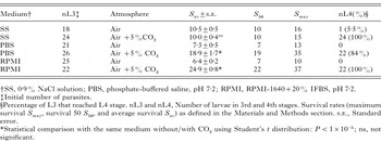
Table 1. Influence of 5% CO2 on M3 and survival of Anisakis simplex larvae incubated in different media
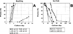
Fig. 1. Effect of pH on the in vitro moulting (A) and survival (B) of Anisakis simplex larvae cultured on RPMI-1640 plus 20% IFBS. (A) Percentage of worms that completed M3 and M4 throughout the culture time at different pHs. (B) Percentage of worms alive throughout the culture time at different pHs.
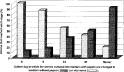
Fig. 2. Effect of pepsin (at 1% p/v) into the RPMI-1640 plus 20% IFBS medium on the in vitro development of Anisakis simplex larvae at pH 4.0.
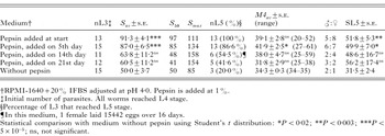
Table 2. Influence of pepsin on the in vitro cultivation of Anisakis simplex larvae at pH 4.0

Table 3. Summary of the data on the in vitro cultivation of Anisakis simplex in RPMI-1640 (+20% IFBS) medium at pH 4.0 added with 1% pepsin
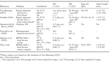
Table 4. Anisakis simplex in vitro culture in the literature
 You have
Access
- 56
- Cited by