Introduction
The spermatheca, an ectodermal accessory female reproductive organ, is variously developed in Hemiptera (Matsuda Reference Matsuda1976). The morphological characteristics of spermathecae have been described by several authors (Dupuis Reference Dupuis1955; Pendergrast Reference Pendergrast1957; Scudder Reference Scudder1959). The results of their studies show great structural and histological diversity of this organ in the studied groups (Schuh and Slater Reference Schuh and Slater1995).
Publications by Kumar (Reference Kumar1962, Reference Kumar1965) and McDonald (Reference McDonald1966) were the first to indicate the importance of the female genital structures (including spermatheca) for classification and phylogeny in Pentatomoidea. This was confirmed later for most of the pentatomoid families (Servadei Reference Servadei1964; Štys Reference Štys1964; Štys and Davidová Reference Štys and Davidová1979; Davidová-Vilimová and Štys Reference Davidová–Vílimová and Štys1980; Schaefer et al. Reference Schaefer, Dolling and Tachikawa1988; Gapud Reference Gapud1991; Kim and Lee Reference Kim and Lee1993, Reference Kim and Lee1995; Schaefer Reference Schaefer1993; Kocorek and Danielczok-Demska Reference Kocorek and Danielczok-Demska2002; Sweet and Schaefer Reference Sweet and Schaefer2002; Pluot-Sigwalt and Lis Reference Pluot-Sigwalt and Lis2008; Stacconi and Romani Reference Stacconi and Romani2011; Candan et al. Reference Candan, Yilmaz, Suludere and Erbey2015).
The spermathecae of selected Scutelleridae species were presented and analysed in the studies on pentatomoidean female genital structures by Kumar (Reference Kumar1962, Reference Kumar1965) and McDonald (Reference McDonald1966).
Additional data on the scutellerid spermathecae can be found in many taxonomic papers: Ahmad and Mushtaq (Reference Ahmad and Mushtaq1977), Ahmad and Moizuddin (Reference Ahmad and Moizuddin1980), Ahmad et al. (Reference Ahmad, Kamaluddin and Moizuddin1980, Reference Ahmad, Moizuddin and Mushtaq1988), Ahmad and Kamaluddin (Reference Ahmad and Kamaluddin1982, Reference Ahmad and Kamaluddin1996), Linnavuori (Reference Linnavuori1982), McDonald and Cassis (Reference McDonald and Cassis1984), Göllner-Scheiding (Reference Göllner-Scheiding1986), Eger (Reference Eger1987, Reference Eger1990, Reference Eger1992), McDonald (Reference McDonald1988), Gaffour-Bensebbane (Reference Gaffour-Bensebbane1990, Reference Gaffour-Bensebbane1991, Reference Gaffour-Bensebbane1993), Candan et al. (Reference Candan, Suludere and Mahmut2007, Reference Candan, Suludere and Güllü2011), Carapezza (Reference Carapezza2009), Tsai et al. (Reference Tsai, Rédei, Yeh and Yang2011), Parveen et al. (Reference Parveen, Ramamurthy, Usmani and Khokhar2013), Eger et al. (Reference Eger, Barcellos and Weiler2015). Fischer (Reference Fischer2001) published a general taxonomic and phylogenetic overview of spermathecal structures within the Scutelleridae, based on material representing 48 species.
The aim of this work is to present and summarise the general view of spermathecal features within the Scutelleridae based on the wider range of specimens including 63 species representing all subfamilies and to establish the significance of spermathecal characteristics for scutellerid classification.
Definition, terminology, and general structure of Scutelleridae spermatheca
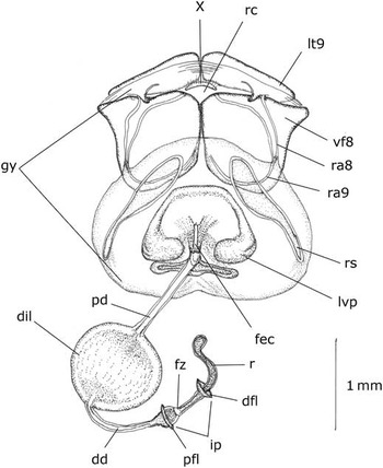
The general structure of scutellerid spermatheca (=receptaculum seminis) is similar to that observed in most Pentatomoidea and is composed of the following parts (Pluot-Sigwalt and Lis Reference Pluot-Sigwalt and Lis2008; Tsai et al. Reference Tsai, Rédei, Yeh and Yang2011) (Fig.1): receptacle, muscular intermediate part, and spermathecal duct. The receptacle [r] (=distal bulb, seminal capsule) functions as a storage place and it is situated on the apical part of the spermatheca. Its wall is more or less sclerotised with the efferent ductules of glandular secretory cells of the spermathecal gland (glandula apicalis).
Pattern of the internal female genitalia in Scutelleridae, Sphaerocoris testudogrisea (De Geer, 1778): dd, distal duct; dfl, distal flange; dil, dilation; fec, fecundation canal; fz, flexible zone; gy, gynatrium; ip, intermediate part; lt9, laterotergites IX; lvp, lateral vaginal pouch; pfl, proximal flange; pd, proximal duct; r, receptaculum; ra8, ramus VIII; ra9, ramus IX; rc, rectum; rs, ring sclerite; vf8, valvifer VIII.
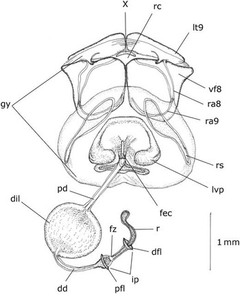
The apical portion of the spermathecal duct is defined as the intermediate part [ip] (=spermathecal pump, pump apparatus, pumping region). This structure regulates the discharge of the sperm. The muscles responsible for the control of sperm movement are attached to two cuticular plate-like flanges [pfl, dfl] (proximal and distal flange). Most scutellerid species have two flanges, but sometimes only one (usually the proximal) is present or both proximal and distal are reduced (rarely). The wall of the intermediate part is sclerotised and pigmented, one fragment (in the medial or basal part) is distinctly paler and is described as a flexible zone [fz] (Pluot-Sigwalt and Lis Reference Pluot-Sigwalt and Lis2008; Tsai et al. Reference Tsai, Rédei, Yeh and Yang2011). Within the lumen of the spermathecal duct two endocuticular structures can be distinguished: the septum (at the level of distal flange) and the fretum (posteriorly to the proximal flange) (Pluot-Sigwalt and Lis Reference Pluot-Sigwalt and Lis2008; Tsai et al. Reference Tsai, Rédei, Yeh and Yang2011). In some studied specimens these structures are clearly visible, more often they are indistinct.
The spermathecal duct [pd, dil, dd] (=ductus receptaculi) is the most differentiated part of scutellerid spermatheca. Such characteristics of the duct as length, diameter or presence and shape of the dilation are very specific for particular taxa and will be described in detail below. The spermathecal opening is connected in most scutellerid species with the longitudinal canal or groove [fec] (=fecundation canal, sclerotised groove), which is variously developed, usually sclerotised and pigmented.
Material and methods
Morphological data
Representatives of all scutellerid subfamilies were studied. Sixty-three species belonging to 46 genera (of a total of 80 genera so far described) were examined. The classification of the family is that presented by Tsai et al. (Reference Tsai, Rédei, Yeh and Yang2011).
The list of material examined is presented below.
Elvisurinae Stål, 1872
-
1. Elvisura irrorata Spinola, 1837 [Senegal, MNHN]
-
2. Coleotichus breddini Schouteden, 1905 [Palau, Koror, BPBM]
-
3. Solenosthedium bilunatum (Lefebvre, 1827) [Turkey, Adana, IRSNB]
-
4. Solenosthedium liligerum (Thunberg, 1783) [Democratic Republic of the Zaire, Libenge, IRSNB]
Eurygastrinae Amyot and Serville, 1843
-
5. Eurygaster austriaca (Schrank, 1776) [Austria, Zeilerberg, ZMHB]
-
6. Eurygaster intergriceps Puton, 1881 [Bulgaria, Choumene, ZMHB]
-
7. Psacasta exanthematica conspersa Germar, 1839 [Bosnia and Herzegovina, Zitomislic, ZHMB]
-
8. Psacasta exanthematica cerinthe (Fabricius, 1787) [Spain, ZHMB]
-
9. Xerobia sculpturata (Stål, 1858) [South Africa, Damara, TMPS]
Hoteinae Carapezza, Reference Carapezza2009
-
10. Deroplax coriacea Schouteden, 1965 [Chad, Oubangui, MRAC]
-
11. Deroplax silphoides (Thunberg, 1783) [Tanzania, Kibonoto, MRAC]
-
12. Hotea acuta Stål, 1865 [Democratic Republic of the Zaire, Oycha, IRSNB]
-
13. Hotea subfasciata (Westwood, 1837) [Cameroon, Johann Albrecht’s Höhe, DEIC]
Odontoscelinae Amyot and Serville, 1843
-
14. Irochrotus montandoni Schouteden, 1903 [Gambia, Bathurst, NHRS]
-
15. Odontoscelis dorsalis (Fabricius, 1798) [Germany, Postdam, Pfauen Island, ZHMB]
-
16. Odontoscelis byrrhus Seidenstücker, 1972 [Turkey, Malatya, ZHMB]
-
17. Rhinolaetia overlaeti Schouteden, 1965 [Democratic Republic of the Zaire, Kafakumba, paratype, MRAC]
Odontotarsinae Mulsant and Rey, 1865
-
18. Alphocoris indutus Stål, 1864 [South Africa, Letaba, TMPS]
-
19. Alphocoris lobulatus Stål, 1873 [Chad, Bebedija, MNHN]
-
20. Morbora hirtula Bergroth, 1904 [Australia, Bundaberg, IRSNB]
-
21. Odontotarsus purpureolineatus (Rossi, 1790) [Austria, Lake Neusiedl, Hackelsberg, ZHMB]
-
22. Phimodera humeralis (Dalman, 1823) [Germany, Warnemunde, ZHMB]
Pachycorinae Amyot and Serville, 1843
-
23. Agonosoma trilineatum (Fabricius, 1781) [Venezuela, Caracas, ZHMB]
-
24. Acantholomidea porosa (Germar, 1839) [United States of America, Louisiana: Pointe Coupee, ZHMB]
-
25. Ascanius hirtipes (Herrich-Schaefer, 1835) [Brazil, ZHMB]
-
26. Camirus conicus (Germar, 1839) [Bolivia, ZHMB]
-
27. Diolcus irroratus (Fabricius, 1775) [Haiti, Port-au-Prince, ZHMB]
-
28. Homaemus proteus (Stål, 1862) [United States of America, Texas, Hidalgo, ZHMB]
-
29. Homaemus aeneifrons (Say, 1824) [United States of America, Colorado, Morrison, ZHMB]
-
30. Missipus variabilis (Spinola, 1852) [Chile, Santiago, ZHMB]
-
31. Pachycoris torridus (Scopoli, 1772) [Costa Rica, Tucurrique, ZHMB]
-
32. Polytes obscurus (Dallas, 1851) [Bolivia, La Paz, ZHMB]
-
33. Symphylus ramivitta Walker, 1868 [Bolivia, Coroico, ZHMB]
-
34. Sphyrocoris obliquus (Germar, 1839) [United States of America, Colorado, Mount Morrison, ZHMB]
-
35. Tetyra pinguis Germar, 1837 [Mexico, Chiapas, ZMPA]
Scutellerinae Leach, 1815
-
36. Anoplogonius nigricollis (Signoret, 1858) [Democratic Republic of the Zaire, Kivu, Kabare, MRAC]
-
37. Augocoris gomesi Burmeister, 1835 [Venezuela, Valencia, ZHMB]
-
38. Augocoris sexpunctatus (Fabricius, 1781) [Argentina, Rosario, ZHMB]
-
39. Calidea bohemani (Stål, 1853) [Democratic Republic of the Zaire, Nioka, MRAC]
-
40. Calidea dregii Germar, 1837 [Democratic Republic of the Zaire, Mpala, MRAC]
-
41. Calidea madagascariensis Signoret, 1861 [Uganda: Butiaba, MRAC]
-
42. Calidea panethiopica Kirkaldy, 1909 [Senegal, KUPC]
-
43. Calliphara nobilis (Linnaeus, 1763) [Indonesia: Sumba, ZMPA]
-
44. Choerocoris variegatus Dallas, 1851 [Australia: Adelaide, ZHMB]
-
45. Choerocoris paganus (Fabricius, 1775) [Australia: Swan River, ZHMB]
-
46. Chrysocoris abdominalis (Westwood, 1837) [Indonesia, Java, ZMPA]
-
47. Cryptacrus comes (Fabricius, 1803) [Democratic Republic of the Zaire, Kapanga, MRAC]
-
48. Cryptacrus novemmaculatus (Signoret, 1851) [Democratic Republic of the Zaire, MRAC]
-
49. Gonaulax grandis (Distant, 1899) [Democratic Republic of the Zaire, Kabwoe, MRAC]
-
50. Gonaulax leroyi Schouteden, 1937 [Democratic Republic of the Zaire, Kibati, MRAC]
-
51. Graptocoris aulicus (Germar, 1837) [Tanzania, Lukuledi, NHRS]
-
52. Lamprocoris lateralis (Guérin-Méneville, 1838) [Taiwan, ZHMB]
-
53. Lampromicra senator (Fabricius, 1803) [Timor, Wienecke, ZMPA]
-
54. Poecolocoris drurarei (Linnaeus, 1771) [China, ZHMB]
-
55. Procilia morgani (White, 1839) [Democratic Republic of the Zaire, Yangambi, IRSNB]
-
56. Scutellera amethystina (Germar, 1839) [Taiwan, Taihanroku, ZHMB]
-
57. Scutiphora pedicellata (Kirby, 1826) [Australia, Narrabri, ZHMB]
-
58. Tetrarthia variegata Dallas, 1851 [Palau, Ngatpang, BPBM]
Sphaerocorini Stål, 1873
-
59. Chiastosternum unicolor (Dallas, 1851) [Democratic Republic of the Zaire, Kapanga, MRAC]
-
60. Sphaerocoris annulus (Fabricius, 1775) [Sudan, Bamako, MNHN]
-
61. Sphaerocoris testudogrisea (De Geer, 1778) [Tanzania, Usambara, DEIC]
-
62. Steganocerus multipunctatus (Thunberg, 1783) [Chad, Bebedjia, Tchad, MNHN]
Tectocorinae Mc Donald and Cassis, 1984
-
63. Tectocoris diopthalmus (Thunberg, 1873) [New Caledonia, ZMPA]
Material for this study was provided by the following institutional collections (with abbreviations used in the text): BMNH – The Natural History Museum, London, United Kingdom; BPBM – Bernice P. Bishop Museum, Honolulu, Hawaii, United States of America; DEIC – Senckenberg German Entomological Institute, Müncheberg, Germany; Deutsches Entomologisches Institut, Eberswalde, Germany; IRSNB – Institut Royal des Sciences Naturelles de Belgique, Brussels, Belgium; MNHN – Muséum National d’Histoire Naturelle, Paris, France; MRAC – Museé Royal de l’Afrique Centrale, Tervuren, Belgium; NHMW – Naturhistorisches Museum Vienna, Austria; NHRS – Naturhistoriska Riksmuseet, Stockholm, Sweden; TMPS – Transvaal Museum, Pretoria, South Africa; ZHMB – Zoological Museum, Humboldt University, Berlin, Germany; ZMAN – Zoölogisch Museum, University of Amsterdam, Amsterdam, the Netherlands; ZMPA – Zoological Institute, Polish Academy of Sciences, Warsaw, Poland; ZSMC – Zoologische Staatssammlung, Munich, Germany.
Spermathecae were observed in dried specimens. The abdomens were treated with 10% KOH. After dissecting and clearing, the spermathecae were placed in glycerine and observed under an Olympus sz-41 light microscope.
The terminology for the spermathecal structures follows that of Plout-Sigwalt and Lis (Reference Pluot-Sigwalt and Lis2008) and Tsai et al. (Reference Tsai, Rédei, Yeh and Yang2011). The following abbreviations were used in the text and figures: dd – distal duct, dfl – distal flange, dil – dilation, fec – fecundation canal, fz – flexible zone, gy – gynatrium, ip – intermediate part, lt9 – laterotergites IX; lvp – lateral vaginal pouch, pfl – proximal flange, pd – proximal duct, r – receptaculum, ra8 – ramus VIII, ra9 – ramus IX, rc – rectum, rs – ring sclerite, vf8 – valvifer VIII.
Results
Morphological descriptions
Elvisurinae Stål, 1872
Elvisura irrorata Spinola, 1837 ( Fig. 2 )
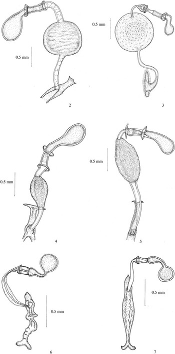
Spermathecae in Scutelleridae. 2. Elvisura irrorata Spinola, 1837 (Elvisurinae). 3. Coleotichus breddini Schouteden, 1905 (Elvisurinae). 4. Solenosthedium bilunatum (Lefebvre, 1827) (Elvisurinae). 5. Solenosthedium liligerum (Thunberg, 1783) (Elvisurinae). 6. Eurygaster austriaca (Schrank, 1776) (Eurygastrinae). 7. Eurygaster intergriceps Puton, 1881 (Eurygastrinae).

Receptacle spherical, sclerotised, connected by a short neck; intermediate part long, with slight constriction in the middle length delimited by two well-developed flanges, the proximal one more expanded; flexible zone short, located in medial and basal part; duct with large, spherical dilation with a strongly pigmented central core; distal and proximal duct almost equal in length with wide diameter; opening simple near Y-shaped, sclerotised area in vaginal wall.
Coleotichus breddini Schouteden, 1905 ( Fig. 3 )
Receptacle spherical, strongly sclerotised connected by a bent neck; intermediate part long, straight, cup-like in basal part, delimited by two, well-developed flanges; flexible zone short, located in basal part; duct dilation large, spherical, strongly sclerotised and pigmented; distal duct short, narrow, the proximal one longer, becoming wider and sclerotised at base; opening simple near ovoid, sclerotised area in vaginal wall.
Solenosthedium liligerum (Thunberg, 1783) ( Fig. 4 );
Solenosthedium bilunatum (Lefebvre, 1827) ( Fig. 5 )
Receptacle long, sclerotised, connected by a neck; intermediate part straight, delimited by two well-developed flanges; flexible zone short, located in basal position; duct dilation elongate, pear-shaped with striated intima; distal duct shorter and wider than proximal; opening simple near ring sclerite and long, sclerotised area in vaginal wall.
Eurygastrinae Amyot and Serville, 1843
Eurygaster austriaca (Schrank, 1776) ( Fig. 6 );
Eurygaster intergriceps Puton, 1881 ( Fig. 7 )
Receptacle sclerotised, spherical, connected by a long, more or less bent neck, cup-like at the base; intermediate part short and straight, proximal flange small, distal flange reduced; flexible zone long occupying almost the whole length; spermathecal duct thin-walled, relatively short, little enlarged in the medial part; opening simple near ring sclerite and long, irregularly sclerotised area in vaginal wall.
Psacasta exanthematica cerinthe (Fabricius, 1787) ( Fig. 8 );
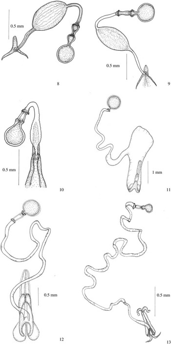
Spermathecae in Scutelleridae. 8. Psacasta exanthematica cerinthe (Fabricius, 1787) (Eurygastrinae). 9. Psacasta exanthematica conspersa Germar, 1839 (Eurygastrinae). 10. Xerobia sculpturata (Stål, 1858) (Eurygastrinae). 11. Hotea acuta Stål, 1865 (Hoteinae). 12. Deroplax coriacea Schouteden, 1965 (Hoteinae). 13. Deroplax silphoides (Thunberg, 1783) (Hoteinae).

Psacasta exanthematica conspersa Germar, 1839 ( Fig. 9 )
Receptacle sclerotised, spherical, connected by a short neck, cup-like at the base; intermediate part straight, proximal flange small, distal flange reduced; flexible zone long, occupying almost the whole length of intermediate part; duct dilation elongate, pear-shaped with striated intima; distal and proximal duct thin-walled, almost equal in length, narrow; opening simple near triangular sclerite and V-shaped, sclerotised area in vaginal wall.
Xerobia sculpturata (Stål, 1858) ( Fig. 10 )
Receptacle slightly sclerotised, spherical; intermediate part straight and short; flexible zone long, occupying almost the whole length of intermediate part; spermathecal duct short, with slightly enlarged thin-walled dilation; distal duct longer than the proximal; opening simple near ring sclerite and V-shaped, sclerotised area in vaginal wall.
Hoteinae Carapezza, Reference Carapezza2009
Deroplax coriacea Schouteden, 1965 ( Fig. 12 );
Deroplax silphoides (Thunberg, 1783) ( Fig. 13 )
Receptacle weakly sclerotised, spherical, without neck; intermediate part straight; proximal flange well developed, distal flange reduced; flexible zone long, occupying almost the whole length of intermediate part; duct dilation absent; spermathecal duct very long, narrow, thin-walled, irregularly coiled; opening near ring V-shaped sclerite and, longitudinal, sclerotised area in vaginal wall.
Hotea acuta Stål, 1865 ( Fig. 11 );
Hotea subfasciata (Westwood, 1837) ( Fig. 14 )
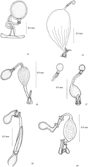
Spermathecae in Scutelleridae. 14. Hotea subfasciata (Westwood, 1837) (Hoteinae). 15. Irochrotus montandoni Schouteden, 1903 (Odontoscelinae). 16. Odontoscelis byrrhus Seidenstücker, 1972 (Odontoscelinae). 17. Odontoscelis dorsalis (Fabricius, 1798) (Odontoscelinae). 18. Rhinolaetia overlaeti Schouteden, 1965 (Odontoscelinae). 19. Morbora hirtula Bergroth, 1904 (Odontotarsinae).

The examined Hotea Amyot and Serville, 1843 species are considerably different, and therefore are described separately:
Hotea acuta Receptacle sclerotised, spherical; intermediate part short, indistinct, only the distal flange present; spermathecal duct thin-walled, distally narrow, becoming wider and sac-like at base; opening simple near V-shaped sclerite and longitudinal sclerotised area in vaginal wall.
Hotea subfasciata Receptacle sclerotised, spherical; intermediate part short, indistinct, only the proximal flange present; spermathecal duct simple, short, dilation absent; opening simple near long, T-shaped, sclerotised area in vaginal wall.
Odontoscelinae Amyot and Serville, 1843
Irochrotus montandoni Schouteden, 1903 ( Fig. 15 )
Receptacle small, spherical, connected by a short, bent neck; intermediate part straight, flexible zone long, occupying almost the whole length of intermediate part; only the proximal flange present, distal flange reduced; spermathecal duct with large, membranous, sac-like dilation; distal duct slightly longer than the proximal; opening simple near ring sclerite and V-shaped, sclerotised area in vaginal wall.
Odontoscelis byrrhus Seidenstücker, 1972 ( Fig. 16 );
Odontoscelis dorsalis (Fabricius, 1798) ( Fig. 17 )
Receptacle slightly sclerotised, spherical; intermediate part short, both proximal and distal flange reduced; flexible zone short, indistinct; spermathecal duct dilation small, elongate, pear-shaped with striated intima; distal and proximal duct thin-walled, almost equal in length, or the distal duct longer than the proximal; opening simple near ring sclerite and V-shaped, sclerotised area in vaginal wall.
Rhinolaetia overlaeti Schouteden, 1965 ( Fig. 18 )
Receptacle sclerotised, spherical, connected by a short neck; intermediate part short, delimited by two flanges, the proximal flange more expanded than the distal one; flexible zone short, occupying almost the whole length of intermediate part; spermathecal duct simple, thin-walled, becoming wider at base; opening simple near ring sclerite and longitudinal, V-shaped, sclerotised area in vaginal wall.
Odontotarsinae Mulsant and Rey, 1865
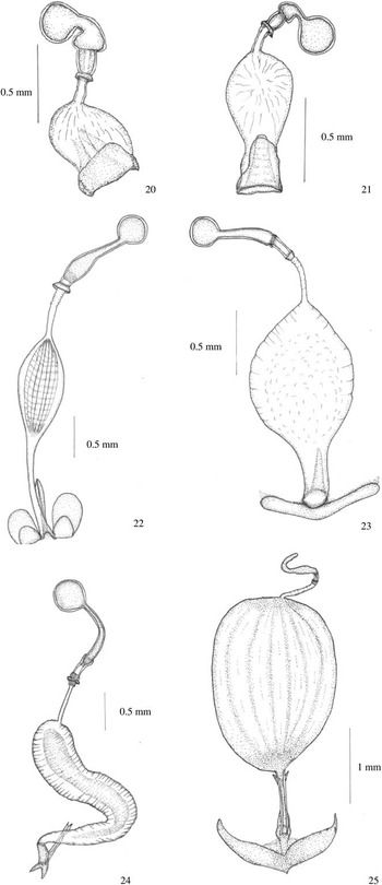
Alphocoris indutus Stål, 1864 ( Fig. 20 );
Spermathecae in Scutelleridae. 20. Alphocoris indutus Stål, 1864 (Odontotarsinae). 21. Alphocoris lobulatus Stål, 1873 (Odontotarsinae). 22. Odontotarsus purpureolineatus (Rossi, 1790) (Odontotarsinae). 23. Phimodera humeralis (Dalman, 1823) (Odontotarsinae). 24. Agonosoma trilineatum (Fabricius, 1781) (Pachycorinae). 25. Ascanius hirtipes (Herrich-Schaefer, 1835) (Pachycorinae).
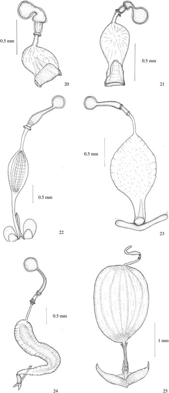
Alphocoris lobulatus Stål, 1873 ( Fig. 21 )
Receptacle sclerotised, distally spherical, connected by a short, bent neck, cup-like at the base; intermediate part straight, short, proximal flange small, distal flange, reduced; flexible zone short, occupying almost the whole length of intermediate part; spermathecal duct thin-walled with saccular dilation in the basal part; distal duct short and narrow; opening wide, near triangular sclerite and V-shaped, sclerotised area in vaginal wall.
Morbora hirtula Bergroth, 1904 ( Fig. 19 )
Receptacle sclerotised, distally spherical, connected by a bent neck; intermediate part straight, delimited by two flanges; flexible zone long, occupying almost the whole length of intermediate part; spermathecal duct with elongate, semi-hyaline dilation; distal and proximal duct short and narrow, almost equal in length; opening simple, near ring sclerite and V-shaped, sclerotised area in vaginal wall.
Odontotarsus purpureolineatus (Rossi, 1790) ( Fig. 22 )
Receptacle sclerotised, distally spherical, connected by a long straight neck, cup-like at the base; intermediate part short, proximal flange well developed, distal one reduced; flexible zone occupying almost the whole length of intermediate part; duct dilation elongate, pear-shaped with striated intima; distal and proximal duct thin-walled, narrow; distal longer than the proximal; opening simple, near semicircular sclerite and long sclerotised area in vaginal wall.
Phimodera humeralis (Dalman, 1823) ( Fig. 23 )
Receptacle sclerotised, distally spherical, connected by a long, straight neck; intermediate part straight, short, proximal flange present, distal flange reduced; flexible zone occupying almost the whole length of intermediate part; spermathecal duct with large, elongate, semi-hyaline, pear-shaped dilation; distal and proximal duct thin-walled, distal duct wide at the base; opening near semicircular sclerite and long sclerotised area in vaginal wall.
Pachycorinae Amyot and Serville, 1843
Agonosoma trilineatum (Fabricius, 1781) ( Fig. 24 )
Receptacle sclerotised, distally spherical, connected by a long, slightly bent neck; intermediate part short, distal flange reduced, proximal flange small; flexible zone long, located in medial part; duct dilation elongate, saccular, gradually narrowing towards the base; distal duct short and narrow; opening with elongate sclerite and long Y-shaped, sclerotised area in vaginal wall.
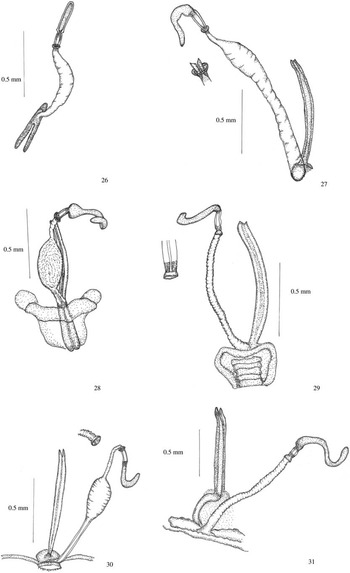
Acantholomidea porosa (Germar, 1839) ( Fig. 26 )
Spermathecae in Scutelleridae. 26. Acantholomidea porosa (Germar, 1839) (Pachycorinae). 27. Camirus conicus (Germar, 1839) (Pachycorinae). 28. Diolcus irroratus (Fabricius, 1775) (Pachycorinae). 29. Homaemus aeneifrons (Say, 1824) (Pachycorinae). 30. Homaemus proteus (Stål, 1862) (Pachycorinae). 31. Sphyrocoris obliquus (Germar, 1839) (Pachycorinae).

Receptacle slightly sclerotised, elongate; intermediate part short, distal flange reduced, proximal flange small; flexible zone long, located in medial part; duct simple with slightly visible dilation in medial part, gradually narrowing towards the base; opening simple, with elongate, V-shaped, sclerotised area in vaginal wall.
Ascanius hirtipes (Herrich-Schaefer, 1835) ( Fig. 25 )
Receptacle sclerotised, elongate and coiled, directly connected to intermediate part; distal flange absent; flexible zone short delimited by proximal flange; duct dilation large, spherical, thin-walled; distal duct short and narrow, the proximal one longer and wider; opening simple, near long Y-shaped, sclerotised area in vaginal wall.
Camirus conicus (Germar, 1839) ( Fig. 27 )
Receptacle slightly sclerotised, elongate and strongly bent, directly connected to intermediate part; distal flange absent; intermediate part moderately long with flexible zone occupying almost the whole length; proximal flange present, indistinctive; spermathecal duct simple, thin-walled, of wide diameter; opening simple, near long sclerotised area in vaginal wall.
Diolcus irroratus (Fabricius, 1775) ( Fig. 28 )
Receptacle slightly sclerotised, elongate and strongly bent, extended at the base; intermediate part short with flexible zone occupying almost the whole length, the distal flange reduced; proximal flange present; duct dilation membranous, small, pear-like; distal duct short and narrow, the proximal one short; opening near long sclerotised area in vaginal wall.
Homaemus aeneifrons (Say, 1824) ( Fig. 29 );
Homaemus proteus (Stål, 1862) ( Fig. 30 )
Receptacle sclerotised, elongate and bent; intermediate part short, flexible zone short with flexible zone occupying almost the whole length delimited by proximal flange; distal flange reduced; spermathecal duct simple or with small, thin walled dilation; distal duct narrow, the proximal one longer; opening simple, near oval sclerite and long sclerotised area in vaginal wall.
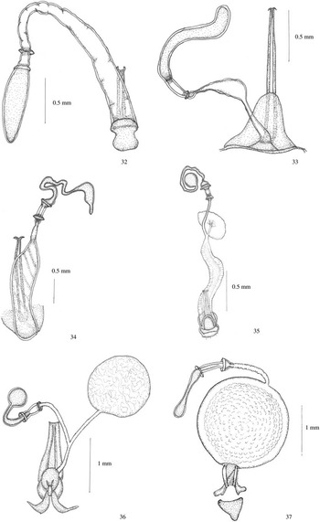
Missipus variabilis (Spinola, 1852) ( Fig. 32 )
Spermathecae in Scutelleridae. 32. Missipus variabilis (Spinola, 1852) (Pachycorinae). 33. Pachycoris torridus (Scopoli, 1772) (Pachycorinae). 34. Polytes obscurus (Dallas, 1851) (Pachycorinae). 35. Symphylus ramivitta Walker, 1868 (Pachycorinae). 36. Tetyra pinguis Germar, 1837 (Pachycorinae). 37. Anoplogonius nigricollis (Signoret, 1858) (Scutellerinae).
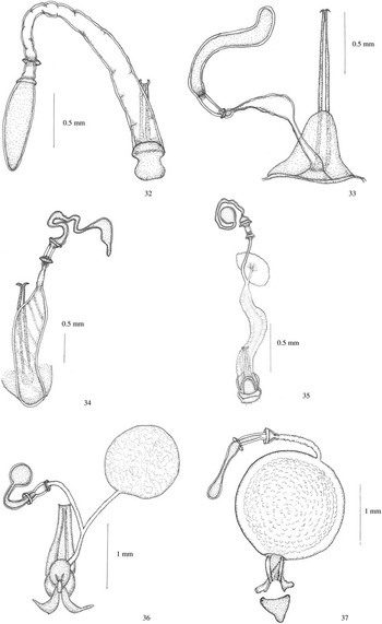
Receptacle sclerotised, elongate; intermediate part short, delimited by two flanges; flexible zone occupying almost the whole length; spermathecal duct simple, thin walled, becoming progressively wider at base; opening simple, near semicircular sclerite and long sclerotised area in vaginal wall.
Pachycoris torridus (Scopoli, 1772) ( Fig. 33 )
Receptacle long, sclerotised and bent; intermediate part delimited by the proximal flange with flexible zone long occupying almost the whole length; distal flange reduced; spermathecal duct simple, thin walled, with small dilation in the middle length; distal duct short, the proximal longer and wider; opening simple, near triangular sclerite and long sclerotised area in vaginal wall.
Polytes obscurus (Dallas, 1851) ( Fig. 34 )
Receptacle sclerotised, elongate, irregularly curved; intermediate part short, delimited by two flanges; flexible zone occupying almost the whole length of intermediate part; spermathecal duct simple, thin walled, distally narrow, becoming progressively wider at base; opening simple, wide, near semicircular sclerite and long sclerotised area in vaginal wall.
Sphyrocoris obliquus (Germar, 1839) ( Fig. 31 )
Receptacle sclerotised, elongate and bent; intermediate part delimited by the proximal flange with flexible zone long occupying almost the whole length; distal flange reduced; spermathecal duct simple, thin walled, narrow; opening connected with near oblong sclerite and long sclerotised area in vaginal wall.
Symphylus ramivitta Walker, 1868 ( Fig. 35 )
Receptacle sclerotised, distally spherical, connected by a long bent neck; intermediate part short, delimited by two flanges; flexible zone long, occupying almost the whole length of intermediate part; spermathecal duct simple, becoming progressively wider at base with saccular diverticulum connected to spermathecal duct in the middle length; opening with semicircular sclerite and long V-shaped, sclerotised area in vaginal wall.
Tetyra pinguis Germar, 1837 ( Fig. 36 )
Receptacle sclerotised, distally spherical, connected by a long, bent neck; intermediate part short, slightly constricted in the middle length delimited by two flanges; flexible zone long, located in medial part; spermathecal duct simple and narrow with saccular, spherical diverticulum connected to spermathecal duct in the middle length; opening with semicircular sclerite and long V-shaped, sclerotised area in vaginal wall.
Scutellerinae Leach, 1815
Scutellerini Leach, 1815
Anoplogonius nigricollis (Signoret, 1858) ( Fig. 37 )
Receptacle elongate distally spherical, connected by a long neck, slightly constricted in the middle length; intermediate part moderately long, delimited by two well-developed flanges, cup-like basally; flexible zone short, located in basal part; spermathecal duct with spherical dilation with a strongly pigmented central core; distal duct longer than the proximal, the proximal one wide, and short; opening simple near triangular, sclerotised area in vaginal wall.
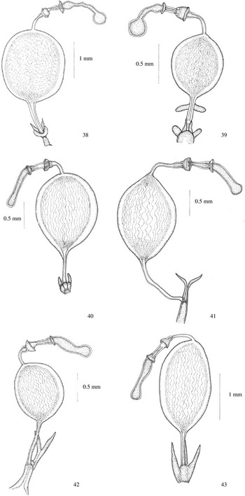
Augocoris gomesi Burmeister, 1835 ( Fig. 38 );
Spermathecae in Scutelleridae. 38. Augocoris gomesi Burmesister, 1835 (Scutellerinae). 39. Augocoris sexpunctatus (Fabricius, 1781) (Scutellerinae). 40. Calidea bohemani (Stål, 1853) (Scutellerinae). 41. Calidea dregii Germar, 1837 (Scutellerinae). 42. Calidea madagascariensis Signoret, 1861 (Scutellerinae). 43. Calidea panethiopica Kirkaldy, 1909 (Scutellerinae).

Augocoris sexpunctatus (Fabricius, 1781) ( Fig. 39 )
Receptacle sclerotised, distally spherical, connected by a long neck, ovoid basally; intermediate part short, delimited by two well-developed flanges; flexible zone short located basally; dilation of spermathecal duct sclerotised, strongly pigmented; distal duct slightly longer than the proximal; opening near semicircular and elongate sclerotised area in vaginal wall.
Calidea bohemani (Stål, 1853) ( Fig. 40 );
Calidea dregii Germar, 1837 ( Fig. 41 );
Calidea madagascariensis Signoret, 1861 ( Fig. 42 );
Calidea panethiopica Kirkaldy, 1909 ( Fig. 43 )
Receptacle sclerotised, elongate, distally spherical, connected by a long neck; intermediate part delimited by two well-developed flanges, cup-like at the base; flexible zone short, located basally; spermathecal duct with sclerotised, spherical or ovoid dilation with a strongly pigmented central core; distal duct longer than the proximal; opening simple nearly elongate approximately V-shaped sclerotised area in vaginal wall.
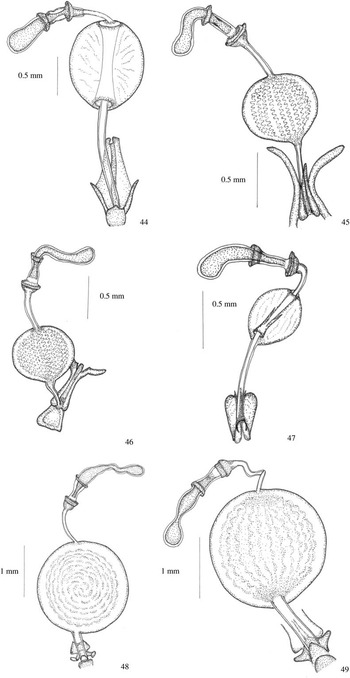
Calliphara nobilis (Linnaeus, 1763) ( Fig. 44 )
Spermathecae in Scutelleridae. 44. Calliphara nobilis (Linnaeus, 1763) (Scutellerinae). 45. Choerocoris variegatus Dallas, 1851 (Scutellerinae). 46. Choerocoris paganus (Fabricius, 1775) (Scutellerinae). 47. Chrysocoris abdominalis (Westwood, 1837) (Scutellerinae). 48. Cryptacrus comes (Fabricius, 1803) (Scutellerinae). 49. Cryptacrus novemmaculatus (Signoret, 1851) (Scutellerinae).

Receptacle sclerotised, distally spherical, connected by moderately short neck; intermediate part short, delimited by well-developed flanges, cup-like at the base; flexible zone short in basal part; dilation ovoid, semi-hyaline with clearly visible of invaginated part of the spermathecal duct; distal duct longer than the proximal; opening simple near elongate, V-shaped sclerotised area in vaginal wall.
Choerocoris variegatus Dallas, 1851 ( Fig. 45 );
Choerocoris paganus (Fabricius, 1775) ( Fig. 46 )
Receptacle sclerotised, elongate, distally spherical, connected by bent neck; intermediate part delimited by two well-developed flanges, cup-like at the base; flexible zone short located in basal part; spermathecal duct with spherical, sclerotised and pigmented dilation; distal and proximal duct almost equal in length; opening simple near elongate, V-shaped sclerotised area in vaginal wall.
Chrysocoris abdominalis (Westwood, 1837) ( Fig. 47 )
Receptacle sclerotised, moderately short, distally spherical; intermediate part delimited by two well-developed flanges, cup-like at the base; flexible zone short, located basally; duct with spherical semi-hyaline dilation, an invaginated part of the spermathecal duct clearly visible; distal duct longer than the proximal; opening simple near long, semicircular sclerotised area in vaginal wall.
Cryptacrus comes (Fabricius, 1803) ( Fig. 48 );
Cryptacrus novemmaculatus (Signoret, 1851) ( Fig. 49 )
Receptacle sclerotised, elongate, distally spherical, connected by long neck; intermediate part pigmented, constricted in the middle length, delimited by two flanges, cup-like at the base; flexible zone short, located basally; duct with large, spherical dilation with a strongly pigmented central core; distal duct narrow, longer than the proximal, the proximal one of wider diameter; opening simple near long, U-shaped sclerotised area in vaginal wall.
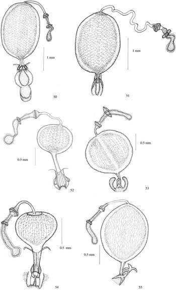
Gonaulax grandis (Distant, 1899) ( Fig. 50 );
Spermathecae in Scutelleridae. 50. Gonaulax grandis (Distant, 1899) (Scutellerinae). 51. Gonaulax leroyi Schouteden, 1937 (Scutellerinae). 52. Graptocoris aulicus (Germar, 1837) (Scutellerinae). 53. Lamprocoris lateralis (Guérin-Méneville, 1838) (Scutellerinae). 54. Scutiphora pedicellata (Kirby, 1826) (Scutellerinae). 55. Lampromicra senator (Fabricius, 1803) (Scutellerinae).
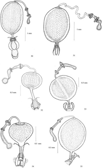
Gonaulax leroyi Schouteden, 1937 ( Fig. 51 )
Receptacle sclerotised, elongate, distally ovoid, connected by long, bent neck; intermediate part constricted in the middle length, delimited by two well-developed flanges, cup-like at the base; flexible zone in the middle length; duct with large, ovoid dilation with a strongly pigmented central core; distal duct narrow and coiled, much longer than the proximal, the proximal one short with wider diameter; opening simple near semicircular, sclerotised area in vaginal wall.
Graptocoris aulicus (Germar, 1837) ( Fig. 52 )
Receptacle sclerotised, spherical distally, connected by long, bent neck; intermediate part long, cup-like at the base, delimited by two well-developed flanges; flexible zone short, in basal part; duct with large, spherical dilation with a strongly pigmented central core; distal and proximal duct almost equal in length, the proximal one shorter with wider diameter; opening simple near U-shaped sclerotised area in vaginal wall.
Lamprocoris lateralis (Guérin-Méneville, 1838) ( Fig. 53 )
Receptacle sclerotised, elongate, distally spherical, connected by long neck; intermediate part long, pigmented, cup-like at the base, delimited by two well-developed flanges, the distal flange more expanded; flexible zone short, basally located; duct with large, spherical, sclerotised dilation; proximal and distal duct short; opening simple near semicircular, sclerotised area in vaginal wall.
Lampromicra senator (Fabricius, 1803) ( Fig. 55 )
Receptacle sclerotised, elongate, distally spherical, connected by long neck; intermediate part long, pigmented, cup-like at the base, delimited by two well-developed flanges; flexible zone short, basally located; duct with large, spherical, sclerotised dilation; distal duct short longer than the proximal; opening simple near Y-shaped, sclerotised area in vaginal wall.
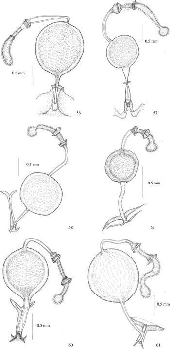
Poecilocoris drurarei (Linnaeus, 1771) ( Fig. 56 )
Spermathecae in Scutelleridae. 56. Poecolocoris drurarei (Linnaeus, 1771) (Scutellerinae). 57. Procilia morgani (White, 1839) (Scutellerinae). 58. Scutellera amethystina (Germar, 1839) (Scutellerinae). 59. Tetrarthia variegata Dallas, 1851 (Scutellerinae). 60. Chiastosternum unicolor (Dallas, 1851) (Scutellerinae). 61. Sphaerocoris annulus (Fabricius, 1775) (Scutellerinae).

Receptacle sclerotised, elongate, distally spherical, connected by long neck; intermediate part straight, long, distinctly cup-like at the base; proximal flange small, the distal flange reduced to small ring; flexible zone short, located in basal part; spermathecal duct with large, spherical dilation with a strongly pigmented central core; proximal and distal duct almost equal in length; opening simple near long, V-shaped sclerotised area in vaginal wall.
Procilia morgani (White, 1839) ( Fig. 57 )
Receptacle sclerotised, elongate, distally spherical, connected by long neck; intermediate part constricted in the middle length, distinctly cup-like at the base; proximal flange well developed, the distal flange short; flexible zone short, located in basal part; spermathecal duct with large, spherical, sclerotised and pigmented dilation; proximal and distal duct almost equal in length, distal duct with wider diameter; opening simple near long, V-shaped sclerotised area in vaginal wall.
Scutellera amethystina (Germar, 1839) ( Fig. 58 )
Receptacle sclerotised, distally spherical, connected by long neck; intermediate part straight, cup-like at the base, delimited by two well-developed flanges; the distal flange more expanded; flexible zone short, indistinct, located in basal part; duct with large, spherical dilation with a strongly pigmented central core; proximal and distal duct almost equal in length and diameter; opening simple near long, Y-shaped sclerotised area in vaginal wall.
Scutiphora pedicellata (Kirby, 1826) ( Fig. 54 )
Receptacle sclerotised, distally spherical, connected by long, slightly bent neck; intermediate part straight, cup-like at the base, delimited by two well-developed flanges; flexible zone short, indistinct, located in basal part; duct with large, spherical dilation with pigmented central core and striated surface; proximal and distal duct almost equal in length; distal duct narrow, proximal wide; opening simple near ring sclerite and long, Y-shaped sclerotised area in vaginal wall.
Tetrarthia variegata Dallas, 1851 ( Fig. 59 )
Receptacle sclerotised, distally spherical, connected by short neck; intermediate part short, pigmented, cup-like at the base, delimited by two well-developed flanges; flexible zone short located in basal part; duct with large, spherical, sclerotised dilation; proximal and distal duct almost equal in length; opening simple near long, V-shaped sclerotised area in vaginal wall.
Sphaerocorini Stål, 1873
Chiastosternum unicolor (Dallas, 1851) ( Fig. 60 )
Receptacle sclerotised, distally spherical, connected by long, slightly bent neck; intermediate part long, pigmented, cup-like at the base, delimited by two well-developed flanges; flexible zone short, located in basal part; duct with large, spherical, strongly pigmented and sclerotised dilation; proximal and distal duct almost equal in length; proximal duct with wider diameter; opening simple near long, Y-shaped sclerotised area in vaginal wall.
Sphaerocoris annulus (Fabricius, 1775) ( Fig. 61 )
Receptacle sclerotised, elongate, distally spherical, connected by long, bent neck; intermediate part, cup-like at the base, delimited by two well-developed flanges; flexible zone short, located in basal part; duct with large, spherical dilation with a strongly pigmented central core; proximal and distal duct narrow, almost equal in length; opening simple near long, V-shaped sclerotised area in vaginal wall.
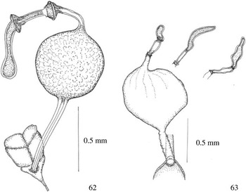
Steganocerus multipunctatus (Thunberg, 1783) ( Fig. 62 )
Spermathecae in Scutelleridae. 62. Steganocerus multipunctatus (Thunberg, 1873) (Scutellerinae). 63. Tectocoris diopthalmus (Thunberg, 1873) (Tectocorinae).

Receptacle sclerotised, elongate, distally spherical, connected by long, neck; intermediate part long, sclerotised, cup-like at the base, delimited by two well-developed flanges; flexible zone short, located in basal part; spermathecal duct with large, spherical dilation with a strongly pigmented central core; proximal and distal duct almost equal in length; opening simple nearly triangular, sclerotised area in vaginal wall.
Tectocorinae McDonald and Cassis, Reference McDonald and Cassis1984
Tectocoris diopthalmus (Thunberg, 1873) ( Fig. 63 )
Receptacle slightly sclerotised, elongate, directly connected to intermediate part, without neck; only the indistinct proximal flange present; distal flange reduced; intermediate part relatively long, flexible zone taking up nearly whole length of intermediate part; spermathecal duct with large, thin-walled, saccular dilation; proximal duct with wide diameter, distal duct shorter, narrow; opening simple near long, V-shaped sclerotised area in vaginal wall.
Discussion
The results presented above show a wide range of spermathecal structures in Scutelleridae. The differences are clearly visible at the suprageneric level and often between separate species.
The primitive (plesiomorphic) pentatomoid spermatheca consists of an apical spherical receptacle; intermediate part delimited by flanges (at least the proximal one) and a simple duct (Gapud Reference Gapud1991; Schaefer Reference Schaefer1993; Pluot-Sigwalt and Lis Reference Pluot-Sigwalt and Lis2008). All examined scutellerid species have spermathecal structures built on this pentatomoidean ground plan (well-developed, more-or-less sclerotised apical receptaculum; intermediate with flanges and flexible zone and spermathecal duct). Based on these plesiomorphic features, several stages of differentiation of the spermathecal structures are observed within the Scutelleridae: from simple spermatheca, similar to the hypothetical plesiomorphic pattern, with globose receptaculum and duct without dilation to the most advanced stage with well developed and complex dilation of the spermathecal duct.
Three types of spermatheca can be distinguished based on the morphological features.
Type I (Figs. 11–19); taxa included: Hoteinae, Odontoscelinae
General structure: spherical receptaculum without neck, distal flange reduced, short intermediate part, spermathecal duct without highly sclerotised complex dilation. This type of spermathecal construction may be regarded as the most primative within the Scutelleridae. Both the shape of the receptaculum (spherical), the short intermediate part and the mostly simple spermathecal duct represent features common with the ground plan model of pentatomoid spermatheca.
Type II (Figs. 6–10, 20–36, 63); taxa included: Eurygastrinae, Odontotarsinae, Pachycorinae, Tectocorinae
General structure: distal flange reduced, intermediate part with long flexible zone, spermathecal duct without highly sclerotised, complex dilation. The spermathecal structures of the species represented by this type are the most differentiated. The common feature of the spermathecae belonging to this type is a relatively short intermediate part with a long flexible zone and an absent or reduced distal flange. The most diverse structure is the spermathecal duct, which is simple without any dilation (most representatives of Pachycorinae), with weakly developed membranous dilation (several species of Eurygastrinae and Odontotarsiane), or with large spherical dilation but weakly sclerotised (Tectocorinae). The state of some characteristics here is difficult to evaluate. The reduction of the distal flange and the simple spermathecal duct may be treated as a plesiomorphy (see the hypothetical model of pentatomoid spermatheca), but on the other hand such simplification may be connected with the regression of some part of the spermatheca, making the plesiomorphy difficult to confirm. Such a situation is observed in Cydnidae (Pluot-Sigwalt and Lis Reference Pluot-Sigwalt and Lis2008), where an extremely simple spermatheca is described (Amaurocoris curtus (Brullé, 1838)), and may be an example of regression. Representatives of the Pachycorinae are included in this type (reduction of the distal flange and long flexible zone) but differ from the rest by possessing an elongate receptaculum without a clearly globose apex (as occurs in the rest of the taxon in this group). This feature is also difficult to evaluate. Compared with the primitive pattern (a globose receptaculum without a neck), it may also represent a more advanced state.
Type III (Figs. 2–5, 37–62); taxa included: Elvisurinae, Scutellerinae
General structure: both proximal and distal flange present, intermediate part with short flexible zone, spermathecal duct with highly sclerotised complex dilation. The type III spermatheca observed in species of the Scutellerinae and Elvisurinae subfamilies represents the most advanced state. A well-developed intermediate part with flanges (proximal and distant), a flexible zone located in the basal position, and distinctly visible spermathecal dilation may be regarded as the synapomorphic features within the family. Spermathecal dilation of Scutellerinae and Elvisurinae is complex and heterogeneous in cross-section; several types can be observed (Tsai et al. Reference Tsai, Rédei, Yeh and Yang2011), but it differs from the spermathecal dilation observed in other scutellerid taxa where it is usually thin-walled, membranous, without the complex of internal structures. Dilation of the spermathecal duct is also observed in many families of Pentatomoidea, i.e., Cydnidae, Pentatomidae, and Thyreocoridae, and shows great diversity. In such a situation it is difficult to estimate the apomorphic or plesiomorphic state of these features and the most possible explanation here may be convergence.
A very important issue for the subfamilial and tribal classification system within the Scutelleridae is fact that the representatives of the Sphaerocorinae and Scutellerinae share apomorphic features (cup-like dilation near the proximal flange). The position of the Sphaerocorinae is still unclear in the classification system within the Scutelleridae; it is included in the Scutellerinae as the Sphaerocorini or treated as a separate subfamily (Göllner-Scheiding Reference Göllner-Scheiding2006). The common spermathecal structures of these two taxa may be the next reason to include this taxon on the tribal level in the Scutellerinae. Generally, the uniform character of spermathecal features may emphasise the monophyletic character of the Scutellerinae.
The next unresolved issue is definitely the relation of the subfamilies Pachycorinae and Hoteinae. The last one was established for the African representatives of the Pachycorinae. Based on the spermathecal structures, it can be seen that the subfamilies Pachycorinae and Tectorinae are more closely related than the Pachycorinae and Hoteinae. Species of Pachycorinae are characterised by deep differences of spermathecal structures. This suggests the polyphyletic character of this taxon. A wider taxonomic survey within this group is necessary.
Conclusions
The results of this study confirm the great diversity of spermathecal structures in Scutelleridae. The analysis presented above, based on the morphological characteristics of spermathecae give a general view of the relationships within the family, but it is impossible to characterise Scutelleridae by considering the spermathecal features only. The evolution of this organ seems to be much more complicated and must be supported by wider studies in various families of Pentatomoidea to define and evaluate the phylogenetic importance of spermathecal characteristics in the classification system.
Acknowledgements
The author is grateful to Professor Jerzy Lis and Dr. Andrzej Wolski (Opole University) for their suggestions and comments on this manuscript. Many thanks are due to the curators of entomological collections for the loan of the material for this study. The linguistic correction of this manuscript was done by the Atominium Specialist Translation Agency.