Complex CHD has become significantly more manageable in developed countries in the last two decades due, in part, to improved surgical methods and the use of prosthetic materials for cardiac conduits and valves. Reference Knirsch and Nadal1 Currently, the leading predisposing factor for infective endocarditis in developed countries is CHD. Reference Johnson, Boyce and Cetta2 The incidence of infective endocarditis in paediatrics is considerably lower than in the adults (0.34–0.64 cases per 100,000 persons per year versus 1.5–6 cases per 100,000 persons per year, respectively). Reference Johnson, Boyce and Cetta2 CHD is present in 35–77% of affected children. Reference Johnson, Boyce and Cetta2,Reference Sun, Lai and Wang3
Diagnosing prosthetic valve infective endocarditis reliably has proven to be challenging. First-line imaging modalities include transthoracic and transesophageal echocardiography. However, these may yield false-negative results. In 2015, the European Society of Cardiology suggested that the sensitivity of the Duke criteria can be improved with the use of advanced cardiovascular imaging modalities such as cardiac MRI, ECG-gated computed tomography angiography (CTA), or 18F-fluorodeoxyglucose positron emission tomography in combination with CT (18FDG-PET/CT) or CT angiography (18FDG-PET/CTA). Reference Habib, Lancellotti and Antunes4 However, there are currently insufficient data, particularly in children, regarding these imaging modalities to make general recommendations. Reference Habib, Lancellotti and Antunes4 We present two cases at our institution with high clinical suspicion of prosthetic valve infective endocarditis where chest CTA and 18FDG-PET/CT, respectively, were used to confirm the diagnosis where echocardiography was inconclusive.
Case reports
Case 1
An 11-year-old female born with Tetralogy of Fallot and pulmonary atresia was admitted to the hospital due to 1 week of fever with an unknown source. Her cardiac surgical history included placement of a palliative aortopulmonary shunt as a newborn, followed by complete repair involving closure of the ventricular septal defect and placement of a conduit between the right ventricle and pulmonary artery at 1 year of age. She subsequently developed severe conduit stenosis and underwent replacement of the conduit with a 16-mm bovine jugular vein conduit containing a venous valve (Contegra®, Medtronic, Minneapolis, MN, United States of America) at 3 years of age. She again developed progressive homograft stenosis and thus underwent transcatheter percutaneous implantation of a bovine jugular venous valve in the pulmonary position (Melody valve®, Medtronic, Minneapolis, MN, United States of America) in the conduit 4 years prior to admission. She was maintained on 81 mg of aspirin daily as anti-thrombotic therapy. Given her fever and cardiac history, prosthetic valve infective endocarditis was suspected. Serial transthoracic echocardiogram did not reveal intracardiac vegetations but did suggest increased stenosis across the valve of the right ventricle to pulmonary artery conduit. Blood cultures drawn upon admission promptly grew methicillin-sensitive Staphylococcus aureus. The diagnosis of prosthetic valve infective endocarditis was, therefore, presumed and she was started on a continuous oxacillin infusion as well as gentamicin and rifampin. Her clinical course was complicated by significant fluid overload with low albumin, ascites, pleural effusion, and sepsis. Transesophageal echocardiogram under sedation was deferred due to clinical instability and it was deemed that the risk of sedation outweighed additional clinical information that might have been gleaned.
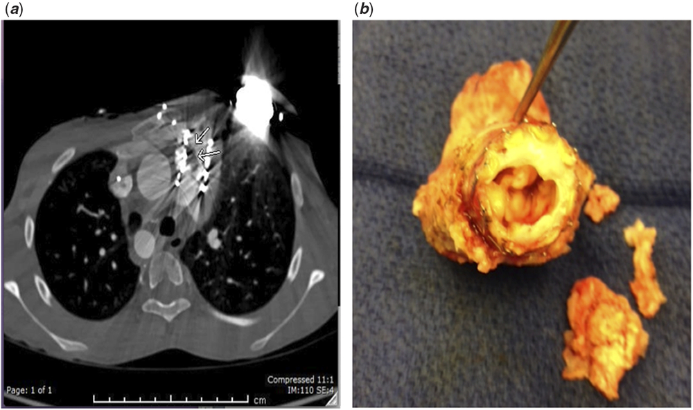
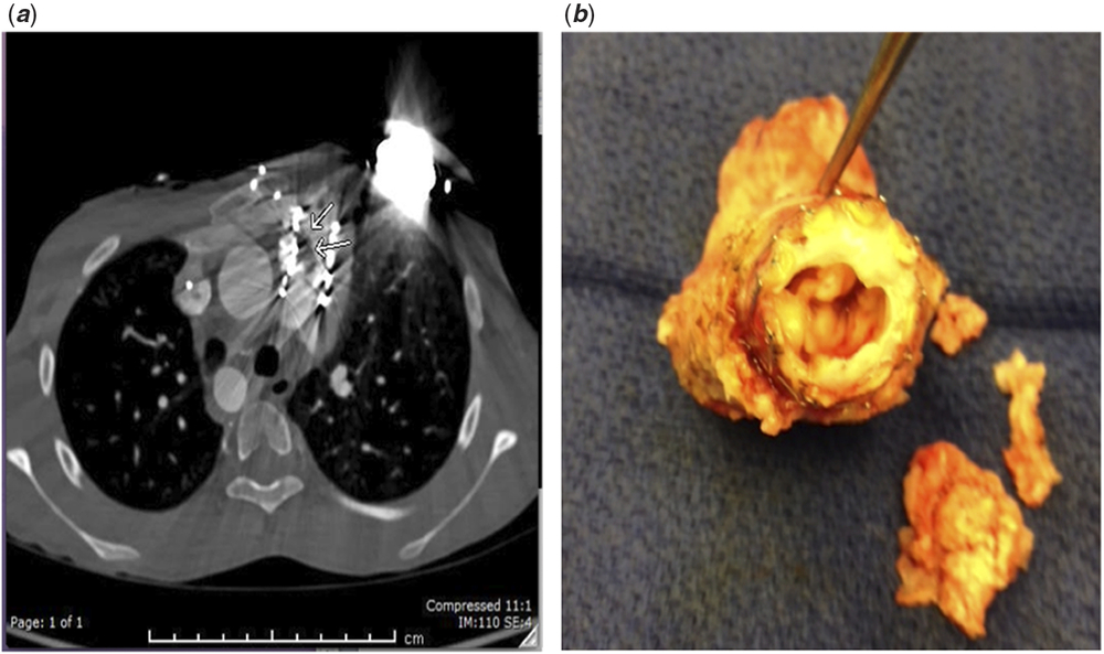
Despite intravenous antibiotic therapy, she had persistently positive blood cultures 6 days into her hospital course. A chest CTA was obtained which revealed a suspected thrombus versus vegetation involving the implanted conduit valve (Fig 1). The patient thus underwent surgical removal of the Melody valve and the Contegra® graft. A large vegetation corresponding to the lesion seen on CTA was confirmed at surgery (Fig 1). A pulmonary homograft was then used as the new right ventricle to pulmonary artery conduit. The patient recovered well after surgery.
( a ) Case 1 – CTA showing a filling defect along the superior and right side of the valve measuring 1.6 × 0.7 cm. ( b ) Case 1 – Image of the Melody valve® with vegetation removed during surgery. CTA=CT angiogram.
Case 2
A 10-year-old male with double outlet right ventricle and d-malposition of the great vessels was hospitalised for intermittent fever lasting 6 weeks. His cardiac history included arterial switch operation, closure of the ventricular septal defect, and placement of a right ventricle to pulmonary artery conduit. He subsequently underwent replacement of the conduit with a GoreTex® conduit (W. L. Gore & Associates, Newark, DE, United States of America) 3 years prior to presentation.
During this admission he had four negative blood cultures, three of which were collected while he was febrile. Transthoracic echocardiogram and chest CTA were both negative for cardiac vegetations and pulmonary emboli. He became afebrile without antibiotic therapy and thus was discharged after 3 days.
He was readmitted the following month with persistent fever and new thigh pain. A MRI study of his femur was negative for abscess and osteomyelitis. A transesophageal echocardiogram was negative for vegetations but did reveal mild conduit stenosis, which was a new finding. Cardiac MRI was requested by the consulting cardiothoracic surgeon to rule out prosthetic valve vegetations and/or surrounding abscess, but was inconclusive.
Multiple serial blood cultures remained negative. An extensive work-up was, therefore, undertaken for fever of unknown origin, including oncology and rheumatology consultations. He underwent a bone marrow biopsy, which was negative. Brucella IgM titer was equivocal and therefore the blood cultures were held for a longer time period. Two of the cultures ultimately grew Gemella spp. 10 days after inoculation. Further history revealed that he had undergone a dental cleaning procedure approximately 2 weeks prior to the onset of fever.
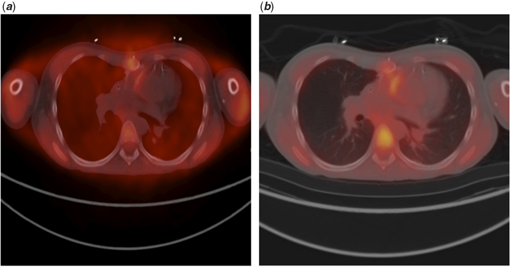
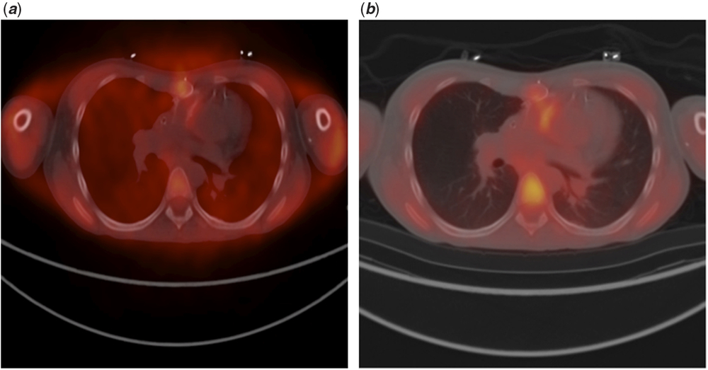
He was started on ceftriaxone and gentamicin for the presumptive diagnosis of prosthetic valve infective endocarditis. In an attempt to confirm the diagnosis, an 18FDG-PET/CT was requested. It showed mild hypermetabolism in the right ventricle to pulmonary artery conduit, suggesting prosthetic valve infective endocarditis (Fig 2). He completed 6 weeks of intravenous ceftriaxone and 2 weeks of intravenous gentamicin and recovered. He was then referred to the hospital where he underwent his last cardiac surgery for further management.
( a ) Case 2 – 18FDG-PET/CT showing mild metabolism at sternotomy, likely due to post-operative changes. Mild hypermetabolism involving the pulmonary artery to right ventricle conduit. ( b ) Case 2 – Attenuated 18FDG-PET/CT image, which corrects for areas of differing tissue density across the image, shows persistent hypermetabolism in the walls of the pulmonary artery to right ventricle conduit. The enhancement of the vertebra is due to normal bone marrow activity. 18FDG-PET/CT=18F-fluorodeoxyglucose positron emission tomography in combination with CT.
Discussion
Prosthetic valve infective endocarditis is being increasingly recognised as a complication in patients after pulmonary valve replacement that can cause significant morbidity and mortality. Reference Robichaud, Hill and Cohen5 One study found an overall incidence of infective endocarditis after pulmonary valve replacement was as high as 333 cases per 100,000 persons-years. Reference Robichaud, Hill and Cohen5 Surgical intervention was required in 53% of those cases. Reference Robichaud, Hill and Cohen5
The modified Duke criteria, currently used to diagnose infective endocarditis, include well-characterised microbiological and echocardiographic findings. However, blood cultures are negative in 23–37% in patients with proven prosthetic heart valve infective endocarditis. Reference Habets, Tanis and van Herwerden6 The sensitivity of transthoracic echocardiogram for the diagnosis of intracardiac vegetations is 70% on native valves and 50% on prosthetic valves, with a specificity of greater than >90%. Reference Meyer, Fischer and Koerfer7 When transthoracic echocardiogram is negative, consideration is given to transesophageal echocardiogram. This is a more invasive procedure that is typically performed under sedation in children and adolescents. Transesophageal echocardiogram has good sensitivity and specificity for diagnosing infective endocarditis but fails to detect life-threatening complications such as mycotic aneurysms and abscesses in up to 30% of patients. Reference Habets, Tanis and van Herwerden6 This is mainly due to acoustic shadowing by the prosthetic heart valve, which may obscure visualisation of adjacent anatomical structures. Reference Habets, Tanis and van Herwerden6 In addition, the implanted pulmonary valve may be in the far field on transesophageal imaging, which reduces the image resolution. Patients with mechanical prosthetic valves, conduits, stents, or ventricular assist devices are also challenging to assess by both echocardiographic modalities due to the potentially significant imaging artefacts caused by these devices. Reference Meyer, Fischer and Koerfer7 Given the limitations of echocardiography in this clinical scenario, both the updated 2015 guidelines on infective endocarditis by the European Cardiology Society and the American Heart Association acknowledge that alternative imaging modalities such as CTA and 18FDG-PET/CT may be useful tools that should be explored further. Reference Habib, Lancellotti and Antunes4,Reference Baddour, Taubert, Gewitz and Fuster8
The majority of the published studies regarding these alternative modes of imaging have been conducted in adults. As such, there remains a paucity of literature on the use of multimodality cardiovascular imaging in the diagnosis of prosthetic valve infective endocarditis in children. A systematic review published in 2017 found 3 studies addressing the value of CTA and 13 studies addressing the value of 18FDG-PET/CT in diagnosis of infective endocarditis in patients with prosthetic valves. Reference Barzouhi, Tanis, Gelder and Vriend9 All of these were done in an adult population and were all classified as low or very low quality according to the GRADE approach. Reference Barzouhi, Tanis, Gelder and Vriend9 These studies showed that when CTA was added to standard diagnostic work-up of suspected infective endocarditis, an overall sensitivity of 100% and specificity of 83% for prosthetic valve infective endocarditis was achieved. Reference Barzouhi, Tanis, Gelder and Vriend9 There was also a change in treatment strategy in 25% of patients with the added use of CTA. Reference Barzouhi, Tanis, Gelder and Vriend9 18FDG-PET/CT had 73–100% sensitivity for diagnosis of infective endocarditis and 71–100% specificity. Reference Barzouhi, Tanis, Gelder and Vriend9 The addition of 18F-FDG-PET/CT to the modified Duke criteria greatly increased sensitivity from 52–70% to 91–97%. Reference Barzouhi, Tanis, Gelder and Vriend9 18FDG-PET/CT also detected unexpected extracardiac complications and additional potential infectious foci in 11–24% of patients. Reference Barzouhi, Tanis, Gelder and Vriend9
To our knowledge, Meyer et al were the first investigators to publish a study of the use of 18FDG-PET/CT for the diagnosis of infective endocarditis in children with CHD. In this retrospective study, five patients with prosthetic valves were diagnosed with prosthetic valve infective endocarditis using 18FDG-PET/CT, of which three were children under 18 years old. All of these patients had an initial transthoracic echocardiogram that was negative for vegetations. The authors concluded that in children with CHD, 18FDG-PET/CT is potentially a useful and reliable tool in the diagnostic algorithm of acute infective endocarditis, to identify septic emboli and for the evaluation of ventricular assist device-related infections. Reference Meyer, Fischer and Koerfer7
Similarly, El Barzouhi et al reported a case of Streptococcus mitis endocarditis of the pulmonary homograft in an adult patient who had undergone the Ross procedure. Reference Barzouhi, Tanis, Gelder and Vriend9 Transthoracic echocardiogram and transesophageal echocardiogram did not show any signs of endocarditis. However, FDG-PET/CT and cardiac CTA showed large vegetations in the pulmonary homograft. Reference Barzouhi, Tanis, Gelder and Vriend9 The authors stated that if clinical suspicion of endocarditis is strong, one should consider additional imaging via CTA and/or 18FDG-PET/CT to assess valves in the pulmonary position, especially in patients that have had prior surgical intervention at this location. Reference Barzouhi, Tanis, Gelder and Vriend9
There are benefits and limitations to each imaging technique. Transesophageal echocardiogram does not employ ionising radiation, but typically requires sedation in the paediatric and adolescent population and, as mentioned above, the image quality may be reduced by artefacts caused by implanted metallic devices. Reference Gomes, Glaudemans and Touw10 CTA does employ ionising radiation and requires the administration of intravenous iodinated contrast, which can be contraindicated in patients with renal abnormalities or contrast allergies. However, it offers rapid acquisition of high-resolution anatomical images and is not as susceptible to metallic artefacts as compared to echocardiography. Reference Gomes, Glaudemans and Touw10 On non-contrast-enhanced CT and CTA images, intracardiac vegetations can appear as irregular, homogenous, hypodense masses on the borderline between the mobile and the fixed portion of mechanic valves or on the leaflets of bioprosthetic valves. Reference Kanza, Allard and Berube11 Areas of calcification, on the other hand, are hyperdense on CT. However, on CTA, the addition of iodine-containing contrast may make distinguishing calcium from the bright appearance of iodine more difficult. Reference Wang, Osborne, Tung, Li and Li12
18FDG-PET/CT can be prone to false-positive results given that increased glucose metabolism can be due to inflammatory as well as infectious states and normal metabolic activity (e.g. bone marrow). Examples of situations that can lead to false-positive results include early post-operative inflammation around the sewing ring of prosthetic valves and unsuccessful suppression of myocardial FDG uptake even in cases of optimal pre-imaging preparation. Reference Schindler13 Mild to moderate amount of FDG uptake around a prosthetic heart valve can also be a normal finding, likely owing to foreign body reaction. Reference Schindler13 False-negative findings may be related to the effects of antibiotic therapy. Reference Schindler13 Therefore, the specificity of 18FDG-PET/CT may be limited in the first 2 months after surgical implantation. Reference Schindler13
Considering the low incidence of paediatric infective endocarditis as compared to adults, it is not likely that a prospective clinical trial will be completed soon. Until such data are available, it is important to gather information on the ability of these studies to identify features of infective endocarditis in children, particularly in those patients with implanted prosthetic materials such as valves and conduits.
The most important limitation of our work is the retrospective nature of case reporting and the lack of a large study population. Additionally, ours represent the relatively few cases in which both transthoracic echocardiogram and transesophageal echocardiogram failed to make the diagnosis of infective endocarditis. Prospective studies in a purely paediatric population are needed to more precisely define the role of each imaging modality in this population.
In summary, the addition of advanced cardiovascular imaging to standard echocardiography in the diagnosis and management of suspected cases of paediatric prosthetic valve infective endocarditis is an evolving field of study that requires further exploration. As approximately one-half of the cases of prosthetic valve infective endocarditis require surgical intervention, it is crucial to obtain confirmatory data when this diagnosis is clinically suspected. The cases discussed here illustrate the adjunctive potential of advanced cardiovascular imaging modalities such as CTA and 18FDG-PET/CT in diagnosing prosthetic valve infective endocarditis when echocardiography is negative or inconclusive.
Acknowledgements
The authors would like to thank Dr Wyman Lai for his critical review of the manuscript.
Financial support
This research received no specific grant from any funding agency, commercial or not-for-profit sectors.
Conflict of interest
None.