Introduction
Skull base osteomyelitis is a rare, life-threatening inflammatory condition that primarily affects immunocompromised, debilitated individuals. It is usually secondary to an infective process in the ear or the paranasal sinuses. It can also be seen subsequent to a fracture of the skull base or after surgical intervention in this area.Reference Ducic1 The causative agent is most commonly bacterial in origin, but invasive fungal species have rarely been isolated.Reference Ferguson2 An insidious clinical presentation makes skull base osteomyelitis difficult to diagnose.Reference Lehrer, Howard and Sypherd3 The condition is associated with a high mortality rate. Improved outcomes depend upon early diagnosis, aggressive medical and/or surgical intervention if necessary, together with the appropriate management of any underlying disorder.Reference Ferguson2, Reference Lehrer, Howard and Sypherd3 The optimum method of investigation for diagnosis of this condition remains controversial, with various reports favouring different imaging modalities.
We present a case of a non-otogenic fungal skull base osteomyelitis in an immunocompromised patient in order to highlight the delay in diagnosis associated with the limitations of routine imaging studies. This is followed by a discussion of the relevant published literature.
Case report
A 70-year-old Asian male presented to the department of medicine with a 1-week history of acute onset headache and vomiting. His past medical history included a cadaveric renal transplant performed two years previously for end stage renal failure secondary to diabetic nephropathy (type II insulin-dependent diabetes) and idiopathic focal segmental glomerulosclerosis confirmed on renal biopsy. He also had a history of secondary hyperparathyroidism, diabetic retinopathy, ischaemic heart disease with a right coronary artery stent, and gout. His regular medications included immunosuppressant cyclosporin 50 mg twice daily and mycophenolate mofetil 500 mg twice daily.
On examination at this time, there were no focal neurological signs evident. Computed tomography (CT) of the patient's head was normal. His white cell count was 6.3 × 10⁹/l and his C-reactive protein (CRP) was 21 mg/l. He was discharged with no changes in his regular medications.
The patient required another hospital admission three months later with further severe headaches, malaise, lethargy, nausea and acute onset confusion. He underwent magnetic resonance imaging (MRI) of his head, with venogram, to exclude intracranial causes of his symptoms. This was reported as unremarkable, with no significant pathology. Due to persistent hyponatraemia consistent with inappropriate antidiuretic hormone secretion, the patient also had CT scans of his abdomen and chest, which were unremarkable. Additional investigations on this admission included cerebrospinal fluid examination, which was also normal. His white cell count was 8.5 × 10⁹/l and his CRP was 36 mg/l. Over the next few days, the patient's state of confusion improved, and he was discharged home with demeclocycline and advice on fluid restriction for his hyponatraemia.
The patient presented seven weeks later with worsening headaches, increasing confusion, left-sided visual loss and unsteadiness. He was found to have swinging pyrexia. He scored 18 out of 30 on a mini mental state examination and 3 out of 10 on an abbreviated mental test. Except for decreased left visual acuity, he did not have any focal neurological signs on examination. The patient's white cell count was 10.5 × 10⁹/l and his CRP was 75 mg/l.
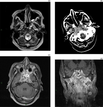
The patient underwent a CT of his head that showed a large nasopharyngeal mass extending into the left pterygopalatine fossa, causing bony destruction of the left sphenoid sinus and posterior left orbit. This was compressing the left optic nerve at the orbital fissure and obstructing the left eustachian tube, with opacification of the left mastoid air cells (Figure 1b). An MRI examination of his head was conducted to further evaluate this nasopharyngeal mass (Figure 1c and d). This demonstrated ill-defined, enhancing material in the nasopharynx and skull base, but no focal mass. Appearances were felt to favour a diffuse skull base infection rather than an infiltrating tumour. At this point, the MRI conducted seven weeks previously was reviewed by the radiology department. It was considered, in retrospect, to show subtle signs of early infection at the inferior portion of the nasopharynx (Figure 1a).
(a) Axial magnetic resonance image (MRI) showing an elevated T2 signal (white arrow) in the left prevertebral muscles, in keeping with early oedema or inflammatory change. (b) Axial computed tomography non-contrast image showing more extensive increased soft tissue material in the nasopharynx (black arrow). (c) Axial and (d) coronal MRI post-contrast images showing extensive enhancing soft tissue in the nasopharynx and skull base (black arrows, (d)), extending intracranially along the cavernous sinus and Meckel's cave (black arrow, (c)). R = right; P = Posterior

The patient underwent a postnasal space examination under general anaesthesia and was found to have fullness in the midline of the posterior nasopharyngeal wall, but no mucosal lesion. This was biopsied and sent for histopathological and microbiological investigations, including tuberculosis and fungal stain. He was commenced on quadruple anti-tuberculosis therapy along with posaconazole, which was continued until the results of his biopsy were available. The patient also had a further lumbar puncture to exclude any meningeal involvement. The results were normal and negative for any growth.
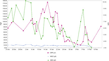
Mycology studies were later available and confirmed the presence of both Aspergillus fumigatus and mucor species. Subsequently, the anti-tuberculosis treatment was stopped, the antifungal medication was changed to voriconazole and the immunosuppressant agent mycophenolate mophetil was discontinued. His inflammatory markers stabilised over the next three weeks (Figure 2). Clinical improvement was evident, with resolution of his swinging pyrexia and consciousness levels.
Inflammatory markers over the patient's seven-week admission period. CRP = C-reactive protein; WCC = white cell count; ANC = absolute neutrophil count

Two weeks later, the patient's Glasgow Coma Scale score acutely decreased to 10 out of 15 (eye response = 3, verbal response = 2 and motor response = 5). An emergency CT scan of his head was carried out. This excluded any cerebral involvement and did not reveal any significant change in the skull base fungal infection. Chest radiographs raised the probability of aspiration pneumonia. The patient passed away two weeks later secondary to aspiration pneumonia.
Discussion
Skull base osteomyelitis is an uncommon infection. It most commonly occurs secondary to ear infections.Reference Chandler, Grobman, Quencer and Serafini4, Reference Grobman, Ganz, Casiano and Goldberg5 It typically presents in patients who have been inadequately treated for necrotising otitis externa. Pseudomonas is the main bacterial pathogen and, less frequently, aspergillus species.Reference Kountakis, Kemper, Chang, DiMaio and Stiernberg6 Skull base osteomyelitis can occasionally originate from a sinonasal source of infection; common micro-organisms include aspergillus, pseudomonas, salmonella and staphylococcus.Reference Grobman, Ganz, Casiano and Goldberg5, Reference Hoistad and Duvall7 Acute pyogenic or fungal skull base osteomyelitis typically produces progressive osteolytic bone destruction, with cranial nerve palsies, vertebral instability and intracranial infectious complications. In a retrospective case series of non-otogenic skull base osteomyelitis, the most prevalent risk factors identified were diabetes mellitus, human immunodeficiency virus infection and long-term steroid use.Reference Ducic1 Clinical features of this condition can be very insidious. Patients usually present with chronic, persistent headache and symptoms related to multiple cranial nerve abnormalities, such as diplopia, blindness, facial paralysis, dysphagia and dysarthria. Lethargy, seizures and coma are late features of the disease.
In patients with suspected cranial neuropathy and signs of infection, imaging of the skull base is best accomplished with MRI. This technique benefits from superior soft tissue discrimination, without the beam hardening artefacts of CT; it is particularly useful for assessing soft tissue planes around the skull base and abnormalities of the medullary bony cavity.Reference Chang, Fischbein and Holliday8, Reference Malone, O'Boynick, Ziegler, Batnitzky, Hubble and Holladay9 Malone et al. described T1 hypointensity and T2 hyperintensity as highly sensitive but non-specific marrow findings of non-otogenic skull base osteomyelitis.Reference Malone, O'Boynick, Ziegler, Batnitzky, Hubble and Holladay9
Clival enhancement on MRI can be seen both with skull base osteomyelitis and malignant processes. The main radiologic differential diagnosis is that of a malignant process, either primary or secondary. Squamous cell carcinoma, lymphoma, nasopharyngeal carcinoma and haematogenous metastases may all have similar MRI findings to skull base osteomyelitis.
Non-neoplastic conditions such as Wegener's granulomatosis, pseudotumour and sarcoidosis may have similar appearances, and biopsy is usually required for differentiation. Particular attention should be paid to the pseudotumoural form of skull base osteomyelitis. This presents as a mass of the skull base and/or a submucosal tumefaction of the nasopharynx, which evokes the diagnosis of a malignant tumour of the nasopharynx or metastatic disease of the skull base.Reference Laccourreye, Halimi, Bonfils and Laccourreye10 There have been less than 20 such cases reported over the last 20 years.Reference Laccourreye, Halimi, Bonfils and Laccourreye10 Atypical skull base osteomyelitis has drawn particular attention in recent skull base society meetings. In recent years, there have been an increasing number of cases reported at the North American Skull Base Society meetings, raising awareness of this lethal condition.Reference Margalit11–Reference Beahm13
Computed tomography findings may be lacking in the early stages of the disease, but CT can later demonstrate bony erosion.Reference Malone, O'Boynick, Ziegler, Batnitzky, Hubble and Holladay9 There have been cases of skull base osteomyelitis reported where initial CT scans failed to demonstrate bony destruction.Reference Malone, O'Boynick, Ziegler, Batnitzky, Hubble and Holladay9, Reference Laccourreye, Halimi, Bonfils and Laccourreye10, Reference Clark, Pretorius, Byren and Milford14, Reference Rowlands, Lekakis and Hinton15 In a case series by Seabold et al., no CT evidence of bone erosion was apparent in 13 out of 35 patients with biopsy-confirmed skull base osteomyelitis.Reference Seabold, Simonson, Weber, Thompson, Harris and Rezai16 Furthermore, bony erosion may fail to resolve for up to two years following treatment,Reference Rubin, Curtin, Yu and Kamerer17 limiting the role for CT in follow up.
Various nuclear medicine imaging techniques have been advocated in suspected skull base osteomyelitis, including gallium-67 scintigraphy, indium-111 white blood cell scans, technetium-99m methylene diphosphonate bone scans and single-photon emission CT.
Technetium radioisotope scans can be helpful for the initial diagnosis, but may remain ‘hot’ for several months following resolution of infection.Reference Seabold, Simonson, Weber, Thompson, Harris and Rezai16 Technetium-99m methylene diphosphonate bone scanning relies upon the ability of the radiolabelled agent to bind to osteoblasts, and can depict as little as a 10 per cent increase in osteoblastic activity. Technetium scans may remain persistently positive long after clinical improvement as a result of on-going bone remodelling.
Gallium scan abnormalities have been shown to be useful for monitoring responses to treatment and evaluating recurrence.Reference Grobman, Ganz, Casiano and Goldberg5 A gallium-67 citrate scan is sensitive but is not specific, because gallium binds to actively dividing cells, including inflammatory cells, tumour cells and osteoblasts. This test is most helpful when used as a monitor following treatment. Gallium scans are superior to technetium scans in monitoring responses to treatment, possibly because the former measure bone remodelling to a lesser degree than the latter and are therefore more sensitive to infection.Reference Alazraki, Fierer and Resnick18
• Early diagnosis of skull base osteomyelitis is difficult due to insidious onset of features and limitations of investigations
• Skull base osteomyelitis should be suspected in immunocompromised patients with subtle radiological abnormalities
• There is controversy regarding the most accurate investigation for early diagnosis; multiple imaging modalities may be required
• Radiological findings can remain positive for a prolonged period, even after long-term treatment
• In the presence of clinical suspicion, an early tissue biopsy should be considered to aid diagnosis
Gallium and technetium scans are complementary to each other in the diagnosis of skull base osteomyelitis: in the first stage of the disease, only the gallium scan is positive and the disease is limited to soft tissues; in the second stage, both scans are positive, with bone involvement limited to the mastoid; and in the third stage, both scans are positive and bone involvement extends to the contralateral side.Reference Benecke19
Technetium and single-photon emission CT scans may have an advantage over CT and MRI techniques in their ability to detect post-operative osteomyelitis. In addition, they may be more useful in the follow up of patients on medical treatment because marrow signal change can persist for up to six months, even after successful treatment.Reference Seabold, Simonson, Weber, Thompson, Harris and Rezai16 A case series study on malignant otitis externa has concluded that CT and/or MRI should be supported by routine single-photon emission CT bone imaging for initial diagnosis.Reference Okpala and Siraj20 However, the anatomical extent of necrotising otitis externa is different to that seen in atypical or central skull base osteomyelitis. There is currently no diagnostic imaging technique for this condition and so biopsy for microbiological culture in cases of suspected skull base osteomyelitis remains essential.Reference Rowlands, Lekakis and Hinton15 Radioisotope bone scanning does, however, have a role in follow up and disease evaluation after medical or surgical treatment.
The case reported here illustrates the limitations of diagnostic modalities, which may delay diagnosis in patients with skull base osteomyelitis. Subtle radiological changes on initial imaging in immunosuppressed patients should be considered with a high index of suspicion.