Introduction
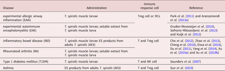
David P. Strachan proposed the ‘hygiene hypothesis’ in 1989, which theorized that the decreased incidence of bacterial and parasitic infections in humans living in developed countries underlies immune system deregulations and increased development of allergic and autoimmune diseases observed in these populations (Strachan, Reference Strachan1989, Reference Strachan2000; Yazdanbakhsh & Matricardi, Reference Yazdanbakhsh and Matricardi2004; Maizels, Reference Maizels2016; Harnett & Harnett, Reference Harnett and Harnett2017; Wu et al., Reference Wu, Wang, Tang and Sun2017; Bach, Reference Bach2018; Caraballo, Reference Caraballo2018). Epidemiological data show that acute or chronic infection by helminths can provide some protection against these diseases (Weinstock et al., Reference Weinstock, Summers, Elliott, Qadir, Urban and Thompson2002; McSorley et al., Reference McSorley, Chaye and Smits2019) in animal models or human beings. Clinical trials indicate that infection with different helminths can provide immunological protection against multiple sclerosis and type 2 diabetes (Tahapary et al., Reference Tahapary, de Ruiter and Martin2015; Charabati et al., Reference Charabati, Donkers, Kirkland and Osborne2020). In recent years, mounting evidence suggests that helminths exert strong immunomodulatory effects on their hosts (Maizels et al., Reference Maizels, Balic, Gomez-Escobar, Nair, Taylor and Allen2004; Varyani et al., Reference Varyani, Fleming and Maizels2017). As a unique inflammation modulator, Trichinella spiralis is known as a zoonotic nematode of intracellular parasitism that infects a wide range of vertebrate hosts, including humans. Humans consume uncooked meat that carries encysted T. spiralis larvae in muscle tissue. Larvae moult and mature into adult worms in the small intestine of the host. The female adult worms release newborn larvae after copulation, which travel through the circulatory system to reach skeletal muscle cells (Fabre et al., Reference Fabre, Beiting, Bliss and Appleton2009; Hernandez-Ancheyta et al., Reference Hernandez-Ancheyta, Salinas-Tobon, Cifuentes-Goches and Hernandez-Sanchez2018). Unlike other intracellular parasites, T. spiralis larvae transform muscle cells into encapsulated nurse cells and occupy the host's muscle cells without killing them, making it one of the most successful parasitic symbiotes, ensuring its survival and immune dialogue with the host by releasing excretory–secretory (ES) products (Hernandez-Ancheyta et al., Reference Hernandez-Ancheyta, Salinas-Tobon, Cifuentes-Goches and Hernandez-Sanchez2018). Trichinella spiralis infection ameliorates immune-mediated diseases in animal models, such as allergies, autoimmune disease, inflammatory bowel disease (IBD), type 1 diabetes, rheumatoid arthritis (RA) and autoimmune encephalomyelitis. The reduced disease symptoms are likely to be mediated by T. spiralis exerting immune effects on T cells, dendritic cells (DCs), natural killer (NK), mast cells and eosinophils (Saunders et al., Reference Saunders, Raine, Cooke and Lawrence2007; Gruden-Movsesijan et al., Reference Gruden-Movsesijan, Ilic, Mostarica-Stojkovic, Stosic-Grujicic, Milic and Sofronic-Milosavljevic2010; Du et al., Reference Du, Tang and Ma2011; Park et al., Reference Park, Cho, Choi, Kim and Yu2011; Cho et al., Reference Cho, Park, Kang, Choi, Ahn and Yu2012; Kuijk et al., Reference Kuijk, Klaver and Kooij2012; Aranzamendi et al., Reference Aranzamendi, de Bruin, Kuiper, Boog, van Eden, Rutten and Pinelli2013a; Sofronic-Milosavljevic et al., Reference Sofronic-Milosavljevic, Radovic, Ilic, Majstorovic, Cvetkovic and Gruden-Movsesijan2013; Zhao et al., Reference Zhao, Liu and Wang2013; Yang et al., Reference Yang, Yang, Wang, Zhan, Gu, Cheng and Zhu2014; Eissa et al., Reference Eissa, Mostafa, Ghazy, El Azzouni, Boulos and Younis2016; Cheng et al., Reference Cheng, Zhu, Wang, Zhuang, Huyan, Sun, Huang, Zhan and Zhu2018; Sun et al., Reference Sun, Li and Yuan2019; Xu et al., Reference Xu, Liu, Yu, Wu and Lu2019a; Xu et al., Reference Xu, Yu, Wu, Liu and Lu2019b) (table 1). Research has shown that T. spiralis differs from other helminths in manipulation of the host immune response not only by well-known characteristics of its life cycle, but also by its inflammation modulation pathway. How the parasite achieves inflammation modulation has not been fully elucidated yet. It is clear that the application of live parasite poses a health risk and ethical issues that need to be avoided. Trichinella spiralis ES products that enable cross-talk between the parasite and the host have attracted a lot of attention (Wang et al., Reference Wang, Wu, Weng, Zheng, Wu and Lv2017). Obtained results indicated that this protein complex mixture from ES products and its particular components (either in native or recombinant form) could largely reproduce the effects of infection on the host immune system (by shifting the immune response from pro- to anti-inflammatory type) (Aranzamendi et al., Reference Aranzamendi, de Bruin, Kuiper, Boog, van Eden, Rutten and Pinelli2013a; Yang et al., Reference Yang, Yang, Wang, Zhan, Gu, Cheng and Zhu2014; Kobpornchai et al., Reference Kobpornchai, Flynn and Reamtong2020). Therefore, almost any research performed in the field seeks to explore the molecular mechanisms and to identify molecules important for creation of the anti-inflammatory milieu achieved by this parasite. This review will generalize the mechanism and focuses on the ES immunomodulatory molecules of T. spiralis that may be important for developing new therapeutics for inflammatory disorders.
Immune-mediated diseases ameliorated by T. spiralis infection or its product.

Contribution of immunology to clearing host inflammation during infection with T. spiralis
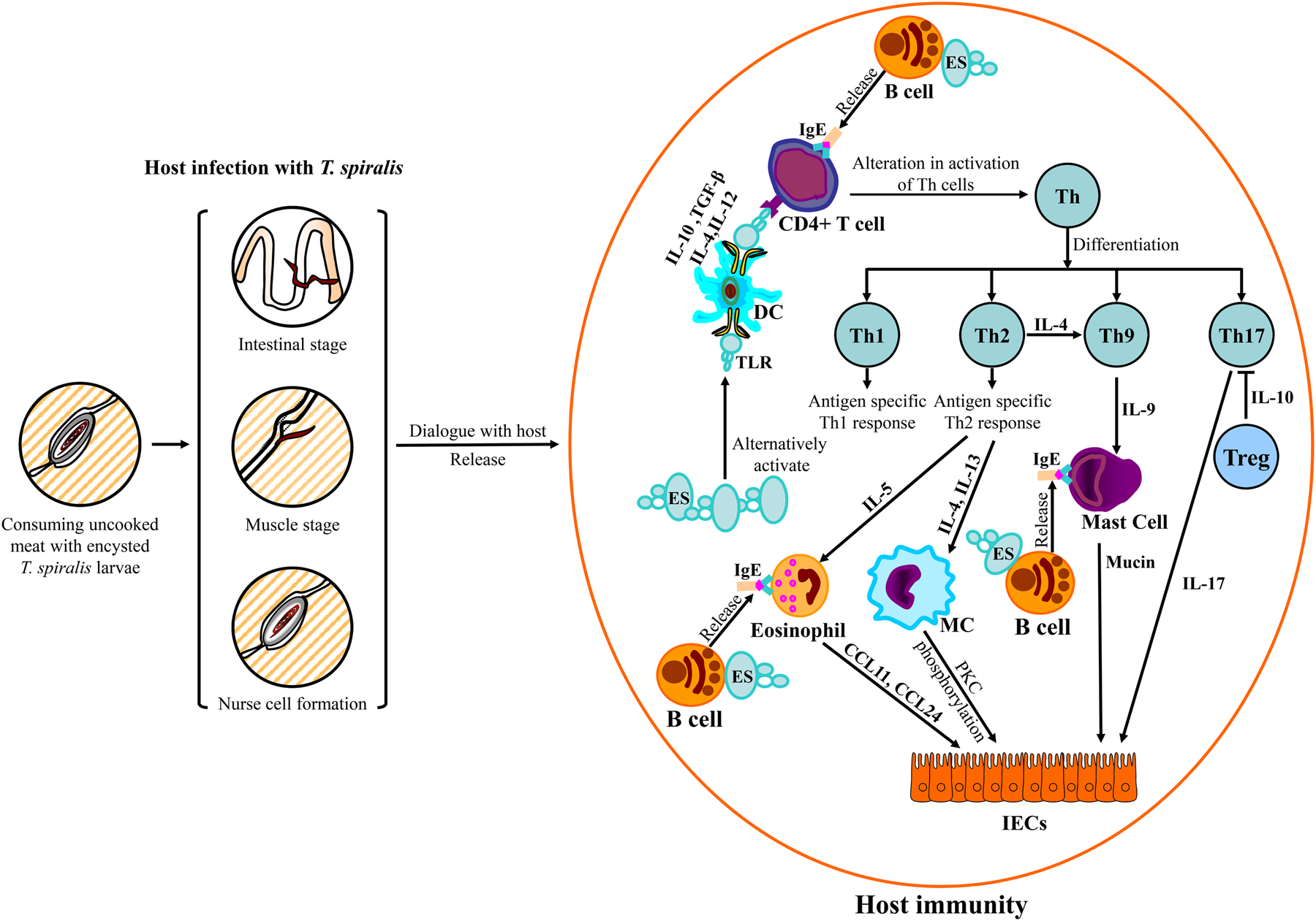
Humans can contract T. spiralis by consuming uncooked meat that carries encysted T. spiralis larvae in muscle tissue. Larvae are released by gastric fluids and migrate to the small intestine of the host, where they moult and mature into adult worms (intestinal stage). The female adult worms release newborn larvae after copulation, which travel through the circulatory system to reach skeletal muscle cells (muscle stage). Trichinella spiralis larvae transform muscle cells into encapsulated nurse cells and establish a niche in muscle tissues (nurse cell formation) (Fabre et al., Reference Fabre, Beiting, Bliss and Appleton2009; Hernandez-Ancheyta et al., Reference Hernandez-Ancheyta, Salinas-Tobon, Cifuentes-Goches and Hernandez-Sanchez2018). The cross-talk between the host and T. spiralis has evolved to maximize the immune modulating efficiency of T. spiralis, which regulates the dominant ascending T helper cell type 2 (Th2) immune responses. Trichinella spiralis alters the host's immune system to ensure its survival by releasing ES molecules (Ilic et al., Reference Ilic, Gruden-Movsesijan and Sofronic-Milosavljevic2012; Wang et al., Reference Wang, Wu, Weng, Zheng, Wu and Lv2017) (fig. 1).
Host–Trichinella spiralis interactions. Trichinella spiralis infection alters and activates host Th cells and leads to an antigen-specific Th1 response and dominant elevation of the Th2 response in the host.

Contribution of immunology to clearing host inflammation in intestinal stage of T. spiralis infection
In intestinal stage of T. spiralis infection, the B cell compartment is affected in T. spiralis-infected individuals and is characterized by an increase in immunoglobulin (Ig)G and IgE in the host (Watanabe, Reference Watanabe2014; Moon et al., Reference Moon, Lee, Soh, Guo, Piao and Quan2018). Allergen-specific IgG can prevent the binding of the allergen to IgE residing on mast cells and basophils. As a result, increasing allergen-specific IgE leads to increased clinical sensitivity, as mast cells become cross-linked with basophils, leading to the upregulation of IgE receptors (Valenta et al., Reference Valenta, Campana and Niederberger2017). Trichinella spiralis infection can protect itself from the host by inducing immune cell of host to produce large amounts of polyclonal nonspecific IgE. By saturating the IgE binding sites on the mast cell and basophils, it inhibits the binding of allergen-specific IgE and ameliorates allergic responses (Gurish et al., Reference Gurish, Bryce, Tao, Kisselgof, Thornton, Miller, Friend and Oettgen2004; Erb, Reference Erb2007). Cytokines produced by B cells modulate the differentiation of CD4+ T cells towards either Th1 or Th2 cells (Harris et al., Reference Harris, Haynes, Sayles, Duso, Eaton, Lepak, Johnson, Swain and Lund2000). Regulatory B (Breg) cells are required for the polarization of the Th cell response towards Th2 (Ronet et al., Reference Ronet, Hauyon-La Torre, Revaz-Breton, Mastelic, Tacchini-Cottier, Louis and Launois2010) (fig. 1). Breg cells participate in the suppression of experimental autoimmune encephalomyelitis (Wolf et al., Reference Wolf, Dittel, Hardardottir and Janeway1996). ES molecules that are secreted from T. spiralis induce Breg cells (Kobpornchai et al., Reference Kobpornchai, Flynn and Reamtong2020); however, the molecules that are involved in this induction remain unclear.
As an important pathogen, T. spiralis-stimulated DCs can provoke regulatory immune responses associated with toll like receptors (TLRs) (Broide, Reference Broide2009; Langelaar et al., Reference Langelaar, Aranzamendi, Franssen, Van Der Giessen, Rutten, van der Ley and Pinelli2009; Yu et al., Reference Yu, Deng, Lu, Jia, Wu and Qi2013; Szkudlapski et al., Reference Szkudlapski, Labuzek, Pokora, Smyla, Gonciarz, Mularczyk, Maluch and Okopien2014; Kim et al., Reference Kim, Park and Yu2015; Wojtkowiak-Giera et al., Reference Wojtkowiak-Giera, Wandurska-Nowak, Derda, Jankowska, Jagodzinski and Hadas2018). Pathogens mainly activate DCs via a pattern recognition receptor (PRR) signalling pathways which play a key role in host cell recognition and response to pathogens (Everts et al., Reference Everts, Smits, Hokke and Yazdanbakhsh2010). DCs are an important antigen-presenting cell with many types of PRRs expressed on the surface of DCs or in DCs to identify and distinguish different pathogen-related antigens (Carvalho et al., Reference Carvalho, Sun, Kane, Marshall, Krawczyk and Pearce2009). Among these PRRs, TLRs are the most important members expressed on the surface of DCs or in DCs (Sun et al., Reference Sun, Wang, Wu, Zhao, Wang, Liu, Song, Wu and Liu2011). Activation of TLRs leads to the recruitment of adaptor proteins, including TIR domain-containing adapter protein 1 (TIRAP1), TIR domain-containing adaptor protein-inducing interferon (TRIF) and myeloid differentiation primary response protein 88 (MyD88). The recruitment of these proteins leads to the activation of nuclear factor kappa B (NF-κB), adaptor protein 1 (AP1) and interferon regulatory factors (IRFs), which results in the production of proinflammatory cytokines. Maintenance of the TLR-dependent signalling pathway is critical for controlling intestinal inflammation, and deregulation of this pathway contributes to various clinical IBD phenotypes (Sun et al., Reference Sun, Wang, Wu, Zhao, Wang, Liu, Song, Wu and Liu2011).
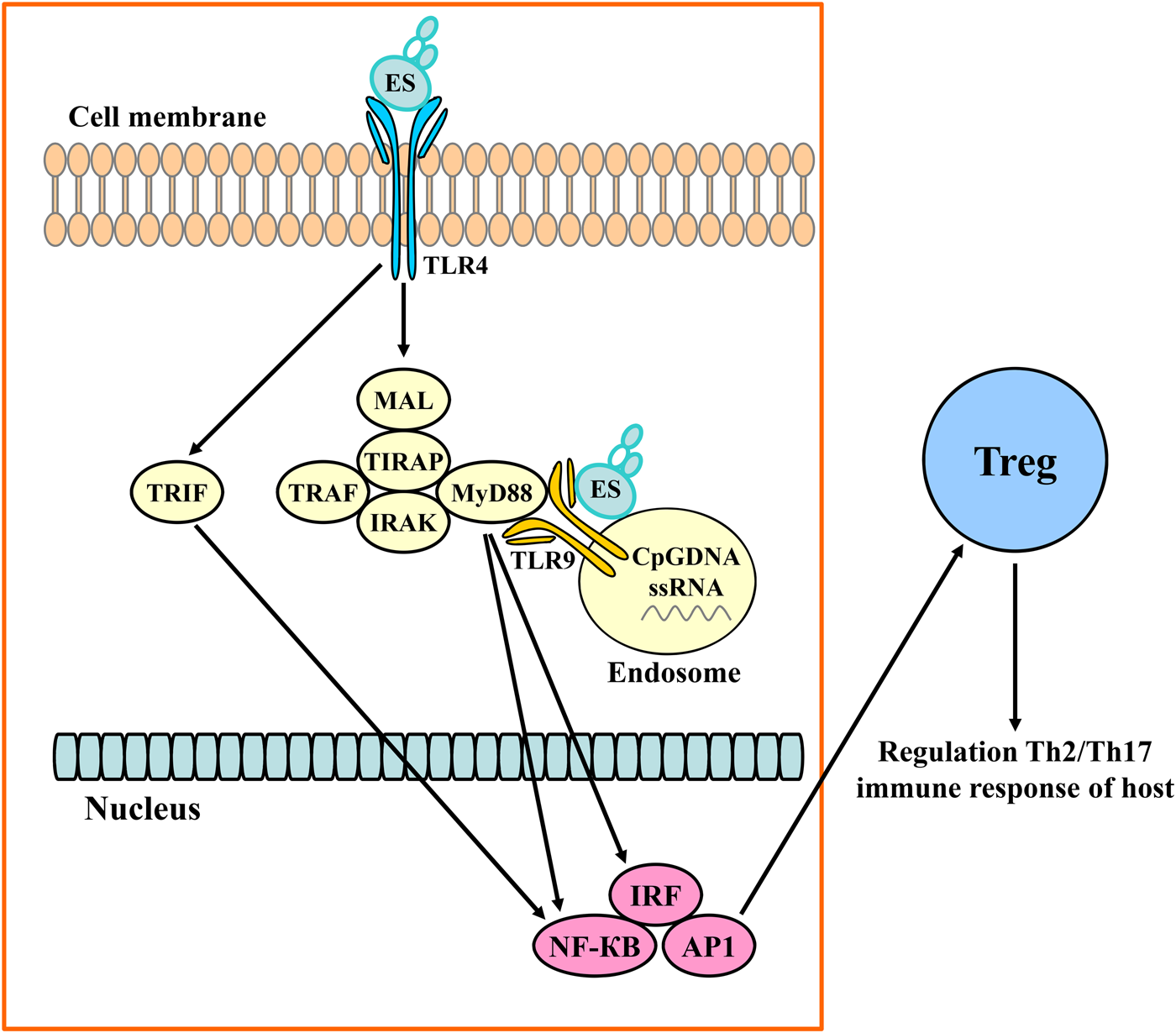
Thirteen mammalian TLR paralogues have now been identified (ten in humans and 12 in mice) (Beiting et al., Reference Beiting, Bliss, Schlafer, Roberts and Appleton2004). TLR1–TLR9 were more studied in the murine disease model correlated with human beings (Yu et al., Reference Yu, Deng, Lu, Jia, Wu and Qi2013; Kim et al., Reference Kim, Park and Yu2015; Sun et al., Reference Sun, Zhao, Wang, Yang, Zhao, Zhan and Zhu2015; Ilic et al., Reference Ilic, Gruden-Movsesijan, Cvetkovic, Tomic, Vucevic, Aranzamendi, Colic, Pinelli and Sofronic-Milosavljevic2018; Wojtkowiak-Giera et al., Reference Wojtkowiak-Giera, Wandurska-Nowak, Derda, Jankowska, Jagodzinski and Hadas2018). Different stages of T. spiralis infection had different impacts on the expression of TLR1–TLR9 (Yu et al., Reference Yu, Deng, Lu, Jia, Wu and Qi2013; Kim et al., Reference Kim, Park and Yu2015).The expression of TLR4 and TLR9 significantly increases in the small intestine after T. spiralis infects the intestine and releases ES molecules compared with TLR1, TLR2 and TLR5 (Kim et al., Reference Kim, Park and Yu2015). Activated TLR9 triggers intracellular signalling, leading to the activation of macrophages, DCs and B cells and the production of cytokines, chemokines and immunoglobulins (Broide, Reference Broide2009; Szkudlapski et al., Reference Szkudlapski, Labuzek, Pokora, Smyla, Gonciarz, Mularczyk, Maluch and Okopien2014). In the adult worm stage of infection, TLR4 expression is upregulated, and the MyD88-dependent signalling pathway is activated (Yu et al., Reference Yu, Deng, Lu, Jia, Wu and Qi2013; Wojtkowiak-Giera et al., Reference Wojtkowiak-Giera, Wandurska-Nowak, Derda, Jankowska, Jagodzinski and Hadas2018). The muscle larvae then inhibit TLR4- and TRIF-dependent signalling pathways by releasing ES molecules (Yu et al., Reference Yu, Deng, Lu, Jia, Wu and Qi2013). TLR4 and TLR9 have different adaptor proteins. TLR4 binds to MyD88/TIRAP and TRIF, while TLR9 binds only to MyD88 (Chen et al., Reference Chen, Szodoray and Zeher2016a). The expression of TRIF is the most important change in Th2-related cytokine gene expression in the host (Xu et al., Reference Xu, Liu and Komai-Koma2004; Piras & Selvarajoo, Reference Piras and Selvarajoo2014) (fig. 2). Therefore, TLR4 may be more closely associated with the Th2 response in individuals who are infected with T. spiralis compared with those of other TLRs (Ilic et al., Reference Ilic, Gruden-Movsesijan, Cvetkovic, Tomic, Vucevic, Aranzamendi, Colic, Pinelli and Sofronic-Milosavljevic2018; Wojtkowiak-Giera et al., Reference Wojtkowiak-Giera, Wandurska-Nowak, Derda, Jankowska, Jagodzinski and Hadas2018).
ES-induced signalling changes in extracellular TLR4 and TLR9. ES molecules that are secreted from T. spiralis selectively activate TLR4 and TLR9. Activated TLR4 induces signals through the activation of the MyD88 adaptor protein and intermediate MyD88 adaptor-like (MAL)/TIRAP, TNF receptor associate factor (TRAF), and interleukin-1 receptor associated kinase (IRAK) signal adaptor protein molecules. Activated TLR9 induces signals through activation of the MyD88 adaptor protein. TLR4- and TLR9-mediated signals induce downstream activation of nuclear factor kappa B (NF-κB), adaptor protein 1 (AP1) and IRF to regulate Th2/Th17 immune responses in Treg cells.

DCs could produce interleukin (IL)-12, which influences the development of Th1 or IL-4 and influences the development of Th2 type of immune response in response to pathogen associated molecular patterns (PAMPS) and damage-associated molecular pattern molecules (Qian & Cao, Reference Qian and Cao2018). ES molecules also stimulate DCs, leading to significantly increased IL-10 and transforming growth factor beta (TGF-β) (Sofronic-Milosavljevic et al., Reference Sofronic-Milosavljevic, Radovic, Ilic, Majstorovic, Cvetkovic and Gruden-Movsesijan2013) (fig. 1). Trichinella spiralis-stimulated DCs provoke regulatory immune responses, thus resulting in the regulatory DC phenotype (Langelaar et al., Reference Langelaar, Aranzamendi, Franssen, Van Der Giessen, Rutten, van der Ley and Pinelli2009). The regulation of DCs occurs in a generalized manner irrespective of their Th1 or Th2 phenotypic affiliation (Mucida & Cheroutre, Reference Mucida and Cheroutre2010). Gruden-Movsesijan et al. (Reference Gruden-Movsesijan, Ilic, Mostarica-Stojkovic, Stosic-Grujicic, Milic and Sofronic-Milosavljevic2010) successfully induced an immune tolerogenic, Th2 response using T. spiralis-sensitized DCs in rats and humans (Ilic et al., Reference Ilic, Gruden-Movsesijan, Cvetkovic, Tomic, Vucevic, Aranzamendi, Colic, Pinelli and Sofronic-Milosavljevic2018). This is a promising tool to avoid the application of live parasites to treat various autoimmune and allergic diseases.
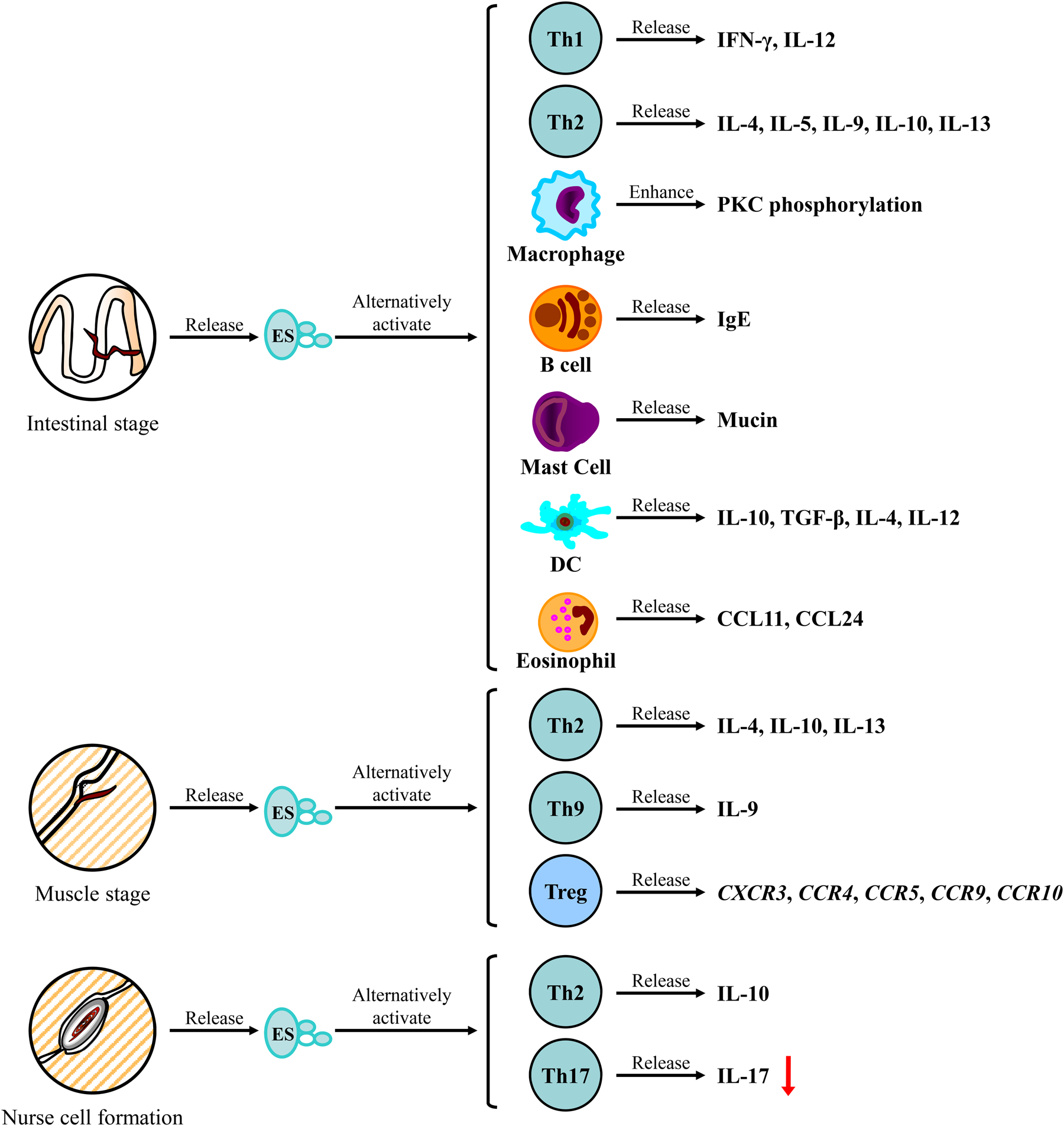
In the early stages of T. spiralis infection, the host also experiences a T helper cell type 1 (Th1) response to protect the host from the parasite, as characterized by increased interferon gamma (IFN-γ) and IL-12 levels in the spleen and serum of the host (Aranzamendi et al., Reference Aranzamendi, Fransen, Langelaar, Franssen, van der Ley, van Putten, Rutten and Pinelli2012; Kang et al., Reference Kang, Cho, Park, Kim, Hong, Lee, Cha, Ock and Yu2012; Ding et al., Reference Ding, Bai, Wang, Shi, Cai, Luo, Liu and Liu2017). The Th1-type immune response is triggered by transcriptional upregulation and increased secretion of proinflammatory cytokines. Increased circulation of cytokines increases immunity by recruiting immune cells to the intestine and increasing the restoration of intestinal epithelial cells (IECs) that are invaded by T. spiralis (Ming et al., Reference Ming, Peng, Zhang, Zhang, Lv, Wang, Cui and Ren2016). Once a T. spiralis adult worm occupies a niche in the enterocytes of the jejunum, a well-characterized type 2 cytokine response (IL-4, IL-5, IL-9, IL-10 and IL-13) orchestrated by CD4+ T cells is elicited in the mesenteric lymph nodes. This cell is subsequently recruited to the gut mucosa and clears inflammation in the host gut tissue (Helmby & Grencis, Reference Helmby and Grencis2003; Sofronic-Milosavljevic et al., Reference Sofronic-Milosavljevic, Ilic, Pinelli and Gruden-Movsesijan2015). Trichinella spiralis infection elicits a Th2 response in the host that is characterized by eosinophilia, high serum IgE levels and high serum levels of IL-4, IL-5 IL-9, IL-10 and IL-13 (Finkelman et al., Reference Finkelman, Shea-Donohue, Morris, Gildea, Strait, Madden, Schopf and Urban2004; Sofronic-Milosavljevic et al., Reference Sofronic-Milosavljevic, Ilic, Pinelli and Gruden-Movsesijan2015). Eotaxin is a fundamental regulator of physiological eosinophil trafficking during healthy and inflamed states (Conroy & Williams, Reference Conroy and Williams2001). Eotaxin-1 (CCL11) and eotaxin-2 (CCL24) are important in intestinal tissue eosinophilia in individuals who are infected with T. spiralis (Bruschi et al., Reference Bruschi, Korenaga and Watanabe2008). The inhibition of eosinophil apoptosis during the later stages of T. spiralis infection is related to the presence of IL-5 (Danilowicz-Luebert et al., Reference Danilowicz-Luebert, O'Regan, Steinfelder and Hartmann2011). Elevated levels of Th2 cytokines activate mast cells to release mucin and induce goblet cell hyperplasia, induce peritoneal macrophages to become activated M2 macrophages and enhance protein kinase C phosphorylation during the intestinal phase of T. spiralis (Kamal et al., Reference Kamal, Wakelin, Ouellette, Smith, Podolsky and Mahida2001; Knight et al., Reference Knight, Pemberton, Robertson, Roy, Wright and Miller2004; Dzik et al., Reference Dzik, Zieliński, Cieśla and Wałajtys-Rode2010). ES products from different stages of T. spiralis alter macrophage phenotypes by inhibiting the production of proinflammatory cytokines, such as tumour necrosis factor alpha (TNF-α), IL-1β, IL-6 and IL-12, which then ameliorate the effects of inflammatory diseases (Akiho et al., Reference Akiho, Blennerhassett, Deng and Collins2002). Trichinella spiralis ES proteins ameliorate intestinal injury by promoting IRF4 and M2-related metabolic regulation via STAT6-dependent transformation of classically activated M1 macrophages to alternatively activated M2 macrophages. M2 macrophages have anti-inflammatory properties, and, thus, ES products may have potential as an immunotherapeutic against inflammatory disorders by inducing M2 macrophages (Han et al., Reference Han, Yu, Zhang, Zhai, Zhang, Meng, Yu, Li and Song2019; Kang et al., Reference Kang, Park, Park, Choi, Lee, Song and Yu2019). The immune response to T. spiralis is a powerful way to reduce inflammation of the host intestine. Trichinella spiralis infection does not trigger apoptotic events and results in significantly increased expression of IL-1β, IL-8, ENA-78, and MCP-2 in IECs (Stadnyk et al., Reference Stadnyk, Dollard and Issekutz2000; Milcheva et al., Reference Milcheva, Petkova, Hurnikova, Janega and Babal2013) (fig. 3). Trichinella spiralis retunes the immune cell repertoire of its host, which further explains why helminths are involved in the immunoregulation of the host and can be applied to treat excessive inflammatory responses (Saunders et al., Reference Saunders, Raine, Cooke and Lawrence2007; Du et al., Reference Du, Tang and Ma2011; Gruden-Movsesijan et al., Reference Gruden-Movsesijan, Ilic, Colic, Majstorovic, Vasilev, Radovic and Sofronic-Milosavljevic2011; Park et al., Reference Park, Cho, Choi, Kim and Yu2011; Cho et al., Reference Cho, Park, Kang, Choi, Ahn and Yu2012; Kuijk et al., Reference Kuijk, Klaver and Kooij2012; Aranzamendi et al., Reference Aranzamendi, de Bruin, Kuiper, Boog, van Eden, Rutten and Pinelli2013a; Sofronic-Milosavljevic et al., Reference Sofronic-Milosavljevic, Radovic, Ilic, Majstorovic, Cvetkovic and Gruden-Movsesijan2013; Zhao et al., Reference Zhao, Liu and Wang2013; Yang et al., Reference Yang, Yang, Wang, Zhan, Gu, Cheng and Zhu2014; Eissa et al., Reference Eissa, Mostafa, Ghazy, El Azzouni, Boulos and Younis2016; Cheng et al., Reference Cheng, Zhu, Wang, Zhuang, Huyan, Sun, Huang, Zhan and Zhu2018; Inclan-Rico & Siracusa, Reference Inclan-Rico and Siracusa2018; Xu et al., Reference Xu, Liu, Yu, Wu and Lu2019a) (fig. 4). Recent studies have shown that Th2-related innate immune responses are regulated by epithelial cells and innate helper T cells, and triggering this response leads to intestinal mucosal restoration (Yang et al., Reference Yang, Yang, Wang, Zhan, Gu, Cheng and Zhu2014; Xu et al., Reference Xu, Liu, Yu, Wu and Lu2019a). Trichinella spiralis infection activates the response in intestinal tuft cells, which are epithelial chemosensory cells found in mucosal tissues and release the alarmin IL-25 and type 2 innate lymphoid cells (ILC2s) that expand upon Trichinella infection and provide the early signals that drive type 2 immunity in the gut (Luo et al., Reference Luo, Chen and Xue2019).
The immune response in different T. spiralis infection stages. Trichinella spiralis infection in the intestine, muscle and nurse cell formation stages leads to the secretion of special excretory–secretory (ES) antigens that activate Th1, Th2, Th9, Th17, and Treg cells, DCs, B, macrophages and mast cells, depending on the stage. Activated cells produce or release cytokines to alter the immune regulation of the host and ensure the survival of T. spiralis through its different life stages.

Trichinella spiralis-induced immune cells involved in remodelling the immune response of the host. T. spiralis ES antigens from all stages induce a network of innate immune cells (DCs, macrophages, mast cells, eosinophils and B-1 cells) and adaptative immune cells (CD4+ T, Th2, Treg, Th9, Th17 and B-2 cells) to release cytokines which leads to an antigen-specific Th1 response and dominant elevation of the Th2 response in the host. Immunomodulation protects against excessive inflammatory responses in allergies, autoimmune diseases, IBD, type 1 diabetes, RA and autoimmune encephalomyelitis.

Contribution of immunology to clearing host inflammation in the muscle stage of T. spiralis infection
In the muscle stage of T. spiralis infection, the Th2 cytokines IL-4, IL-10 and IL-13 are upregulated (Akiho et al., Reference Akiho, Blennerhassett, Deng and Collins2002). The muscle stage also induces a Th9 response by producing high levels of IL-9, which mediates the immune response by increasing the levels of circulating IgE antibodies (Beiting et al., Reference Beiting, Gagliardo, Hesse, Bliss, Meskill and Appleton2007; Ilic et al., Reference Ilic, Worthington, Gruden-Movsesijan, Travis, Sofronic-Milosavljevic and Grencis2011, Reference Ilic, Gruden-Movsesijan and Sofronic-Milosavljevic2012). The research indicates that IL-9 is a potent Th2 adjuvant and has an important role in the induction of a Th2-type response. During T. spiralis infection of muscle cells, the expression of the chemokine receptors CXCR3, CCR4, CCR5, CCR9 and CCR10 correlates with increased Treg cell recruitment to infected muscle cells (Ahn et al., Reference Ahn, Kang, Kim and Yu2016) (fig. 3). Trichinella spiralis infection upregulates TLR9 expression, which then stimulates DCs to produce IL-10 and promote the induction of Treg cells (Kim et al., Reference Kim, Park and Yu2015). In addition, T. spiralis regulates the host's Th2 and Th17 immune responses in mice by releasing ES molecules into the small intestine and muscle tissue. Hence, TLR9 may be closely associated with Treg cell-mediated immune responses in individuals who are infected with T. spiralis (Aranzamendi et al., Reference Aranzamendi, Sofronic-Milosavljevic and Pinelli2013b; Kim et al., Reference Kim, Park and Yu2015).
Long-term T. spiralis infection in muscle tissue induces immunomodulation and is mainly characterized by a Th2 response in which it is generally beneficial to the host to repair or prevent tissue damage that results from inflammation (Fabre et al., Reference Fabre, Beiting, Bliss and Appleton2009). The parasite orchestrates a long-lasting molecular cross-talk with the host through muscle larvae excretory-secretory products (ES L1). Those products can successfully modulate parasite-specific immune responses as well as responses to unrelated antigens (either self or non-self in origin), providing an anti-inflammatory milieu and maintaining homeostasis (Sofronic-Milosavljevic et al., Reference Sofronic-Milosavljevic, Ilic, Pinelli and Gruden-Movsesijan2015).
Contribution of immunology to clearing host inflammation in nurse cells formation of T. spiralis infection
After nurse cells form, the anti-inflammatory cytokine IL-10 remains upregulated, while the inflammatory pathology mediator IL-17 is downregulated (fig. 3), which balances the T cell response and limits inflammation in the host (Beiting et al., Reference Beiting, Bliss, Schlafer, Roberts and Appleton2004; Elliott & Weinstock, Reference Elliott and Weinstock2012). The ES antigens released by T. spiralis during each of its developmental stages induce different mediators to increase the populations of Treg cells in the host (Ilic et al., Reference Ilic, Gruden-Movsesijan and Sofronic-Milosavljevic2012). Trichinella spiralis leverages this immunomodulation to prevent its eradication by an excessive immune response, as excessive inflammatory responses by the host may lead to organ damage (Kahl et al., Reference Kahl, Brattig and Liebau2018). Treg cells control Th2-dominated diseases, such as allergies (McKay, Reference McKay2009). The induction of Treg cells by T. spiralis is important in remodelling the immune response of the host (Gruden-Movsesijan et al., Reference Gruden-Movsesijan, Ilic, Colic, Majstorovic, Vasilev, Radovic and Sofronic-Milosavljevic2011; Park et al., Reference Park, Cho, Choi, Kim and Yu2011; Aranzamendi et al., Reference Aranzamendi, de Bruin, Kuiper, Boog, van Eden, Rutten and Pinelli2013a). The immune response suppresses the differentiation of Th17 cells through the interaction of Foxp3 and RORγt, which are transcription factors that are expressed in Treg and Th17 cells, respectively (Elliott & Weinstock, Reference Elliott and Weinstock2012; Fasching et al., Reference Fasching, Stradner, Graninger, Dejaco and Fessler2017). Th17 cells, which express high levels of IL-17, participate in the progression of many autoimmune diseases (Bedoya et al., Reference Bedoya, Lam, Lau and Larkin2013). Trichinella spiralis inhibits the formation of Th17 cells, thus downregulating the expression of IL-17 in its host (Ahn et al., Reference Ahn, Kang, Kim and Yu2016; Steel et al., Reference Steel, Faniyi and Rahman2019).
Trichinella spiralis infection is characterized by a Th2 response (Saunders et al., Reference Saunders, Raine, Cooke and Lawrence2007). ES products released by the different life stages of T. spiralis are also involved in the Th2 response (Liu et al., Reference Liu, Wang and Bai2014; Cvetkovic et al., Reference Cvetkovic, Sofronic-Milosavljevic, Ilic, Gnjatovic, Nagano and Gruden-Movsesijan2016; Guo et al., Reference Guo, Sun, Gu, Wang, Huang and Zhu2016). A study showed the Th2 response can influences the development of autoimmune responses (Cho et al., Reference Cho, Park, Kang, Choi, Ahn and Yu2012), so that T. spiralis or its products can be applied in controlling the levels of inflammation in infected tissues.
Application of T. spiralis or its products to modulate autoimmune diseases
Trichinella spiralis infection is characterized by a Th2 response, and this Th2 response is critical in controlling the levels of inflammation in infected tissues (Saunders et al., Reference Saunders, Raine, Cooke and Lawrence2007). Trichinella spiralis modulates the host immune system and influences the development of autoimmune responses (Cho et al., Reference Cho, Park, Kang, Choi, Ahn and Yu2012).
Application of T. spiralis or its products to modulate IBD
Trichinella spiralis infection has therapeutic potential in IBD with Th1-driven intestinal inflammation (Zhao et al., Reference Zhao, Liu and Wang2013). Trichinella spiralis infection inhibits epithelial and crypt cell destruction and sustains mast cell stabilization, which prevents mucosal mast cell hyperplasia and exacerbates motor responses against intestinal inflammation (Serna et al., Reference Serna, Porras and Vergara2006). ES products from adult T. spiralis (AES) alleviate IBD with acute inflammatory colonic disease in mice induced by dextran sodium sulphate, which was mediated by inhibition of Th1/Th17 proinflammatory cytokines and an increase in the production of anti-inflammatory Th2/Treg regulatory cytokines in the lymphocytes of colon, mesenteric lymph node and spleen of mice treated with AES (Yang et al., Reference Yang, Yang, Wang, Zhan, Gu, Cheng and Zhu2014; Kang et al., Reference Kang, Park, Park, Choi, Lee, Song and Yu2019). Motomura et al. (Reference Motomura, Wang, Deng, El-Sharkawy, Verdu and Khan2009) showed that T. spiralis antigens activate Th2 and Treg cells, leading to the production of IL-13 and TGF-β, respectively, subsequently reducing Th1-mediated inflammatory responses. Therefore, T. spiralis and its associated antigens may have bidirectional immunomodulatory effects, and T. spiralis antigens may provide a novel strategy to retune the immune cell repertoire and ameliorate colonic inflammation by expanding the Treg cell population and activating the Th2 response.
Application of T. spiralis or its products to modulate experimental allergic airway inflammation (EAAI)
Infection with T. spiralis confers protection against EAAI (Kang et al., Reference Kang, Park, Cho, Park, Jang, Yang, Jang, Kim and Yu2014). Trichinella spiralis has an immunosuppressive effect through the inhibition of the Th1-related cytokine IFN-γ and the Th17-related cytokine IL-17, promotion of the Th2-related cytokines IL-4, IL-5 and IL-13, release of the Treg-related cytokines IL-10 and TGF-β, induction of regulatory cells and increase in allergen-specific IgE and eosinophils (Conroy & Williams, Reference Conroy and Williams2001; Watanabe, Reference Watanabe2014; Moon et al., Reference Moon, Lee, Soh, Guo, Piao and Quan2018)
Application of T. spiralis or its products to modulate experimental autoimmune encephalomyelitis (EAE)
Trichinella spiralis infection is accompanied by increased production of IL-4 and IL-10, which lead to reduced levels of IFN-γ and IL-17 in EAE immune response in mice (Kuijk et al., Reference Kuijk, Klaver and Kooij2012; Sofronic-Milosavljevic et al., Reference Sofronic-Milosavljevic, Radovic, Ilic, Majstorovic, Cvetkovic and Gruden-Movsesijan2013). The decreased production of IFN-γ and IL-17 correlates with the alleviation of EAE symptoms. In addition, the amelioration of EAE symptoms is also attributed to the upregulation of Treg cells, which is a common immune response in individuals who are infected with T. spiralis (Beiting et al., Reference Beiting, Bliss, Schlafer, Roberts and Appleton2004; Gruden-Movsesijan et al., Reference Gruden-Movsesijan, Ilic, Mostarica-Stojkovic, Stosic-Grujicic, Milic and Sofronic-Milosavljevic2010; Kuijk et al., Reference Kuijk, Klaver and Kooij2012).
Application of T. spiralis or its products to modulate type 1 diabetes mellitus
Type 1 diabetes mellitus is a Th1 inflammatory disease. Helminth infections induce a Th2 immune response by altering the production of IL-4 (Hubner et al., Reference Hubner, Shi and Torrero2012). Trichinella spiralis infection reduces the development of diabetes by inducing the Th2 response, which in turn reduces the Th1-mediated destruction of beta cells (Saunders et al., Reference Saunders, Raine, Cooke and Lawrence2007; Wang et al., Reference Wang, Wu, Weng, Zheng, Wu and Lv2017).
Application of T. spiralis or its products to modulate rheumatoid arthritis
Th17 cells play an important role in the pathogenesis of RA (Kotake et al., Reference Kotake, Yago, Kobashigawa and Nanke2017). Th17 cell activity is dampened by a variety of regulatory T cells that express the transcription factor Foxp3 (Tahmasebinia & Pourgholaminejad, Reference Tahmasebinia and Pourgholaminejad2017). Treg cells suppress effector T cells either directly via cell-to-cell contact or via the secretion of TGF-β and IL-10 (Li et al., Reference Li, Seth, Gray, Bayston, Mahida and Wakelin1998). Trichinella spiralis antigens have anti-arthritic potential by upregulating Foxp3+ Treg cells, which leads to reduced production of IL-17 and increased production of IL-10 (Eissa et al., Reference Eissa, Mostafa, Ghazy, El Azzouni, Boulos and Younis2016; Cheng et al., Reference Cheng, Zhu, Wang, Zhuang, Huyan, Sun, Huang, Zhan and Zhu2018).
Application of T. spiralis or its products in therapy of tumour model
Trichinella spiralis muscle larva ES proteins inhibit the proliferation of human H446 small cell lung cancer cells and induce apoptosis by activating mitochondrial apoptosis pathways (Luo et al., Reference Luo, Yu, Xie, Li, Su, Zhao and Du2017). The A200711 protein of T. spiralis induces apoptosis in H7402 hepatocellular carcinoma cells (Wang et al., Reference Wang, Liu and Sun2013). Trichinella spiralis infection reduces B16-F10 melanoma growth and small cell lung cancer metastasis through changes in cytokine profiles of the individuals, including CXCL9, CXCL10 and CXCL13 in mice (Kang et al., Reference Kang, Jo, Cho, Yu, Leem, Song, Ock and Cha2013). The mechanism is still unknown.
Therapy using live T. spiralis may be difficult and unethical to patients with autoimmune diseases. Instead, molecules that are secreted by T. spiralis may provide an alternative strategy by generating antigen-specific immunoregulation (Abdoli, Reference Abdoli2019; Maizels, Reference Maizels2020).
Molecules released by T. spiralis
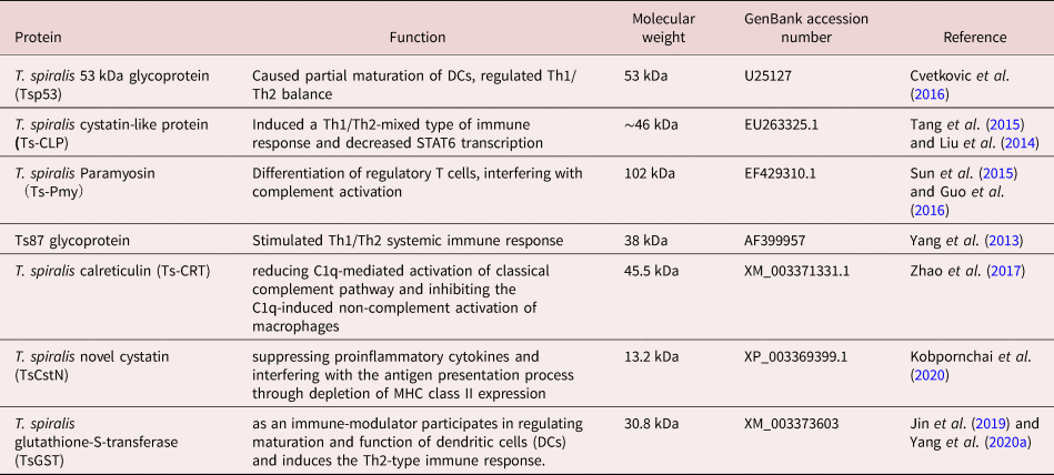
Trichinella spiralis can survive for long periods in the host, and this is mediated by the parasite's ability to modulate the host immune response through the secretion of immunomodulatory molecules (Yang et al., Reference Yang, Yang, Gu, Wang, Zhao and Zhu2013; Liu et al., Reference Liu, Wang and Bai2014; Sun et al., Reference Sun, Zhao, Wang, Yang, Zhao, Zhan and Zhu2015; Tang et al., Reference Tang, Liu, Wang, Yu, Shi, Boireau, Cozma, Wu and Liu2015; Cvetkovic et al., Reference Cvetkovic, Sofronic-Milosavljevic, Ilic, Gnjatovic, Nagano and Gruden-Movsesijan2016; Guo et al., Reference Guo, Sun, Gu, Wang, Huang and Zhu2016) (table 2). ES products released by the different life stages of T. spiralis are involved in acute inflammatory responses and modulate molecular cross-talk between the parasite and host (Sun et al., Reference Sun, Wang, Wu, Zhao, Wang, Liu, Song, Wu and Liu2011; Kang et al., Reference Kang, Park, Park, Choi, Lee, Song and Yu2019).
Molecules released by T. spiralis.

It is important to identify and characterize T. spiralis immunomodulatory molecules using genomic, proteomic and molecular methods, as they may provide a promising strategy to control and prevent parasite infection, as well as serve as novel immunomodulatory therapeutic drugs for immunopathological diseases (Bai et al., Reference Bai, Wu and Wang2012; Cvetkovic et al., Reference Cvetkovic, Ilic, Sofronic-Milosavljevic and Gruden-Movsesijan2014).
L1 products (T. spiralis 53 kDa glycoprotein Tsp53) found in T. spiralis induce the proliferation of naïve T cells and their polarization towards Th2 and anti-inflammatory responses. Tsp53 induces the production of IL-4 and IL-10 in T cells (Du et al., Reference Du, Tang and Ma2011; Cvetkovic et al., Reference Cvetkovic, Sofronic-Milosavljevic, Ilic, Gnjatovic, Nagano and Gruden-Movsesijan2016; Nascimento Santos et al., Reference Nascimento Santos, Carvalho Pacheco, Silva Pinheiro and Alcantara-Neves2017); therefore, Tsp53 is an important anti-inflammatory molecule. Recombinant Tsp53 (rTsP53) downregulates the proinflammatory cytokines TNF-α, IL-6, IL-12 and iNOS and elevates production of the regulatory factors IL-10 and TGF-β1 by inducing the transition of macrophages from M1 to M2 (Chen et al., Reference Chen, Tang, Liang, Yang, Wu, Hu, Li, Zeng and Ma2016b). rTsP53 has anti-inflammatory properties in mice by rescuing rTsP53-treated mice from 2,4,6-trinitrobenzene sulphonic acid-induced IBD (Du et al., Reference Du, Tang and Ma2011). Thus, rTsP53 has therapeutic potential for inflammatory disorders. Trichinella spiralis cystatin-like protein (Ts-CLP) induces the production of IL-10. IL-10 inhibits TNF-α production, which is a key feature in IBD pathogenesis; therefore, parasitic cystatins may be potential candidates for IBD therapy (Liu et al., Reference Liu, Wang and Bai2014; Tang et al., Reference Tang, Liu, Wang, Yu, Shi, Boireau, Cozma, Wu and Liu2015). Trichinella spiralis paramyosin (Ts-Pmy) modulates the host immune system by stimulating DCs to differentiate CD4+ T cells into regulatory T cells (Sun et al., Reference Sun, Zhao, Wang, Yang, Zhao, Zhan and Zhu2015; Guo et al., Reference Guo, Sun, Gu, Wang, Huang and Zhu2016). Ts87, a novel Ts87 gene from the cDNA library of adult T. spiralis, balances the Th1/Th2 response and downregulates IL-17 expression (Yang et al., Reference Yang, Yang, Gu, Wang, Zhao and Zhu2013). Further research is required on how Ts-Pmy and Ts87 may be applied to treat inflammatory diseases. As new identified molecular from T. spiralis, T. spiralis calreticulin (Ts-CRT) can bind to host complement C1q, which not only reduces C1q-mediated activation of classical complement pathway but also inhibits the C1q-induced non-complement activation of macrophages (Zhao et al., Reference Zhao, Shao, Chen, Sun, Sun, Huang, Zhan and Zhu2017). Trichinella spiralis novel cystatin (TsCstN) elicits an anti-inflammatory property by suppressing proinflammatory cytokines and interfering with the antigen presentation process through depletion of MHC class II expression (Kobpornchai et al., Reference Kobpornchai, Flynn and Reamtong2020). As an immune-modulator, T. spiralis glutathione-S-transferase (TsGST) participates in regulating maturation and function of DCs and induces the Th2-type immune response of its host (Jin et al., Reference Jin, Yang, Liu, Shi, Cai, Luo, Liu and Bai2019; Yang et al., Reference Yang, Liu and Bai2020a). The three molecules from T. spiralis might be a potential molecular target for antiinflammation.
In addition to the above-mentioned protein molecules, it has recently been discovered that extracellular vesicles (EVs) also have immunomodulatory effects. EVs are small membrane-bound structures secreted by helminths which mediate immune evasion during parasite infection (Yang et al., Reference Yang, Liu and Liu2020b). EVs produced by T. spiralis muscle larvae can carry immunomodulatory proteins and have the capacity independently to induce regulatory responses (Kosanović et al., Reference Kosanović, Cvetković, Gruden-Movsesijan, Vasilev, Svetlana, Ilić and Sofronić-Milosavljević2019).
In the in vivo application of T. spiralis immunomodulatory molecules, the induction of Th2 and regulatory cytokines is important for creating anti-inflammatory responses. The recombinant antigens with high-quality T. spiralis glycans which affect the anti-inflammatory environment and can interfere with the development of inflammatory diseases will instead of native antigen from ES of adult T. spiralis or muscle larvae and make is a generalized and cheap therapeutics for inflammatory disorders. A better understanding of the functions of T. spiralis immunomodulatory molecules will be important for developing new therapeutics for inflammatory disorders, including allergies and autoimmune diseases (Cvetkovic et al., Reference Cvetkovic, Ilic, Sofronic-Milosavljevic and Gruden-Movsesijan2014; Kahl et al., Reference Kahl, Brattig and Liebau2018).
Prospective applications of T. spiralis-derived compounds
The relationship between humans and T. spiralis can be considered mutualistic rather than typical parasitism (Szkudlapski et al., Reference Szkudlapski, Labuzek, Pokora, Smyla, Gonciarz, Mularczyk, Maluch and Okopien2014). Live helminth therapy is encouraged to maximize the immune efficiency of helminths; however, it may be difficult and unethical to suggest helminth therapy to patients. Instead, generating antigen-specific immunoregulation through molecules that are secreted by T. spiralis may provide an alternative strategy (Sofronic-Milosavljevic et al., Reference Sofronic-Milosavljevic, Ilic, Pinelli and Gruden-Movsesijan2015; Smallwood et al., Reference Smallwood, Giacomin, Loukas, Mulvenna, Clark and Miles2017; Abdoli, Reference Abdoli2019; Maizels, Reference Maizels2020). For example, rTsP53, Ts87, Ts-CLP and Ts-Pmy can serve as prospective immunotherapeutic for inflammatory disorders. These T. spiralis-derived compounds would form an ideal basis for therapeutic application in the treatment of autoimmune diseases, including allergies, Crohn's colitis, ulcerative colitis, type 1 diabetes and autoimmune encephalomyelitis.
Acknowledgements
We would like to thank OIE Collaborating Center on Foodborne Parasites in the Asian-Pacific Region for providing literature search. The work was funded by grants from National Key Research and Development Program of China (2016YFD0500700).
Financial support
This work was supported by the National Key Research and Development Program of China (2016YFD0500700).
Conflicts of interest
None.
Ethical standards
All authors were in accordance with the ethical standards of the institutional research committee of Jilin University for a review article.