Introduction
Paramphistomiasis is one of the most pathogenic, gastrointestinal parasitic diseases of domesticated animals, which causes heavy losses to the livestock industry (Khani et al., Reference Khani, Tanveerl, Maqbool and Masood2008). In subtropical and tropical areas, the infection leads to economic losses related to mortality and low productivity (Spence et al., Reference Spence, Fraser, Dettman and Battese1992, Reference Spence, Fraser and Chang1996; Kilani et al., Reference Kilani, Guillot and Chermette2003). Death due to immature paramphistomes is very high, and may be as high as 80–90% in domesticated ruminants (Ilha et al., Reference Ilha, Loretti and Reis2005; Khani et al., Reference Khani, Tanveerl, Maqbool and Masood2008). In Egypt, Paramphistomum microbothrium is considered to be one of the most important species of paramphistomes (Hiekal & Hilali, Reference Hiekal and Hilali1993). Although treatment for adult fluke has no direct benefit to the animal, it may reduce the source of infection for the snail intermediate host. This then reduces the size of the next generation of infective fluke larvae on pasture. The treatment of this disease has been carried out with different fasciolicidal compounds, all showing weak efficacy. Therefore, the search for alternative paramphistomicidal drugs is warranted.
Artemether is a lipid-soluble methylether of dihydroartemisinin, and the most widely used antimalarial (Haynes, Reference Haynes2006). Artemisinin is a naturally occurring sesquinterpene lactone which contains an endoperoxide bridge essential for activity. It is obtained from the leaf of qinghao, or sweet wormwood Artemesia annua (White, Reference White1996). The discovery of artemisinin and establishment of its antimalarial activity by Chinese scientists represented one of the great discoveries in medicine in the second half of the 20th century. The Chinese prepared dihydroartemisinin, artemether and artesunate in the 1970s (Krishna et al., Reference Krishna, Bustamanate, Haynes and Staines2008). Artemether was first synthesized and used as an antimalarial in China (Klayman, Reference Klayman1985; White, Reference White1996). The compound was believed to be the most rapidly acting of all antimalarial drugs, and was both orally and parenterally effective. Recently, artemether has been used in artemisinin-based combination treatments in malaria-endemic countries which were experiencing high levels of resistance to currently used antimalarial drugs, as recommended by the World Health Organization & UNICEF (2003). On the other hand, artemether has shown a broad spectrum of activity against Schistosoma japonicum, S. mansoni (Utzinger & Keiser, Reference Utzinger and Keiser2004), Clonorchis sinensis, Opisthorchis viverrini (Keiser et al., Reference Keiser, Xiao, Xue, Chang, Odermatt, Tesana, Tanner and Utzinger2006c), Echinostoma caproni (Keiser et al., Reference Keiser, Brun, Fried and Utzinger2006a), Fasciola hepatica (Keiser et al., Reference Keiser, Shu-Hua, Tanner and Utzinger2006b; Keiser & Morson, Reference Keiser and Morson2008) as well as F. gigantica (Shalaby et al., Reference Shalaby, El Namaky and Kamel2009). The ultrastructural tegumental damage caused by artemether involved disruptions, loss of definition, vacuolation and lysis. By scanning electron microscopy, marked swelling of the tegument was noted (Xiao et al., Reference Xiao, Hotez and Tanner2000).
Since the integrity of the trematode tegument is essential for nutritive, immunoprotective and osmoregulatory functions (Hanna, Reference Hanna1980; Threadgold, Reference Threadgold1985; Skuce & Fairweather, Reference Skuce and Fairweather1989), a scanning electron microscopic study was undertaken to assess, for the first time, whether artemether had any effect on attachment organs and tegument of adult P. microbothrium following incubation in vitro.
Materials and methods
Drug
Artemether was obtained from Kunming Pharmaceutical Cooperation (Kunming, PR China).
Effect of artemether on adult P. microbothrium
Adult worms of P. microbothrium were collected from the rumen of cattle slaughtered in a Cairo abattoir, and were identified according to the method of Sey & Abdel-Rahman (Reference Sey and Abdel-Rahman1975). Under sterile conditions in a laminar flow cabinet, worms were washed in several changes of warm (37°C), sterile complete RPMI 1640 culture medium containing antibiotics (penicillin, 50 IU ml− 1; streptomycin, 50 μg ml− 1). They were subsequently transferred to fresh culture medium containing 50% (v/v) heat-denatured rabbit serum; 2% (v/v) rabbit red blood cells, as recommended by Ibarra & Jenkins (Reference Ibarra and Jenkins1984); and artemether at three different concentrations: 10, 20 and 30 μg ml− 1. Dilutions were made from a stock solution of artemether at 10 mg ml− 1, prepared with 60% (v/v) dimethyl sulphoxide (DMSO). The whole worms were incubated for 24 h at 37°C in an atmosphere of 5% CO2. Solvent control worms were incubated for 24 h in RPMI 1640 culture medium containing 0.18% (v/v) DMSO. Normal control worms were fixed immediately following the initial washing. Six worms were examined for each concentration.
Scanning electron microscopy (SEM)
Following incubation, adult worms of P. microbothrium were fixed intact for 12 h in a 3:1 mixture of 4% (w/v) glutaraldehyde in 0.12 m Millonig's buffer, pH 7.4 and 1% aqueous osmium tetroxide. Worms were washed repeatedly in double-distilled water, dehydrated through acetone, critical point dried in carbon dioxide, fixed to aluminium stubs, and coated with gold–palladium. The worms were viewed in a Jeol scanning electron microscope (Jeol Corp., Mitaka, Japan) operated at 15 kV.
Results
SEM of normal fresh and control flukes
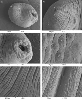
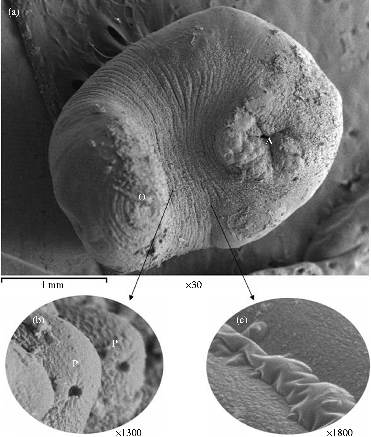
Adult P. microbothrium is pear-shaped with a broadly rounded posterior end and narrower anterior end (fig. 1a). The oral aperture is terminally positioned surrounded by the oral sucker, the genital pore lies ventrally in the middle of the anterior third of the body (fig. 1b), and the acetabulum is subterminally positioned (fig. 1c). The anterior third of the body has clearly visible concentric aggregations of dome-shaped papillae (fig. 1d). These papillae are very densely arranged towards the oral end, but decrease gradually toward the middle of the body before disappearance. Around the acetabular aperture, the tegument has folds irregularly directed with prominent domed papillae arranged singly or in groups of a few between the folds (fig. 1c). The tegument covering the body is transversely folded or ridged (fig. 1e), the folds being closer and more numerous near the anterior end, decreasing in number posteriorly. At higher magnification, the tegumental folds form microridges, which anastomose and interdigitate in a complex pattern (fig. 1f). No significant differences were observed between fresh and control flukes incubated for 24 h in solvent (0.18% (v/v) DMSO).
Scanning electron micrographs (SEM) of normal fresh flukes. (a) Entire worm (ventral view) showing broadly rounded posterior end and narrower anterior end. O, oral sucker; G, genital pore; A, acetabulum. (b) Anterior third of the body, showing concentric aggregations of dome-shaped papillae densely arranged towards the oral end. O, oral sucker; G, genital pore. (c) Posterior third of the body, showing subterminally positioned acetabulum with domed papillae. A, acetabulum. (d) Anterior third of the body, at higher magnification, showing clearly visible aggregations of dome-shaped papillae. P, papilla. (e) The tegument, showing folds or ridges, closer to each other near the anterior end of the fluke. (f) The tegumental folds, at higher magnification, showing microridges, which anastomose and interdigitate in a complex pattern.

Effects of artemether
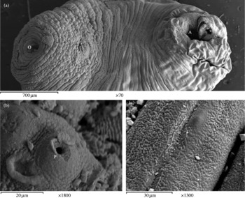
After 24 h of incubation with 10 μg ml− 1 artemether, tegumental damage of both anterior and posterior ends of the fluke had occurred in the majority of examined specimens (fig. 2a). The oral sucker was distorted and retracted, and the papillae surrounding the oral aperture were ruptured. Most of the papillae at the anterior third of the fluke's body appeared to have burst, causing a number of pits (fig. 2b). The tegument at the acetabular region was more swollen than normal so that the papillae appeared to be sunken (fig. 2a). At the mid-body region, the tegument had a slightly swollen appearance (fig. 2c).
Scanning electron micrographs of adult P. microbothrium following 24 h of incubation in 10 μg ml− 1 artemether. (a) Entire worm (ventral view) revealing damage of both anterior and posterior ends, distortion of the oral sucker, and tegumental swelling at the acetabular region so that the papillae appeared to be sunken. O, oral sucker; G, genital pore; A, acetabulum. (b) A ruptured papilla (under high magnification). P, papilla. (c) The tegumental swelling at the mid-body region.

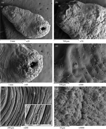
After 24 h of incubation with 20 μg ml− 1 artemether, the tegumental disruption became more pronounced, and both oral sucker and acetabulum were severely distorted (fig. 3a–c). The tegumental swelling was similar to that described for the 10 μg ml− 1 concentration, except that it extended to the anterior third of the body. The papillae appeared to be submerged by the tegument, which gave the surface a smooth appearance. The tegument was characterized by a number of pits caused by rupture of papillae (fig. 3d). At the mid-body region, the tegument over the transverse folds had been stripped off to expose the basal lamina beneath (fig. 3e and f). At higher magnification, erosion of the surface had occurred to such an extent that no microridges remained, only a mass of fibrous structures (fig. 3g).
Scanning electron micrographs of adult P. microbothrium following 24 h of incubation in 20 μg ml− 1 artemether. (a) Entire worm (ventral view) showing pronounced tegumental disruption and distortion of both oral sucker and acetabulum. O, oral sucker; G, genital pore. (b) The tegumental swelling at the anterior third of the body. O, oral sucker; G, genital pore. (c) The severely distorted acetabulum. A, acetabulum. (d) The anterior third of the body, at high magnification, showing a smooth appearance of the tegument, which was marked by a number of pits caused by rupture of papillae. P, papilla. (e, f) The mid-body region. In this specimen, the tegument over the transverse folds had been stripped off to expose the basal lamina beneath. (g) A higher power SEM of the tegumental folds, showing erosion of the surface to such an extent that no microridges remained.

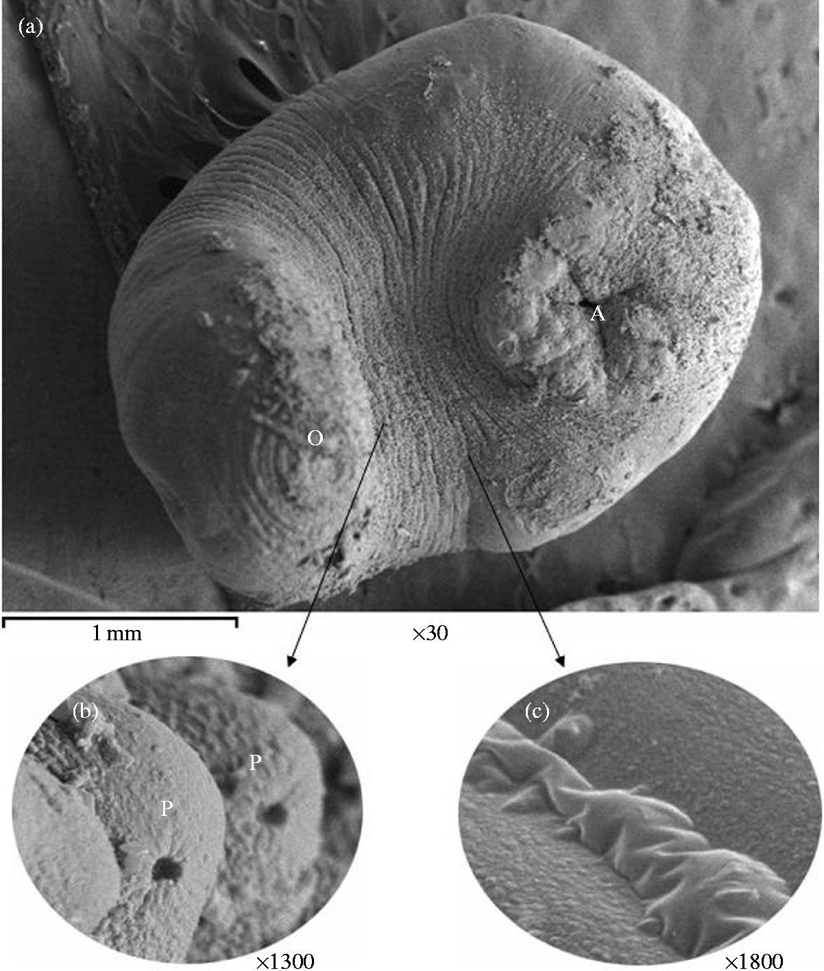
After 24 h of incubation with 30 μg ml− 1 artemether, in almost all specimens examined, gross swellings of the fluke's body, clearly visible to the naked eye, were observed. Damage to both oral sucker and acetabulum was so extreme that little recognizable structure remained (fig. 4a). Their positions were still apparent, but erosion of the surface had occurred. At the mid-body region, the papillae appeared to have burst, causing rounded holes (fig. 4b), and the areas between the transverse folds were swollen (fig. 4c).
Scanning electron micrographs of adult P. microbothrium following 24 h of incubation in 30 μg ml− 1 artemether. (a) Entire worm (ventral view) showing extreme damage of both oral sucker and acetabulum, as well as erosion of the surface. O, oral sucker; A, acetabulum. (b, c) A high power SEM of the mid-body region showing perforation of papillae and tegumental swelling in areas between the transverse folds. P, papilla.
Discussion
Previous studies have shown that artemether possesses a broad spectrum of activity against various trematodes (Utzinger & Keiser, Reference Utzinger and Keiser2007). In this study we have, for the first time, assessed the effect of artemether on P. microbothrium. Differences in response to artemether action were concentration dependent. The tegument swelling was believed to be part of the general response of the fluke to a stress situation, representing an attempt by the fluke to replace damaged surface membrane (Stitt & Fairweather, Reference Stitt and Fairweather1993). The swollen appearance of Paramphistomum tegument was due to a swelling of the basal infolds of the tegumental syncytium, the swelling became progressively more severe with higher concentration of artemether. Neither in vitro nor in vivo studies of drug action on Paramphistomum tegument have been carried out. Therefore, the results of artemether investigations against other helminths were applied for the evaluation and analysis of its effect on P. microbothrium in vitro. Such tegumental swelling induced by artemether has been described for different schistosome species (Xiao et al., Reference Xiao, Chollet, Utzinger, Matile, Mei and Tanner2001; Utzinger et al., Reference Utzinger, Xiao, Tanner and Keiser2007), F. gigantica (Shalaby et al., Reference Shalaby, El Namaky and Kamel2009) and F. hepatica (Keiser et al., Reference Keiser, Shu-Hua, Tanner and Utzinger2006b; Keiser & Morson, Reference Keiser and Morson2008). Moreover, a similar pattern of tegumental swelling of adult F. hepatica has been described following incubation with a number of anthelmintics, as recently summarized (Hegazi et al., Reference Hegazi, Abd El-Hady and Shalaby2007), and appeared to be a common feature of drug-treated parasites. The swelling of the tegument was possibly due to the disruption of ion pumps present on the apical plasma membrane (Skuce et al., Reference Skuce, Anderson and Fairweather1987). This action might be linked with the postulated action of artemether, including an interference with parasite sarcoplasmic/endoplasmic reticulum Ca2+-ATPase PfATP6 (SERCA), and disruption of mitochondrial function of the parasite (Golenser et al., Reference Golenser, Waknine, Krugliak, Hunt and Grau2006). Consequently, gross swellings of the body of the fluke, clearly visible to the naked eye, were observed in almost all specimens examined at 30 μg ml− 1 artemether. The damage that artemether caused to the tegumental surface would undoubtedly disrupt many of the physiological processes associated with the tegument, including osmoregulation, protection, secretion and synthesis (Halton, Reference Halton2004). Besides, the damage of the tegumental folds of the acetabular region might disrupt its function in drawing the rumen wall tissue of the host into the acetabular cavity (Nollen & Nadakavukaren, Reference Nollen and Nadakavukaren1974) resulting in a weak hold for the parasite.
In vitro studies on biologically related trematodes, F. hepatica and schistosomes, suggested that haem played a role in the mechanism of action of artemether (Xiao et al., Reference Xiao, Chollet, Utzinger, Matile, Mei and Tanner2001). The peroxide bond undergoes reductive activation by haem released by haemoglobin digestion by the parasite, producing carbon-centred free radicals (Golenser et al., Reference Golenser, Waknine, Krugliak, Hunt and Grau2006). Trematodes must ingest host haemoglobin for their nutrition, resulting in pigment formation in their intestines (Kloetzel & Lewert, Reference Kloetzel and Lewert1966). Therefore, it was reasonable to suggest that artemether could also be activated within trematodes by haem or other iron-compounds, producing toxic compounds and/or free iron radicals, as suggested by Butler et al. (Reference Butler, Gilbert, Hulme, Irvine, Renton and Whitwood1998).
The rupture of sensory papillae induced by artemether, in the present study, has also been observed in specimens of a closely related trematode, Cotylophoron cotylophorum, treated in vitro with praziquantel, after 30 h of exposure (Veerakumari & Munuswamy, Reference Veerakumari and Munuswamy1999). Domed papillae commonly occur in trematodes, and they have a sensory function (Tandon & Mattra, Reference Tandon and Mattra1982). These papillae, when present near the mouth, could be involved in contact reception during food detection or feeding; those found on the general body surface might function in recording pressure changes as the tegument stretches (Bennett, Reference Bennett1975). The domed papillae in the region of the genital pore probably have a specific sensory function during pairing for sexual reproduction (Bakke, Reference Bakke1976a, Reference Bakkeb). The damage that artemether causes to the papillae would undoubtedly disrupt many of these sensory functions.
In this study, the tegument of P. microbothrium has been shown to be very susceptible to the action of artemether. Furthermore, this drug induced profound changes to the tegument at relatively low concentrations. The ability of artemether to cause tegumental damage means that it effectively destroys the parasite's first and main line of defence, and also allows the drug potential access to other, internal tissues of the fluke, which will lead to more widespread damage. The use of artemether offers a new dimension and potential for control of such a neglected infectious disease in ruminants, at a time when paramphistomiasis has emerged as an important cause of productivity loss. Further studies of the in vitro effects of artemether on P. microbothrium are clearly warranted, also in vivo studies should be done and other peroxides tested. In addition, it would be interesting to see whether juvenile worms are also affected.