1. Introduction
Rare earth elements (REE) are reliable geochemical tracers and have been used successfully in oceanographic studies of redox conditions. Significantly, because of the substitution of REE for Ca, marine carbonates and phosphate particulates are often considered to be the archives of seawater or pore-fluid REE signals (Jarvis et al. Reference Jarvis, Burnett, Nathan, Almbaydin, Attia, Castro, Flicoteaux, Hilmy, Husain and Qutawnah1994; Shields & Stille, Reference Shields and Stille2001; Jiang et al. Reference Jiang, Zhao, Chen, Yang, Yang and Ling2007; Hood & Wallace, Reference Hood and Wallace2015; Tostevin et al. Reference Tostevin, Wood, Shields, Poulton, Guilbaud, Bowyer, Penny, He, Curtis, Hoffmann and Clarkson2016 b; Wallace et al. Reference Wallace, Hood, Shuster, Greig, Planavsky and Reed2017; Zhu & Jiang, Reference Zhu and Jiang2017). However, because of the complexity of the controlling factors involved in this process, interpretations without careful assessment can be equivocal. Earlier studies usually utilized a leaching procedure to extract the REE. However, Tostevin et al. (Reference Tostevin, Shields, Tarbuck, He, Clarkson and Wood2016 a) recently proved that this method might be compromised by clay or Fe oxide contaminations during digestion. An alternative approach is to use laser ablation – inductively coupled plasma – mass spectrometry (LA-ICP-MS) to directly analyse solid samples. The advantages of LA-ICP-MS analysis include comparatively easy measurements of multiple elements with low detection limits and the ability to determine micrometric trace-element variations that are undetectable in bulk-rock analyses. In fact, LA-ICP-MS has been demonstrated to be a powerful tool in understanding the superposition of geological events, the formation of mineral deposits and even several fundamental questions concerning the evolution of the Earth system (Bright et al. Reference Bright, Cruse, Lyons, MacLeod, Glascock and Ethington2009; Large et al. Reference Large, Halpin, Danyushevsky, Maslennikov, Bull, Long, Gregory, Lounejeva, Lyons, Sack, McGoldrick and Calver2014; Auer et al. Reference Auer, Reuter, Hauzenberger and Piller2017; Wallace et al. Reference Wallace, Hood, Shuster, Greig, Planavsky and Reed2017; Zhou et al. Reference Zhou, Wang, Fu, Ye, Wang, Su, Wang, Ge, Liang and Wei2017; Zhu et al. Reference Zhu, Wang, Ye, Wang, Huang, Zhu and Yang2019).
The goal of this study is to evaluate the effectiveness of using LA-ICP-MS for REE analysis of a lower Cambrian phosphate nodule preserved in a drill core from the Daotuo manganese deposit in Songtao County, northeastern Guizhou, South China. To our knowledge, this is the first lower Cambrian nodule reported from core material, which minimizes the influence of surface weathering. Previous geological and geochronological investigations of South China have provided an ideal framework for the P-enriched Niutitang Formation, such as the identification of the depositional condition and palaeolatitude (Chen et al. Reference Chen, Zhou, Fu, Wang and Yan2015 a; Yeasmin et al. Reference Yeasmin, Chen, Fu, Wang, Guo and Guo2017). Here, we emphasize that a combination of in situ LA-ICP-MS mapping and quantitative LA-ICP-MS spot analyses can provide unique information to determine the evolutionary history of a target.
2. Geological setting
The South China Craton, which consists of the Yangtze and Cathaysia blocks, developed a thick succession of well-studied Neoproterozoic and lower Cambrian strata (Wang & Li, Reference Wang and Li2003; Figs 1, 2). Palaeogeographic reconstruction of the upper Ediacaran Yangtze Block has revealed three sedimentary facies: (1) the shallow platform in the NW composed of carbonate layers of thickness > 100 m (the Dengying Formation); (2) the equivalent deep-water basin in the SE represented by the Liuchapo/Laobao cherts; and (3) a transitional slope characterized by mixed lithologies (Fig. 1; Steiner et al. Reference Steiner, Li, Qian, Zhu and Erdtmann2007). During early Cambrian time, the platform setting suffered widespread drowning and it subsequently evolved into a muddy shelf (Yeasmin et al. Reference Yeasmin, Chen, Fu, Wang, Guo and Guo2017). The Niutitang Formation, which overlies the Dengying/Liuchapo Formation, is dominated by high total organic carbon (TOC) (up to 15%) black shales (Zhai et al. Reference Zhai, Wu, Ye, Zhang and An2016). This black shale sequence also hosts discontinuous phosphate nodule, barite, Ni–Mo–PGE (platinum-group elements)–Au sulphide and V-rich deposits (Xu et al. Reference Xu, Lehmann, Mao, Qu and Du2011; Lehmann et al. Reference Lehmann, Frei, Xu and Mao2016). The polymetallic unit was once considered to be close to the Ediacaran–Cambrian boundary (Horan et al. Reference Horan, Morgan, Grauch, Coveney, Murowchick and Hulbert1994; Mao et al. Reference Mao, Lehmann, Du, Zhang, Ma, Wang, Zeng and Kerrich2002), but more recent radiometric ages of 532.3 ± 0.7 Ma (Jiang et al. Reference Jiang, Pi, Heubeck, Frimmel, Liu, Deng, Ling and Yang2009), 522.7 ± 4.9 Ma (Wang et al. Reference Wang, Shi, Jiang and Zhang2012), 522.3 ± 3.7 Ma and 524.2 ± 5.1 Ma (Reference Chen, Zhou, Fu, Wang and YanChen et al. 2015 a) from tuff beds at the base of the Niutitang Formation suggest that the polymetallic sulphide ore is much younger. Xu et al. (Reference Xu, Lehmann, Mao, Qu and Du2011) reported an Re–Os age of 521 ± 5 Ma for the polymetallic unit, which agrees well with the biostratigraphic Tommotian stage (or Stage 2–3). Stratigraphically downward, U–Pb ages of 536.3 ± 5.5 Ma (Chen et al. Reference Chen, Wang, Qing, Yan and Li2009), 542.1 ± 5.0 Ma and 542.6 ± 3.7 Ma (Chen et al. Reference Chen, Zhou, Fu, Wang and Yan2015 a) from the underlying Liuchapo Formation shift the position of the Ediacaran–Cambrian boundary within the Liuchapo Formation.
Palaeogeographic map showing the distribution of various ore deposits on the Yangtze Block during early Cambrian time (McKerrow et al. Reference McKerrow, Scotese and Brasier1992; Lehmann et al. Reference Lehmann, Frei, Xu and Mao2016; Yeasmin et al. Reference Yeasmin, Chen, Fu, Wang, Guo and Guo2017). P – phosphorite.

Generalized litho- and bio-stratigraphy of the upper Ediacaran–lower Cambrian strata in South China (Wang et al. Reference Wang, Shi, Jiang and Zhang2012).

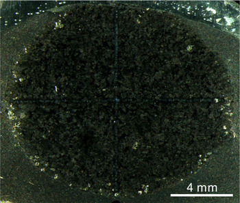
The Daotuo drill site, where we obtained the nodule sample, is located in Songtao County, c. 90 km NW of Tongren City (Fig. 1). The core covers a complete succession from the Sturtian-aged Tiesi’ao Formation to the lower Cambrian Bianmachong Formation. The phosphate nodule investigated in this study was preserved in the basal part of the Niutitang Formation, just below the polymetallic sulphide ore horizon (Figs 2, 3).
Phosphate nodule thin-section under reflected light.

3. Methods
LA-ICP-MS was used to provide both imaging of the nodule and core-to-rim quantitative spot analysis. The REE contents were quantitatively measured at the Ministry of Education, Key Laboratory of Orogenic Belts and Crustal Evolution, School of Earth and Space Sciences, Peking University. The instruments used include a 193-nm excimer LA system (COMPexPro 102) and an Agilent 7500ce ICP-MS. Masses of 139La, 140Ce, 141Pr, 146Nd, 147Sm, 153Eu, 157Gd, 159Tb, 163Dy, 165Ho, 166Er, 169Tm, 172Yb and 175Lu were determined and calibrated through internal standardization of 43Ca, assuming 48.02% CaO (Jarvis et al. Reference Jarvis, Burnett, Nathan, Almbaydin, Attia, Castro, Flicoteaux, Hilmy, Husain and Qutawnah1994). The NIST 610 standard was used as a reference. Although this NIST glass is not matrix matched to the phosphate sample, it offers evident advantages compared with various matrix-matched materials (Auer et al. Reference Auer, Reuter, Hauzenberger and Piller2017). Two other quality-control standards were analysed: NIST 612 and NIST 614. In total, 66 and 67 spots (60 μm in size) in the horizontal and vertical directions of the oval nodule were ablated, respectively. The GLITTER software (version 4.4.2) was used to obtain time-averaged REE concentrations. Reference materials ran alongside the samples were within 10% of the reported values for each element.
The resulting REE abundances were normalized to Post-Archaean Average Shale (PAAS; Taylor & McLennan, Reference Taylor and McLennan1985) to remove the odd–even effect of element distributions and to produce curves in which enrichments and depletions are apparent. Because of excess La in seawater, the conventional Ce/Ce* calculation, Ce/Ce* = 2CeN/(LaN + PrN), can lead to incorrect Ce anomalies. The Ce/Ce* values presented here were therefore calculated geometrically by extrapolating back from Pr and Nd, that is, Ce/Ce* = CeN/(PrN × PrN/NdN), as suggested by Lawrence et al. (Reference Lawrence, Greig, Collerson and Kamber2006).
The imaging experiment was conducted at the Key Laboratory of Petroleum Geochemistry, Research Institute of Petroleum Exploration and Development, using an Analyte Excite 193-nm excimer LA system coupled with an iCAP Q ICP-MS. A detailed description of the operating parameters can be found in Wang et al. (Reference Wang, Zhang, Ye, Wang, Zhou and Su2016), including the laser energy, spot size, scan rate, gas flow and radio frequency power. LA-ICP-MS imaging of an approximately 0.9 cm2 area covering one-quarter of the total nodule was obtained to display two-dimensional maps of element distributions. The raw LA-ICP-MS data were evaluated by means of factor analysis to assess the inter-element relationships. After calibration of variables and extraction of principal components, the factor axes were optimized using the Varimax rotation method.
4. Results
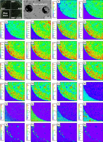
As illustrated in Figure 4, the LA-ICP-MS mapping reveals that the nodule is highly enriched in Ca, P, Sr and REE, but is depleted in Al, Si, Sc, Ti and Zr compared with its shale matrix. Notably, the REE distributions exhibit a concentric structure with moderate REE contents in the inner zone and elevated levels in the outer zone. Other elements – Fe, S, As and Se – are only concentrated between the nodule and the surrounding shale. Factor analysis of the raw LA-ICP-MS dataset led to the extraction of three components, explaining 64.1% of the total variance (Fig. 5; online Supplementary Tables S1 and S2, available at http://journals.cambridge.org/geo).
Optic, electronic (BSE) and LA-ICP-MS images of the studied nodule. Note that the white dashed lines indicate the routes of spot analysis and the white box represents the mapping area. The scales of LA-ICP-MS images are counts-per-second (CPS).

Principal component analysis based on the raw LA-ICP-MS dataset.

For quantitative spot analysis, the concentrations of most of the REE as well as the calculated Ce anomalies exhibit systematic variations in both the horizontal and vertical directions of the nodule (Fig. 6; online Supplementary Table S3). Specifically, Ce anomalies decrease gradually from the centre to the edge (Ce/Ce* = 0.8 to 0.92), while the REE contents generally increase towards the rim. This feature is significant for light REE (LREE) and MREE, but is not significant for heavy REE (HREE). The PAAS-normalized REE patterns are characterized by striking MREE enrichments (DyN/SmN = 0.61 to 1.31), with no apparent variation along the transections (Figs 6, 7).
Horizontal (A01–A66) and vertical (B01–B67) profiles of Ce/Ce*, ΣREE and DyN/SmN. See Figure 4 for trails of the spots.

PAAS-normalized REE patterns of phosphate nodule.

5. Discussion
5.a. Principal component analysis
Principal component analysis (PCA), which enables dimension reduction of multivariate datasets, is widely used to understand element enrichments and their governing processes in geological environments (e.g. Gregory et al. Reference Gregory, Large, Halpin, Lounejeva Baturina, Lyons, Wu, Danyushevsky, Sack, Chappaz, Maslennikov and Bull2015; Ahm et al. Reference Ahm, Bjerrum and Hammarlund2017; Ye et al. Reference Ye, Wang, Wang, Zhai, Wu and Zhang2020). PCA enables the investigation of how different variables vary with each other, and the grouping of them into principal components. The first component in our PCA model includes two groups: PC1 (Ca, P and Sr) and negative PC1 (Al, Si, Sc, Ti and Zr). The former represents carbonate fluorapatite (CFA), while the latter corresponds to detrital components of the black shale host, such as quartz, rutile, zircon and other silicate minerals. The comparable distributions of Sr and Ca indicate extensive substitution of Sr2+ for Ca2+ with no change in charge balance (Figs 4, 5; Chakhmouradian et al. Reference Chakhmouradian, Reguir, Zaitsev, Couëslan, Xu, Kynický, Mumin and Yang2017).
Component 2 consists of the entire REE series, which share very similar physical and chemical properties. This uniformity arises from the nature of their electronic configurations, resulting in a primarily stable oxidation state. The small differences in this set of elements can be attributed to a steady decrease in ionic radius with increasing atomic number (i.e. the lanthanide contraction). REE can reside in CFA through the substitution of REE3+ for Ca2+ or through direct attachment to crystal surface (Jarvis et al. Reference Jarvis, Burnett, Nathan, Almbaydin, Attia, Castro, Flicoteaux, Hilmy, Husain and Qutawnah1994; Reynard et al. Reference Reynard, Lecuyer and Grandjean1999). Fleet et al. (Reference Fleet, Liu and Pan2000) found that both the ninefold-coordinated Ca1 site and the sevenfold-coordinated Ca2 site can accommodate significant amounts of REE. The REE signatures retained by CFA are believed to be critical for determining the environmental dynamics, as discussed in the following section.
Component 3 includes Fe, S, As and Se, which are typical elements within the structure of pyrite. In organic-rich deposits, syngenetic and diagenetic pyrites have been recognized as sinks of several trace metals (Large et al. Reference Large, Halpin, Danyushevsky, Maslennikov, Bull, Long, Gregory, Lounejeva, Lyons, Sack, McGoldrick and Calver2014; Gregory et al. Reference Gregory, Large, Halpin, Lounejeva Baturina, Lyons, Wu, Danyushevsky, Sack, Chappaz, Maslennikov and Bull2015). The incorporation of As into pyrite can occur in two different ways: (1) the substitution of As− for S in the S unit; or (2) the substitution of As3+ for Fe2+ (Reich & Becker, Reference Reich and Becker2006; Deditius et al. Reference Deditius, Utsunomiya, Renock, Ewing, Ramana, Becker and Kesler2008; Neumann et al. Reference Neumann, Scholz, Kramar, Ostermaier, Rausch, Berner and Immenhauser2013). Selenium concentration of pyrite is primarily regulated by its substitution for S. Experimental studies have shown that up to 99.5% Se in solution could be taken up by pyrite precipitation, demonstrating the high affinity of Se for Fe sulphides (Diener et al. Reference Diener, Neumann, Kramar and Schild2012). Notably, the pyrites formed around the periphery of our nodule are most likely the consequence of late-stage sulphate reduction and void filling (Fig. 3).
5.b. Interpretations of REE patterns
5.b.1. Ce anomaly
Cerium is the only REE that undergoes redox transformation under low-temperature conditions. The oxidation of dissolved Ce3+ to form insoluble Ce4+ takes place in the modern oxygenated water column through coating of organic matter and/or Mn–Fe oxides, resulting in lower Ce concentrations in the deep ocean relative to La and Pr, which is expressed as a negative Ce anomaly. Under reducing conditions, the insoluble Ce4+ is converted back into soluble Ce3+, which behaves similarly to the other REEs. In this case, little or no inter-element fractionation is identified (Elderfield & Sholkovitz, Reference Elderfield and Sholkovitz1987; Moffett, Reference Moffett1990; Alibo & Nozaki, Reference Alibo and Nozaki1999; Bau & Koschinsky et al. Reference Bau and Koschinsky2009).
Since phosphate nodules grow beneath the sediment–water interface, it is reasonable to assume that the systematic variations in Ce/Ce* and the total REE contents (ΣREE) are the result of progressive diagenetic alternation. Shields & Stille (Reference Shields and Stille2001) proposed that the diagenetic reaction can be evaluated using plots of Ce/Ce* versus ΣREE and DyN/SmN because diagenetic REE exchange with host sediments would erase the original negative Ce anomaly and induce greater REE enrichment and MREE arching. Accordingly, Ce/Ce* is expected to correlate positively with ΣREE and negatively with DyN/SmN. However, as shown in Figure 8, neither of these relationships was observed for the studied sample. Instead, there is a negative correlation between Ce/Ce* and ΣREE. If diagenesis did play a role in producing the elevated REE concentrations, the nodule rim, which underwent severe diagenetic addition of REE, should have more muted Ce anomalies compared with its core. Such a scenario contradicts our data, where the most prominent negative Ce anomaly is present in the rim concurrent with the relatively higher REE abundances (Fig. 6); other explanations for these variations are therefore required.
Cross-plots of Ce/Ce* versus ΣREE and DyN/SmN.

Based on observations of the modern environment, two hypotheses are proposed to account for the Ce/Ce* and ΣREE profiles of the nodule. First, we note that there could be a significant difference between the inner and outer parts in terms of the exposure time to pore water. As CFA crystallites grew, the inner part would be impermeable and more closed, while the outer part was still in contact with ambient fluid, resulting in the incorporation of REE into the rim (Ilyin Reference Ilyin1998; Zhu et al. Reference Zhu, Jiang, Yang, Pi, Ling and Chen2014). It should be emphasized that, unlike the late diagenetic modification discussed above, this process must occur at a very early stage when pore water was not completely isolated from seawater and possessed a prominent negative Ce anomaly. The outer zone, which experienced a stronger exchange with the pore water, therefore exhibits lower Ce/Ce* (i.e. is more oxygenated). This hypothesis also explains why the core-to-rim increasing trend is less pronounced for HREE than for LREE and MREE (Fig. 4; online Supplementary Table S3). REE mainly exist as carbonate complexes in seawater. The stability of REE – carbonate ion complexes and the opportunity for particulate adsorption vary inversely through the REE series (Koeppenkastrop & De Carlo, 1992; Sholkovitz, Reference Sholkovitz1992; Sholkovitz et al. Reference Sholkovitz, Landing and Lewis1994). As a consequence, LREE and MREE with higher mobilities were preferentially bonded to the CFA surface, whereas HREE were retained in the solution.
Second, the changes in Ce/Ce* and ΣREE may be related to changes in the water depth. Modern coastal and marine surface waters generally have little or no negative Ce anomalies, but the Ce/Ce* values decrease steadily with increasing depth to form a more typical Ce deficit. The REE contents also exhibit systematic variations with increasing water depth (Alibo & Nozaki, Reference Alibo and Nozaki1999; Deng et al. Reference Deng, Ren, Guo, Cao, Wang and Liu2017). The observed Ce anomalies are therefore considered to be a function of bathymetry. Importantly, this assumption holds true only if the pore water always had a good connection to the overlying seawater during precipitation of CFA.
5.b.2. MREE enrichment
Most of our analysed spots are markedly enriched in MREE (Fig. 7). Several models have been suggested to explain the mechanisms of this pattern. One possibility is that the MREE are preferentially taken up and are substituted for Ca in the CFA lattice. The partition coefficients of the REE between apatite and melt exhibit a convex-upwards shape, indicating that apatite accommodates MREE more readily than LREE or HREE (Reynard et al. Reference Reynard, Lecuyer and Grandjean1999; Klemme & Dalpé, Reference Klemme and Dalpé2003). However, seawater-like REE distributions are known to be preserved in many modern and ancient P-enriched deposits (Toyoda & Tokonami, Reference Toyoda and Tokonami1990; Jiang et al. Reference Jiang, Zhao, Chen, Yang, Yang and Ling2007; Zhu et al. Reference Zhu, Jiang, Yang, Pi, Ling and Chen2014; Xin et al. Reference Xin, Jiang, Yang, Wu and Pi2015; Zhai et al. Reference Zhai, Wu, Ye, Zhang and An2016); the substitution model alone may therefore not explain all of the MREE enrichments.
Another possibility is that this pattern represents the REE characteristics of organic matter. Several studies have revealed that organic colloids can dominate REE retention in aqueous systems (Elderfield et al. Reference Elderfield, Upstill-Goddard and Sholkovitz1990; Sholkovitz, Reference Sholkovitz1992; Stolpe et al. Reference Stolpe, Guo and Shiller2013). An extraction experiment by Freslon et al. (Reference Freslon, Bayon, Toucanne, Bermell, Bollinger, Chéron, Etoubleau, Germain, Khripounoff, Ponzevera and Rouget2014) demonstrated that sedimentary organic compounds from different environments share similar MREE-enriched patterns. The remineralization of organic particles in highly productive areas or during diagenesis is capable of imparting its signature to the dissolved load. Authigenic CFA precipitated in equilibrium with the evolved fluid would record the organic REE patterns accordingly. Based on the apparent black opaque appearance of our sample, the REE distributions are at least partially controlled by organic component. However, some organic-poor phosphorites from this time are also enriched in MREE (Xin et al. Reference Xin, Jiang, Yang, Wu and Pi2015; Zhu & Jiang, Reference Zhu and Jiang2017), suggesting that there are other REE sources in addition to the organic matter. Furthermore, the REE compositions of kerogen in the Niutitang black shales show diverse patterns instead of a single MREE bulge, which might be related to different contributions of marine biomass (Pi et al. Reference Pi, Liu, Shields-Zhou and Jiang2013).
The reduction of Fe oxides can also be a potential driver of elevated MREE concentrations. In sediments off the coast of Peru and on the California margin, the coincidence of pore water exhibiting the MREE bulge with the peak of dissolved Fe production suggests that Fe oxides are the carriers of this MREE signal (Johannesson & Zhou, Reference Johannesson and Zhou1999; Haley et al. Reference Haley, Klinkhammer and McManus2004). Accordingly, MREE enrichment is sometimes considered to be the diagnostic pattern for ferruginous environments (e.g. Kim et al. Reference Kim, Torres, Haley, Kastner, Pohlman, Riedel and Lee2012; Chen et al. Reference Chen, Algeo, Zhao, Chen, Cao, Zhang and Li2015 b; Ye et al. Reference Ye, Wang, Wang, Zhai, Wu and Zhang2020). For example, MREE enrichments of the Mesoproterozoic Xiamaling carbonate concretions (Liu et al. Reference Liu, Tang, Shi, Zhou, Zhou, Shang, Li and Song2019) and the Ediacaran Doushantuo cap carbonates (Wu et al. Reference Wu, Jiang, Palmer, Wei and Yang2019) are both attributed to metal oxide reduction. Since carbonate samples generally have low TOC, it is reasonable to exclude organic matter as a candidate. But for our nodule, assigning the REE pattern to individual sources is still challenging. Future research, such as sequential leaching experiment, may provide new insights into the causes of such MREE enrichment pattern.
Interestingly, among these hypotheses regarding MREE enrichment, some researchers have speculated that seawater of pre-Cenozoic oceans was itself MREE-enriched (Grandjean-Lécuyer et al. Reference Grandjean-Lécuyer, Feist and Albarède1993; Ilyin, Reference Ilyin1998; Lécuyer et al. Reference Lécuyer, Reynard and Grandjean2004; Emsbo et al. Reference Emsbo, McLaughlin, Breit, du Bray and Koenig2015). Ilyin (Reference Ilyin1998) found that almost all Proterozoic–Cambrian phosphorites have a so-called old-phosphorite REE type, which contains a negative Ce anomaly and remarkable HREE depletion. However, this secular variation idea was strongly criticized by Shields & Webb (Reference Shields and Webb2004), given that contemporaneous calcites have retained REE signals similar to the modern seawater pattern. They claimed that this HREE deficiency and other non-seawater-like features were likely derived from post-depositional exchange or non-quantitative uptake of REE.
Here, we propose that the conflicting views about seawater REE composition can be reconciled through a chemically stratified model (Fig. 9). A growing amount of evidence has demonstrated that Precambrian and early Cambrian oceans were characterized by extreme spatial heterogeneity and stratification (Li et al. Reference Li, Love, Lyons, Fike, Sessions and Chu2010; Poulton et al. Reference Poulton, Fralick and Canfield2010; Jin et al. Reference Jin, Li, Algeo, Planavsky, Cui, Yang, Zhao, Zhang and Xie2016; Zhang et al. Reference Zhang, Wang, Wang, Bjerrum, Hammarlund, Costa, Connelly, Zhang, Su and Canfield2016; Hammarlund et al. Reference Hammarlund, Gaines, Prokopenko, Qi, Hou and Canfield2017). Because of the redox control on REE behaviour, the REE patterns of ancient seawater should be comparable to those of modern pore water, which has been verified to record discernible REE patterns within a fixed respiration sequence (i.e. oxic, nitrogenous, manganous, ferruginous, sulphidic and methanic; Haley et al. Reference Haley, Klinkhammer and McManus2004; Canfield & Thamdrup, Reference Canfield and Thamdrup2009; Kim et al. Reference Kim, Torres, Haley, Kastner, Pohlman, Riedel and Lee2012; Li et al. Reference Li, Cheng, Algeo and Xie2015). Marine minerals precipitated in different locations of the ocean would therefore carry different REE signals (that is to say, the ferruginous condition mentioned above might not have been restricted to pore water). In the modern oceans, the massive sedimentation of carbonates occurs on tropical and subtropical continental shelves, referred to as the carbonate factory (Bosscher & Schlager, Reference Bosscher and Schlager1992). The REE distributions of these carbonates are inherited from the oxic surface waters. In contrast, the depositions of contemporaneous phosphorites are more constrained to marginal settings beneath highly productive upwelling currents (as discussed further in the following section), although some may suffer reworking and winnowing. With regard to early Cambrian oceans, such environments could be close to the oxygen-deficient manganous and/or ferruginous or even sulphidic zone (Pufahl & Hiatt, Reference Pufahl and Hiatt2012). Indeed, the stratification of REE has been identified in oceans during Proterozoic to early Cambrian time (Planavsky et al. Reference Planavsky, Bekker, Rouxel, Kamber, Hofmann, Knudsen and Lyons2010; Hood & Wallace, Reference Hood and Wallace2015; Tostevin et al. Reference Tostevin, Wood, Shields, Poulton, Guilbaud, Bowyer, Penny, He, Curtis, Hoffmann and Clarkson2016 b; Wu et al. Reference Wu, Jiang, Palmer, Wei and Yang2019). A fundamental change of marine redox state during late Phanerozoic time might have ultimately led to a uniform seawater REE pattern.
A conceptual model for Precambrian and early Cambrian oceans (Canfield & Thamdrup, Reference Canfield and Thamdrup2009; Li et al. Reference Li, Cheng, Algeo and Xie2015). The left panel represents the evolution of REE within different redox zones (modified from Deng et al. Reference Deng, Ren, Guo, Cao, Wang and Liu2017).

5.c. Mechanisms of phosphogenesis
The main realms of modern phosphorite formations are the major upwelling systems along continental margins, such as on the western coasts of Peru and Chile (Manheim et al. Reference Manheim, Rowe and Jipa1975; Burnett, Reference Burnett1977; Glenn & Arthur, Reference Glenn and Arthur1988). The initial enrichments of P in these settings are orchestrated by a complex interplay between several processes, including organic matter degradation, Fe oxide pumping and microbial activity.
The role of microbes in phosphogenesis has particularly attracted the attention of scientists in recent years. For sediments beneath the Benguela upwelling area off the coast of Namibia, Schulz & Schulz (Reference Schulz and Schulz2005) reported the co-occurrence of a narrow horizon of CFA, a spike in the dissolved phosphate content and an aggregation of giant sulphide-oxidizing bacteria Thiomargarita namibiensis. T. namibiensis is known to be capable of accumulating polyphosphate intracellularly under oxic conditions, then hydrolysing the polyphosphate and releasing phosphate when the surrounding water becomes anoxic (Schulz & Schulz, Reference Schulz and Schulz2005). Moreover, Goldhammer et al. (Reference Goldhammer, Brüchert, Ferdelman and Zabel2010) found that sulphide-oxidizing bacteria can collect 33P-labelled phosphate into their cells and catalyse nearly instantaneous conversion of phosphate to apatite. Brock & Schulz-Vogt (Reference Brock and Schulz-Vogt2011) investigated parameters that could stimulate the decomposition of polyphosphate in a marine Beggiatoa strain, and concluded that sulphide exposure would trigger phosphate release by Beggiatoa. Overall, under alternating oxic and anoxic regimes, sulphur bacteria appear to have a remarkable effect on focusing pore-water phosphate. Such a condition could easily have been achieved in Ediacaran–Cambrian oceans, in which oxidant supply and oxygen level were relatively unstable, fostering frequent redox oscillations, vigorous microbial activity and subsequent pore-water phosphate build-up.
Indeed, filamentous microfossils that resemble modern sulphide-oxidizing bacteria were reported from the Ediacaran Doushantuo Formation in South China (Bailey et al. Reference Bailey, Corsetti, Greene, Crosby, Liu and Orphan2013). These fossils contain opaque inclusions that represent putative relict S globules. Although such fossils are not observed in our nodule, their findings reflect that sulphur bacteria, which are known to mediate CFA precipitation in modern environments, might have been present in phosphogenic settings during the Ediacaran–Cambrian period.
6. Conclusions
In summary, the observed core-to-rim Ce/Ce* variations of a phosphate nodule from the basal part of the Niutitang Formation are interpreted to represent REE exchange with pore water at a very early stage or they might be correlated with increasing water depth during progressive growth of the nodule. The rare presence of pyrite within the nodule indicates that CFA was precipitated before extensive sulphate reduction. Causes of the MREE enrichment are still enigmatic. Potential mechanisms include preferential MREE substitution for Ca, degradation of organic matter and deposition under ferruginous environments. It is likely that the observed MREE patterns are the result of a combination of these mechanisms. Phosphate nodules deposited on continental margins during early Cambrian time were subject to a mid-depth chemical zonation with high organic loading from the surface waters. Such a scenario is consistent with the idea that transient redox switches can play an important role in phosphate accumulation.
Supplementary Material
To view supplementary material for this article, please visit https://doi.org/10.1017/S0016756820000850
Acknowledgements
We thank Fang Ma for laboratory assistance. Financial support was provided by the National Key Research and Development Program of China (2017YFC0603101), the National Science and Technology Major Project of the Ministry of Science and Technology of China (2016ZX05004001), the National Natural Science Foundation of China (41530317, 41872125), the Strategic Priority Research Program of the Chinese Academy of Sciences (XDA14010101) and the Scientific Research and Technological Development Project of CNPC (2016A-0200). This paper benefited greatly from the comments of Malcolm Wallace, Gerald Auer and an anonymous reviewer.
Declaration of Interest
The authors declare no competing interests.