Introduction
Functional neuroanatomical models of post-traumatic stress disorder (PTSD) have been shaped by findings of decreased activation of the medial prefrontal cortex (mPFC) and increased activation of the amygdala in response to trauma- or fear-related stimuli (for reviews see Shin et al. Reference Shin, Orr, Carson, Rauch, Macklin, Lasko, Peters, Metzger, Dougherty, Cannistraro, Alpert, Fischman and Pitman2004, Reference Shin, Rauch and Pitman2006; Liberzon & Martis, Reference Liberzon and Martis2006; Etkin & Wager, Reference Etkin and Wager2007; Francati et al. Reference Francati, Vermetten and Bremner2007; Liberzon & Sripada, Reference Liberzon and Sripada2008). Medial prefrontal hyporesponsivity in PTSD has been demonstrated in various types of functional neuroimaging studies (Liberzon & Sripada, Reference Liberzon and Sripada2008), including those that involve script-driven imagery, which is a technique that uses brief autobiographical narratives of personal life events (i.e. ‘scripts’) as stimuli. During script-driven imagery, participants recall and visualize these events while psychophysiologic (Pitman et al. Reference Pitman, Orr, Forgue, de Jong and Claiborn1987, Reference Pitman, Orr, Forgue, Altman, de Jong and Herz1990; Orr et al. Reference Orr, Lasko, Metzger, Berry, Ahern and Pitman1998) and/or brain responses are measured. Studies of brain responses during traumatic versus neutral imagery in PTSD groups, relative to trauma-exposed, non-PTSD groups, have revealed diminished activation in the mPFC, including the anterior cingulate cortex (ACC), medial frontal gyrus (MFG) and subcallosal gyrus (Bremner et al. Reference Bremner, Narayan, Staib, Southwick, McGlashan and Charney1999a; Shin et al. Reference Shin, McNally, Kosslyn, Thompson, Rauch, Alpert, Metzger, Lasko, Orr and Pitman1999; Lanius et al. Reference Lanius, Williamson, Densmore, Boksman, Gupta, Neufeld, Gati and Menon2001, Reference Lanius, Frewen, Girotti, Neufeld, Stevens and Densmore2007; Liberzon et al. Reference Liberzon, Britton and Phan2003; Lindauer et al. Reference Lindauer, Booij, Habraken, Uylings, Olff, Carlier, den Heeten, van Eck-Smit and Gersons2004; Britton et al. Reference Britton, Phan, Taylor, Fig and Liberzon2005).
Neuroimaging studies of PTSD have also found similar patterns of brain activation in response to generic (non-personalized), trauma-related stimuli. Diminished mPFC activation has been shown in response to such stimuli as trauma-related sounds and pictures (Bremner et al. Reference Bremner, Staib, Kaloupek, Southwick, Soufer and Charney1999b; Yang et al. Reference Yang, Wu, Hsu and Ker2004; Hou et al. Reference Hou, Liu, Wang, Li, Liang, He, Liu, Zhang, Li and Jiang2007), combat-related words (Shin et al. Reference Shin, Whalen, Pitman, Bush, Macklin, Lasko, Orr, McInerney and Rauch2001), and abuse-related words (Bremner et al. Reference Bremner, Vermetten, Vythilingam, Afzal, Schmahl, Elzinga and Charney2004). Similarly, mPFC hyporesponsivity in PTSD has been associated with trauma-unrelated emotional stimuli, such as aversive photographs (Phan et al. Reference Phan, Britton, Taylor, Fig and Liberzon2006) and task-irrelevant, threat-related pictures (Kim et al. Reference Kim, Chey, Chung, Bae, Khang, Ham, Yoon, Jeong and Lyoo2008), in addition to fearful facial expressions, which have also been associated with amygdala hyperresponsivity in PTSD (Rauch et al. Reference Rauch, Whalen, Shin, McInerney, Macklin, Lasko, Orr and Pitman2000; Shin et al. Reference Shin, Wright, Cannistraro, Wedig, McMullin, Martis, Macklin, Lasko, Cavanagh, Krangel, Orr, Pitman, Whalen and Rauch2005; Williams et al. Reference Williams, Kemp, Felmingham, Barton, Olivieri, Peduto, Gordon and Bryant2006). Decreased activation in ventromedial PFC during extinction recall, another trauma-unrelated condition in which extinguished conditioned stimuli were presented in the absence of the fear stimulus, has also been demonstrated in PTSD (Milad et al. Reference Milad, Pitman, Ellis, Gold, Shin, Lasko, Zeidan, Handwerger, Orr and Rauch2009).
A question that has received relatively little attention is whether patterns of mPFC hyporesponsivity and amygdala hyperresponsivity in PTSD extend to trauma-unrelated, yet stressful, personal stimuli. An initial neuroimaging study that addressed this question used script-driven imagery and functional magnetic resonance imaging (fMRI) to examine neural responses to the recollection and imagery of both trauma-related and other emotional (i.e. sad and anxious) personal life events in a cohort of trauma-exposed civilians with versus without PTSD (Lanius et al. Reference Lanius, Williamson, Hopper, Densmore, Boksman, Gupta, Neufeld, Gati and Menon2003). Those with PTSD exhibited significantly less ACC activation during both trauma-related and trauma-unrelated emotional recollections.
In the current study, we used positron emission tomography (PET) and script-driven imagery in a cohort of male and female veterans with PTSD to determine whether mPFC hyporesponsivity and amygdala hyperresponsivity could be observed during the recollection and imagery of trauma-unrelated stressful personal life events compared to neutral personal life events. The current study was part of a larger script-driven imagery PET study in which we also examined regional cerebral blood flow (rCBF) responses to trauma-related versus neutral scripts (Shin et al. Reference Shin, Orr, Carson, Rauch, Macklin, Lasko, Peters, Metzger, Dougherty, Cannistraro, Alpert, Fischman and Pitman2004). In this previous study of 36 Vietnam veterans, including both male combat veterans (MCVs) and female nurse veterans (FNVs), we found decreased rCBF in MFG in the PTSD group for the traumatic versus neutral imagery comparison. In addition, rCBF changes in MFG were inversely correlated with rCBF changes in the amygdala. For the traumatic condition, PTSD symptom severity was inversely correlated with rCBF in MFG and positively correlated with rCBF in the right amygdala.
The present research consisted of a further analysis of the data obtained from 35 of the 36 participants in the previous study. Participants recalled and imagined trauma-unrelated stressful, neutral and traumatic personal life events in separate conditions. We compared rCBF during the trauma-unrelated stressful versus neutral imagery conditions, and also the trauma-unrelated stressful versus traumatic conditions. Participants' psychophysiologic data acquired during script-driven imagery were also analyzed to quantify arousal during the trauma-unrelated stressful imagery scans. Based on previous findings (Lanius et al. Reference Lanius, Williamson, Hopper, Densmore, Boksman, Gupta, Neufeld, Gati and Menon2003; Shin et al. Reference Shin, Orr, Carson, Rauch, Macklin, Lasko, Peters, Metzger, Dougherty, Cannistraro, Alpert, Fischman and Pitman2004), we predicted that the Vietnam veterans with current PTSD, compared to those without a history of PTSD, would exhibit diminished activation in medial prefrontal regions (i.e. MFG and ACC) during the trauma-unrelated stressful condition compared to the neutral condition. Given that Lanius et al. (Reference Lanius, Williamson, Hopper, Densmore, Boksman, Gupta, Neufeld, Gati and Menon2003), using fMRI, reported similar patterns of decreased anterior cingulate activation during traumatic and non-traumatic emotional recollections and imagery in PTSD, we did not necessarily expect to find significant differences between the trauma-unrelated stressful and traumatic imagery conditions.
Regarding the amygdala, heightened responsivity has been more consistently linked to the processing of external (e.g. pictures) as opposed to internal (e.g. mental imagery) stimuli in healthy populations (Lane et al. Reference Lane, Reiman, Ahern, Schwartz and Davidson1997; Damasio et al. Reference Damasio, Grabowski, Bechara, Damasio, Ponto, Parvizi and Hichwa2000; Phan et al. Reference Phan, Wager, Taylor and Liberzon2002), whereas mPFC activation does not seem to depend on the external versus internal nature of affective stimuli (Reiman et al. Reference Reiman, Lane, Ahern, Schwartz, Davidson, Friston, Yun and Chen1997). Based on our focus on the neural processing of script-driven imagery and previous results in PTSD (Lanius et al. Reference Lanius, Williamson, Hopper, Densmore, Boksman, Gupta, Neufeld, Gati and Menon2003; Shin et al. Reference Shin, Orr, Carson, Rauch, Macklin, Lasko, Peters, Metzger, Dougherty, Cannistraro, Alpert, Fischman and Pitman2004), we did not necessarily expect to find significant between-group differences in amygdala activation in the trauma-unrelated stressful versus neutral imagery comparison.
Method
Participants
Presented here are previously unanalyzed data from our earlier study (Shin et al. Reference Shin, Orr, Carson, Rauch, Macklin, Lasko, Peters, Metzger, Dougherty, Cannistraro, Alpert, Fischman and Pitman2004). One of the non-PTSD participants included in the original report was not included in the statistical analyses described here because she did not complete the PET scans for the trauma-unrelated stressful imagery condition.
Participants were 35 right-handed Vietnam veterans who had served in combat (MCVs) or as nurses in the combat theater (FNVs) and met DSM-IV criterion A for PTSD, as assessed with the Clinician-Administered PTSD Scale (CAPS; Weathers et al. Reference Weathers, Keane and Davidson2001). Presence or absence of the categorical PTSD diagnosis was determined according to DSM-IV diagnostic criteria (APA, 1994). Seventeen participants (seven MCV and 10 FNV; age 51.71±3.46 years) had current PTSD at the time of study (PTSD group), and 18 participants (nine MCV and nine FNV; age 53.22±2.78 years) had never had PTSD (non-PTSD group). Level of education was not significantly different between the PTSD group and the non-PTSD group [16.67±2.42 v. 16.91±1.65 years; t(33)=0.35, p=0.73]. Consistent with their diagnoses, the PTSD group exhibited significantly higher CAPS total scores than the non-PTSD group [73.9±20.5 v. 4.4±5.7; t(33)=13.8, p<0.001]. On the depression subscale of the Symptom Checklist-90-Revised (SCL-90-R; Derogatis, Reference Derogatis1983), the PTSD group exhibited higher scores than the non-PTSD group [1.68±0.96 v. 0.42±0.80; t(33)=4.2, p<0.001].
Axis I co-morbidity was assessed using the Structured Clinical Interview for DSM-IV (SCID; First et al. Reference First, Spitzer, Gibbon and Williams1995). Participants showed the following current co-morbidity: in the PTSD group, major depressive disorder (MDD; n=8), panic disorder (n=3), social phobia (n=2), specific phobia (n=2), binge eating disorder (n=1), and somatoform disorder (n=1); in the non-PTSD group, dysthymia (n=2), specific phobia (n=1), and somatoform disorder (n=1). All participants were without a history of head injury, neurological disorders, or other major medical conditions. No participant was taking psychotropic or cardiovascular medication at the time of study, and all urine drug screens were negative.
Each participant provided written informed consent. The Institutional Review Boards of the Massachusetts General Hospital (MGH) and the Veterans Affairs Medical Center, Manchester, NH approved this study.
Procedures
These were as described previously (Rauch et al. Reference Rauch, van der Kolk, Fisler, Alpert, Orr, Savage, Fischman, Jenike and Pitman1996; Shin et al. Reference Shin, McNally, Kosslyn, Thompson, Rauch, Alpert, Metzger, Lasko, Orr and Pitman1999, Reference Shin, Orr, Carson, Rauch, Macklin, Lasko, Peters, Metzger, Dougherty, Cannistraro, Alpert, Fischman and Pitman2004).
Script-driven imagery
Prior to PET scanning, participants provided written descriptions of two neutral, two Vietnam-related traumatic, and two trauma-unrelated stressful personal events (Pitman et al. Reference Pitman, Orr, Forgue, de Jong and Claiborn1987). For the trauma-unrelated stressful condition, personal events were not related to the index traumatic event and the participants' PTSD symptoms. Examples include going through a divorce; work-related stress, such as being removed from an occupational position; and getting caught driving a family member's car with no permission and no driver's license. Unlike the traumatic events, which were assessed on the basis of whether they met criterion A for PTSD, the trauma-unrelated stressful events were not formally assessed for this criterion. Scripts describing each event in the second-person, present tense were prepared by study staff and tape-recorded in a neutral voice for playback in the PET scanner. Each participant was studied in two scans per condition. Before each scan, participants were instructed to close their eyes and imagine the described event as vividly as possible, as if they were actually participating in it. After the script was played, oxygen-15-labeled carbon dioxide ([15O]CO2), administered via inhalation, began. PET data acquisition occurred for 60 s, during which participants continued to recall and imagine the event while inhaling the [15O]CO2. The order of script conditions was counterbalanced across participants, and PET scans were separated by at least 10 min to allow for radiation decay.
Psychophysiologic responses
Heart rate (HR), skin conductance (SC) and left lateral frontalis electromyographic (EMG) data were recorded with a modular instrument system (Coulbourn Instruments, USA) in the MGH PET laboratory according to established procedures (Pitman et al. Reference Pitman, Orr, Forgue, de Jong and Claiborn1987, Reference Pitman, Orr, Forgue, Altman, de Jong and Herz1990; Orr et al. Reference Orr, Lasko, Metzger, Berry, Ahern and Pitman1998; Shin et al. Reference Shin, Orr, Carson, Rauch, Macklin, Lasko, Peters, Metzger, Dougherty, Cannistraro, Alpert, Fischman and Pitman2004). Psychophysiologic measurements were recorded for 30 s before (baseline), 60 s during (imagery), and 30 s after (recovery) each PET scan. The mean value during the baseline period was subtracted from the mean value during the imagery period for each scan, yielding ‘response’ (i.e. change) scores. HR, SC and EMG responses during both scans within a condition were averaged.
[15O]CO2 PET data acquisition
In brief, PET data were gathered by a 15-slice, whole-body tomograph (Scanditronix PC4096; General Electric Medical Systems, USA). The camera produced contiguous slices 6.5 mm apart, with axial resolution at 6.0-mm full-width at half-maximum (FWHM; axial field 97.5 mm). Each participant was fitted with a thermoplastic custom-molded head holder, an overlying face mask attached to a vacuum, and nasal cannulae, which delivered the [15O]CO2 at a concentration of 80 mCi/l (2960 MBq/l); the flow rate was 2 l/min. Because of the design of the mask and vacuum system, only a small fraction of the [15O]CO2 delivered was inhaled. Each participant's head was aligned in the scanner relative to the canthomeatal line, and transmission measurements were made using an orbiting pin source.
Statistical analysis
Psychophysiologic analysis
Two-factor multivariate analysis of variance (MANOVA) was performed for the three simultaneous psychophysiologic responses (HR, SC and EMG), with diagnosis (current PTSD versus no lifetime PTSD) as a between-subjects factor and condition (trauma-unrelated stressful versus neutral imagery) as a within-subjects repeated measure. Univariate ANOVAs were also conducted for the HR, SC and EMG responses separately for (1) the trauma-unrelated stressful versus neutral imagery comparison and (2) the trauma-unrelated stressful versus traumatic imagery comparison. The criterion for statistical significance was p<0.05, two-tailed (unless indicated otherwise).
PET analysis
To maintain methodological consistency with the original report (Shin et al. Reference Shin, Orr, Carson, Rauch, Macklin, Lasko, Peters, Metzger, Dougherty, Cannistraro, Alpert, Fischman and Pitman2004), statistical parametric mapping analysis of the PET data was conducted with the SPM99 computer software package (Wellcome Department of Cognitive Neurology, UK; Friston et al. Reference Friston, Frith, Liddle and Frackowiak1991). Images were motion-corrected, normalized, and smoothed using a two-dimensional (2D) Gaussian filter of 10-mm width (FWHM). At each voxel, the PET data were normalized using the global mean and fit to a linear statistical model using the method of least squares. Hypotheses were tested as contrasts in which linear compounds of the model parameters were evaluated using t statistics, which were then transformed into z scores.
We conducted separate voxelwise tests of script condition for (1) the trauma-unrelated stressful versus neutral imagery comparison and (2) the trauma-unrelated stressful versus traumatic imagery comparison within each diagnostic group. We also conducted separate voxelwise tests of the differences between diagnostic groups on the trauma-unrelated stressful versus neutral contrast (i.e. diagnosis×condition interaction) and the trauma-unrelated stressful versus traumatic comparison. Significance thresholds were set in accordance with the original report: p<0.001, uncorrected (z>3.09) for a priori regions of interest (ROIs; i.e. mPFC and amygdala); and a more conservative significance threshold of p<0.00001, uncorrected (z>4.27) for regions for which we had no a priori prediction. A random-effects model was used for PET data analysis. To replicate the methods in the Shin et al. (Reference Shin, Orr, Carson, Rauch, Macklin, Lasko, Peters, Metzger, Dougherty, Cannistraro, Alpert, Fischman and Pitman2004) report, the results from a fixed-effects model are described briefly.
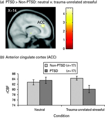
Functional ROIs were also defined around loci of greater rCBF changes in the PTSD group relative to the non-PTSD group for the trauma-unrelated stressful versus neutral comparison that were identified in predicted regions by the random-effects voxelwise analyses, namely the ACC, Montreal Neurological Institute (MNI) coordinates +14, +34, +28. Functional ROIs were defined as activation clusters around these peak coordinates with a significance threshold of p<0.001 using the MarsBaR SPM-based toolbox (http://marsbar.sourceforge.net/; Brett et al. Reference Brett, Anton, Valabreque and Poline2002). From the ROI, we extracted rCBF values per condition per participant and displayed the condition means for each group in Fig. 1. Because of technical difficulties, we were unable to extract ROI data from one non-PTSD participant.
(a) Regional cerebral blood flow (rCBF) decreases during the trauma-unrelated stressful versus neutral comparison in participants with post-traumatic stress disorder (PTSD; n=17) compared to non-PTSD participants (n=18). rCBF data are thresholded at p<0.001, shown in sagittal perspective [Montreal Neurological Institute (MNI)×coordinate=14], and superimposed on a standard T1 template (SPM99; Wellcome Department of Cognitive Neurology, UK). (b) Normalized rCBF data extracted from anterior cingulate cortex (ACC; z=4.33; MNI coordinates +14, +34, +28) for the PTSD (n=17) and non-PTSD (n=17) groups. Error bars represent standard error of the mean (s.e.m.).

We examined whether the inverse functional relationship between the mPFC and amygdala previously found in the traumatic versus neutral imagery comparison in the PTSD group (Shin et al. Reference Shin, Orr, Carson, Rauch, Macklin, Lasko, Peters, Metzger, Dougherty, Cannistraro, Alpert, Fischman and Pitman2004) would also be found in the trauma-unrelated stressful imagery versus neutral comparison. Voxelwise correlational analyses were conducted using (1) the rCBF difference scores extracted from the mPFC functional ROI and (2) the whole-brain voxelwise trauma-unrelated stressful versus neutral contrast maps for each participant. We also completed a voxelwise ‘covariates only’ analysis in the PTSD group only, to determine whether the significant correlations between PTSD symptom severity and rCBF in the amygdala and mPFC found for trauma-related stimuli in Shin et al. (Reference Shin, Orr, Carson, Rauch, Macklin, Lasko, Peters, Metzger, Dougherty, Cannistraro, Alpert, Fischman and Pitman2004) would extend to the trauma-unrelated stressful stimuli. For these replication analyses, we focused specifically on the amygdala and mPFC.
Results
Psychophysiologic responses
Trauma-unrelated stressful versusneutral comparison
A significant diagnosis×condition interaction was observed for the multivariate ANOVA for HR, SC and EMG responses. Overall, PTSD participants showed higher physiological responses during trauma-unrelated stressful versus neutral imagery, compared to non-PTSD participants [F(1, 53)=3.0, p<0.05, half-sided].
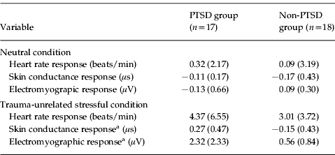
In the univariate analyses, there were significant main effects of diagnosis for SC [F(1, 33)=7.26, p=0.01] and EMG responses [F(1, 33)=7.57, p=0.01], with the PTSD group showing greater overall responses than the non-PTSD group. There were also significant main effects of condition, with greater responses during trauma-unrelated stressful versus neutral imagery across groups, for HR [F(1, 33)=17.50, p<0.001], SC [F(1, 33)=4.02, p=0.05] and EMG responses [F(1, 33)=19.77, p<0.001]. More importantly, there was a significant diagnosis×condition interaction for EMG response [F(1, 33)=9.04, p=0.005] and also a trend towards significance for SC response [F(1, 33)=3.29, p=0.08]. Inspection of the means indicated that EMG and SC response differences between the trauma-unrelated stressful and neutral conditions were greater in the PTSD group than in the non-PTSD group (Table 1).
Psychophysiologic responses to neutral and trauma-unrelated stressful imagery scripts
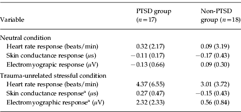
PTSD, Post-traumatic stress disorder.
Data are given as mean (standard deviation).
a p<0.05.
Trauma-unrelated stressful versus traumatic comparison
There were significant main effects of diagnosis for SC [F(1, 33)=11.88, p=0.002] and EMG responses [F(1, 33)=13.33, p=0.001], with the PTSD group showing greater overall responses than the non-PTSD group. There were no significant main effects of condition or diagnosis×condition interactions for HR, SC or EMG responses.
PET results
Trauma-unrelated stressful versus neutral comparison: within-diagnostic group results
PTSD participants demonstrated significant decreases in frontopolar gyrus (+12, +68, 0, z=3.74, k=123 voxels) and ACC (0, +40, +24, z=3.31, k=21 voxels). The fixed-effects analysis revealed significant decreases in the PTSD group in ACC (+10, +46, +8, z=4.07; and +12, +36, +28, z=4.58). The PTSD group demonstrated no significant rCBF increases. The non-PTSD group showed no significant rCBF decreases or increases.
Trauma-unrelated stressful versus neutral comparison: diagnosis×condition interactions
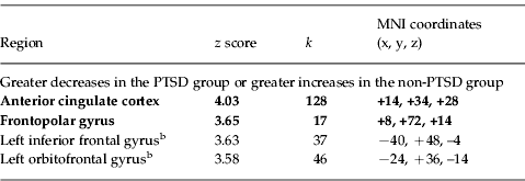
The voxelwise tests for diagnosis×condition interaction identified a significant locus in the ACC (Table 2, Fig. 1). Data extracted from the functional ROI showed a greater rCBF decrease (i.e. deactivation, or lower rCBF in the trauma-unrelated stressful imagery condition than in the neutral imagery condition) in the PTSD group relative to the non-PTSD group [F(1, 32)=23.40, p<0.001; Fig. 1]. The voxelwise tests revealed no regions with greater rCBF increases (i.e. higher rCBF in the trauma-unrelated stressful imagery condition than in the neutral imagery condition) in the PTSD group, or greater rCBF increases or decreases in the non-PTSD group. Fixed-effects analyses for the diagnosis×condition interaction revealed a similar significant deactivation in ACC (+12, +36, +28, z=4.33, k=122 voxels), and also a significant deactivation in MFG (+8, +60, 0, z=3.63, k=110 voxels). To evaluate whether depressive co-morbidity contributed to these findings, we provisionally removed participants with current MDD (n=8 in the PTSD group) and dysthymia (n=2 in the non-PTSD group) and repeated the diagnosis×condition interaction analyses. For the random-effects analysis, the ACC deactivation remained significant (+12, +36, +28, z=4.16, k=186 voxels); for the fixed-effects analysis, the ACC (+10, +38, +28, z=4.63, k=210 voxels) and MFG (+12, +62, –2, z=3.34, k=46 voxels) deactivations also remained significant.
Voxelwise random-effects analyses: regional cerebral blood flow (rCBF) findings for trauma-unrelated stressful versus neutral imagery: diagnosis×condition interactionsFootnote a
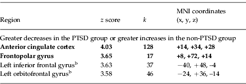
MNI, Montreal Neurological Institute; PTSD, post-traumatic stress disorder; k, number of voxels reported by SPM.
a A priori regions of interest (ROIs) are shown in bold.
b z score below statistical significance threshold for unpredicted regions, but listed for completeness.
Trauma-unrelated stressful versus traumatic comparison
The voxelwise tests of the trauma-unrelated stressful versus traumatic imagery comparison yielded no statistically significant within-group rCBF changes or diagnosis×condition interactions in a priori ROIs.
Medial frontal/amygdala correlations
rCBF change scores derived from the statistically significant ACC deactivation in the trauma-unrelated stressful versus neutral contrast were not found to be correlated with rCBF changes in the amygdala within the PTSD group.
Symptom severity correlations
No significant correlations were detected between rCBF during trauma-unrelated stressful imagery and symptom severity (i.e. CAPS scores) within the PTSD group.
Discussion
The results of the present study extend previous findings of diminished recruitment of mPFC during mental imagery of traumatic events in persons with PTSD (Bremner et al. Reference Bremner, Narayan, Staib, Southwick, McGlashan and Charney1999a; Shin et al. Reference Shin, McNally, Kosslyn, Thompson, Rauch, Alpert, Metzger, Lasko, Orr and Pitman1999, Reference Shin, Orr, Carson, Rauch, Macklin, Lasko, Peters, Metzger, Dougherty, Cannistraro, Alpert, Fischman and Pitman2004; Lanius et al. Reference Lanius, Williamson, Densmore, Boksman, Gupta, Neufeld, Gati and Menon2001; Liberzon et al. Reference Liberzon, Britton and Phan2003; Lindauer et al. Reference Lindauer, Booij, Habraken, Uylings, Olff, Carlier, den Heeten, van Eck-Smit and Gersons2004; Britton et al. Reference Britton, Phan, Taylor, Fig and Liberzon2005) to include imagery of personal events that were not trauma-related but were nevertheless experienced as stressful. The PFC, which has extensive connections with other cortical and subcortical regions, plays a crucial role in cognitive processing and emotion regulation and is highly sensitive to stress exposure (Arnsten, Reference Arnsten2009). Mild acute stress is linked to deficits in prefrontal cognitive abilities, and chronic, prolonged stress has been found to cause architectural changes such as decreased dendritic branching in the mPFC (Arnsten & Goldman-Rakic, Reference Arnsten and Goldman-Rakic1998; Radley et al. Reference Radley, Sisti, Hao, Rocher, McCall, Hof, McEwen and Morrison2004). Veterans with current PTSD, compared to veterans with no lifetime PTSD, exhibited rCBF decreases in the mPFC during the recollection of trauma-unrelated stressful (compared to neutral) personal life events. These findings of mPFC hyporesponsivity to emotional, non-trauma-specific symptom provocation in PTSD are in line with Lanius et al.'s (Reference Lanius, Williamson, Hopper, Densmore, Boksman, Gupta, Neufeld, Gati and Menon2003) findings using emotional, autobiographical probes in fMRI. Medial prefrontal cortical deficiency, amygdala hyperresponsivity, and hippocampal deficiency have been proposed as components of a neurocircuitry model of PTSD (Shin et al. Reference Shin, Rauch and Pitman2006; Shin & Liberzon, Reference Shin and Liberzon2010). mPFC is also implicated in fear-conditioning models of PTSD (Rauch et al. Reference Rauch, Shin and Phelps2006; Milad et al. Reference Milad, Pitman, Ellis, Gold, Shin, Lasko, Zeidan, Handwerger, Orr and Rauch2009). Within such models, mPFC abnormalities observed in PTSD may be linked to failure to extinguish responses to emotionally salient stimuli, and this failure may underlie the re-experiencing and hyperarousal symptoms of PTSD.
The results are consistent with the findings of Shin et al. (Reference Shin, Orr, Carson, Rauch, Macklin, Lasko, Peters, Metzger, Dougherty, Cannistraro, Alpert, Fischman and Pitman2004) in the same participants by showing that veterans with PTSD compared to veterans without PTSD exhibited greater relative rCBF decreases during trauma-unrelated stressful versus neutral recollections. This finding is consistent with a recent study showing heightened physiologic reactivity to threat in PTSD for both idiographic traumatic imagery and standard anger, panic and physical danger imagery conditions (McTeague et al. Reference McTeague, Lang, Laplante, Cuthbert, Shumen and Bradley2010).
The mPFC rCBF changes during trauma-unrelated stressful imagery for the PTSD group were not inversely correlated with rCBF changes in the amygdala/peri-amygdaloid cortex. It may be the case that, because the trauma-unrelated stressful life events were less severe than the traumatic events that elicited the PTSD, they elicited less variability in amygdala. We also found that mPFC changes in response to trauma-unrelated stressful imagery were not correlated with symptom severity in the PTSD group. Phan et al. (Reference Phan, Britton, Taylor, Fig and Liberzon2006) similarly did not detect significant correlations between neural responses to aversive generic pictures in PTSD and clinical symptoms. Differential amygdala activity was not associated with PTSD in either the current study or the previous study of imagery of trauma-unrelated, emotional personal events by Lanius et al. (Reference Lanius, Williamson, Hopper, Densmore, Boksman, Gupta, Neufeld, Gati and Menon2003), and has not been consistently demonstrated during traumatic imagery (for review, see Liberzon & Sripada, Reference Liberzon and Sripada2008).
The present results are potentially confounded by Axis-I co-morbidity in the PTSD group. However, the key results remained significant even after temporarily excluding participants with co-morbid depression. In an fMRI script-driven imagery study by Lanius et al. (Reference Lanius, Frewen, Girotti, Neufeld, Stevens and Densmore2007), a group with co-morbid PTSD and MDD showed greater activation than a PTSD-only group in ACC regions. However, in another study, reduced mPFC activity in response to fearful versus neutral faces was exhibited in a co-morbid PTSD-depression group compared to a PTSD-only group (Kemp et al. Reference Kemp, Felmingham, Das, Hughes, Peduto, Bryant and Williams2007). Despite their conflicting findings, these studies demonstrate the importance of controlling for depression when studying brain function in PTSD samples.
It may be tempting to conclude that because the PTSD participants showed an rCBF and psychophysiologic response pattern during recollection of trauma-unrelated stressful life events similar to the pattern they showed during imagery of the traumatic events that caused their PTSD, this pattern of responding to emotionally negative life events represents a constitutional risk factor, rather than a result of the traumatic event that caused the PTSD. Such a conclusion, however, may not be warranted. A limitation of this study is that we do not have reliable data regarding the number of trauma-unrelated stressful events that were experienced before versus after the index trauma. However, even in persons who experienced their most stressful, non-traumatic event before their traumatic event, brain changes caused by the latter may have altered the manner in which they recalled or responded to subsequent mental imagery of the former. Therefore, the current research design leaves unanswered whether the presently observe deficient function of medial prefrontal structures is a pre-existing risk factor for the development of PTSD after trauma exposure or an acquired sign of the disorder. Resolving this dilemma calls for prospective longitudinal research designs or twin studies (Pitman et al. Reference Pitman, Gilbertson, Gurvits, May, Lasko, Metzger, Shenton, Yehuda and Orr2006; Shin et al. Reference Shin, Rauch and Pitman2006). In studies of twin pairs discordant for trauma exposure, traumatic personal stimuli are unavailable in half of the sample. However, the present results suggest that future neuroimaging studies of PTSD in trauma-discordant twin pairs may usefully include trauma-unrelated probes, such as script-driven imagery of non-traumatic but emotionally stressful life events, as reported here and by Lanius et al. (Reference Lanius, Williamson, Hopper, Densmore, Boksman, Gupta, Neufeld, Gati and Menon2003).
In conclusion, veterans with PTSD exhibited rCBF decreases in mPFC during the recollection of stressful personal events that were unrelated to their index traumatic experience. Functional neuroanatomical models of PTSD must account for dysfunctional brain responses to stress-related, personal emotional stimuli that are not trauma specific.
Acknowledgments
This study was supported by Merit Review grants from the Veterans Affairs Medical Research Service (Drs Pitman, Carson and Orr); an Award from the National Alliance for Research on Schizophrenia and Depression, Great Neck, NY (Dr Shin); and grant MH60219 from the National Institute of Mental Health, Bethesda, MD (Dr Rauch). We are grateful to the participants. We also thank S. Barrow, A. Loring and S. Weise for technical assistance, and J. C. Britton for comments on a previous version of this manuscript.
Declaration of Interest
Dr Rauch received funded research through MGH for Brain Stimulation Therapy from Medtronics, Inc.; funded research through MGH for VNS from Cyberonics; and funded research through MGH on anxiolytic action from Cephalon. He also received honoraria from Novartis for consultation on emerging treatments; Neurogen for his participation as a consultant on emerging trends in anxiety associated with insomnia; Sepracor for his consultation on fear/conditioning/extinction; Primedia for his participation in developing a CE activity; and Medtronics, Inc for his attendance of the Advisory Board meeting on the Anatomy and Neuroscience of anxiety and depression. The financial disclosures for Dr Dougherty are as follows: Current: Medtronic (Research and Consulting/Honoraria – significant); Eli Lilly (Research and Consulting/Honoraria – significant); Brand Ideas (Consulting/Honoraria – modest); McNeil (Research and Consulting/Honoraria – significant); Reed Elsevier (Consulting/Honoraria – modest); Cyberonics (Research – significant); More than 1 year ago: Jazz Pharmaceuticals (Consulting/Honoraria – modest); Wyeth (Consulting/Honoraria – modest); Bristol–Myers Squibb (Consulting/Honoraria – modest); Northstar Neuroscience (Research and Consulting/Honoraria – significant); Forest (previous Research – significant); Trancept Pharmaceuticals (Consulting/Honoraria – modest); Cephalon (previous Research – modest); Cyberonics (Consulting/Honoraria – significant); Trancept Pharmaceuticals (Consulting/Honoraria – modest); JK Associates, Inc. (Consulting/Honoraria – modest); American Psychiatric Publishing, Inc. (Consulting/Honoraria – modest); Advocate Health and Hospitals Corporation (Consulting/Honoraria – modest); DHHS/NIH (Consulting/Honoraria – modest); Leerink Swann LLC (Consulting/Honoraria – modest); Oxford University Press (Consulting/Honoraria – modest); Professional Practice Group/Psychiatry Syracuse (Consulting/Honoraria – modest); Y&R Inc. DBA Sudler & Henessy (Consulting/Honoraria – modest).