The burden of compromised pregnancy
Pregnancy is a highly vulnerable period for both the mother and her unborn child. Complications during this time can adversely affect both maternal health and fetal development. The World Health Organization estimates that 830 women die each day due to preventable causes related to pregnancy and childbirth, while more than 6 million perinatal deaths occur each year.Reference Alkema, Chou and Hogan 1 , 2 Adverse intrauterine conditions are not only major drivers of short-term neonatal morbidity and mortality, but also impose serious risks for childhood and adult health, such as an increased incidence of various cardiometabolic diseases at adulthood.Reference Bernstein, Horbar, Badger, Ohlsson and Golan 3 – Reference Gluckman, Hanson, Cooper and Thornburg 5 For instance, fetal development in utero can be highly sensitive to external perturbations and environmental stressors, such as parental socioeconomic disadvantages, even more so than to changes in the length of gestation itself.Reference Eiríksdóttir, Ásgeirsdóttir and Bjarnadóttir 6 , Reference Usynina, Grjibovski, Odland and Krettek 7 Adverse conditions in utero may then trigger adaptive mechanisms to protect the developing fetus against the suboptimal intrauterine environment at the expense of increasing risk of disease in later life.Reference Gluckman, Hanson, Cooper and Thornburg 5 , Reference Liu, Basso and Kramer 8 Even though complications of pregnancy present a substantial burden on public health across the world, there is a lack of understanding of the underlying pathways, partly due to the multifactorial nature of these syndromes and partly due to the plethora of ethnic, social and economic confounding factors involved. Equally, there is a shortfall of effective prognostic, diagnostic and treatment options for some disorders, such as for preeclampsia. This calls for the development of improved animal models, in which to carry out more targeted research to isolate mechanisms and to thereby design potential therapeutic interventions.Reference Say, Chou and Gemmill 9 – Reference Ngan Kee 11
Increased uteroplacental vascular resistance: effects on maternal and fetal health
In mammals, advancing gestation is associated with an increase in uteroplacental blood flow to sustain the demands of the growing fetus.Reference Meschia 12 , Reference Trudinger, Giles and Cook 13 Hence, fetal weight is closely linked to the extent of the physiological increase in uteroplacental blood flow.Reference Meschia 12 – Reference Ferrell 14 This highlights the importance of the well-perfused placenta in determining the appropriate transplacental exchange of nutrients and blood gases in healthy pregnancy.Reference Meschia 12 – Reference Ferrell 14 Therefore, increased vascular resistance in the uteroplacental vascular bed can directly slow fetal growth and compromise fetal development by impairing uteroplacental blood flow, limiting adequate oxygen and nutrient delivery to the growing young.Reference Poston 15 , Reference Lang, Baker and Braems 16 In a human study with low-risk participants that were matched for age, socioeconomic background, ethnicity, health and nutritional access, intrauterine growth restriction (IUGR) was associated with indices of low uteroplacental blood flow.Reference Gaillard, Steegers, Tiemeier, Hofman and Jaddoe 17 , Reference Hennington and Alexander 18 This strongly indicates that altered placental vascular function is more important in determining fetal growth than other factors, such as maternal nutrition per se.Reference Gaillard, Steegers, Tiemeier, Hofman and Jaddoe 17 , Reference Hennington and Alexander 18 The same study also identified reduced placental perfusion as a risk factor for adverse cardiovascular and metabolic conditions in later life, which could not be explained by differences in birth weight.Reference Gaillard, Steegers, Tiemeier, Hofman and Jaddoe 17 , Reference Godfrey 19 This suggests that the placenta is also key in determining developmental origins of disease with lifelong consequences on offspring health, both in conjunction with and independent of its effects on fetal growth.Reference Gaillard, Steegers, Tiemeier, Hofman and Jaddoe 17 , Reference Godfrey 19
In parallel with human clinical studies, abnormal uteroplacental vascularization has been found in many animal models of compromised pregnancy, and uteroplacental perfusion is reduced in many ovine models of suboptimal pregnancy.Reference Lang, Baker and Braems 16 , Reference Reynolds, Caton and Redmer 20 , Reference Swanson and David 21 Similarly, interventions that restored uteroplacental blood flow could improve the severity of fetal growth restriction (FGR).Reference Lang, Baker and Braems 16 , Reference Reynolds, Caton and Redmer 20 , Reference Swanson and David 21 Consistent results have been found in rodent models of IUGR, which have been exploited significantly in this field of research due to their rapid generational turnover and the haemochorial nature of their placentation that resembles the human situation.Reference Swanson and David 21 – Reference Kulandavelu, Whiteley, Bainbridge, Qu and Adamson 25
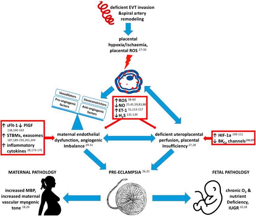
An increase in vascular resistance in the uteroplacental vascular bed can also have profound adverse effects on maternal physiology. Most directly, this is by perturbing the high capacitance–low resistance uteroplacental vascular bed, promoting an increase in maternal total peripheral vascular resistance, raising maternal cardiac afterload and thereby contributing to an increase in maternal arterial blood pressure with advancing gestational age.Reference VanWijk, Kublickiene, Boer and VanBavel 26 In addition, increased vascular tone in the uteroplacental vascular bed can trigger the placenta to enter into a state of oxidative stress, aggravating placental malperfusion and dysfunctionReference Zamudio, Wu and Ietta 27 , Reference Schoots, Gordijn, Scherjon, van Goor and Hillebrands 28 (Fig. 1). This can cause the placenta to become a source of circulating vasoactive factors that eventually cause widespread systemic maternal vascular dysfunction, as occurs in preeclampsiaReference Burton and Jauniaux 29 – Reference Woods 32 (Fig. 1). Pathways involved in this pathogenesis include the activation of the renin-angiotensin system, stimulation of prostaglandins, release of various anti-angiogenic factors, and altered synthesis of, and reactivity to, several gasotransmitters and vasoconstrictors.Reference Intapad, Warrington and Spradley 24 , Reference Woods 32 – Reference Mata-Greenwood, Blood and Sands 35 For example, in an interesting study, Woods et al. demonstrated that reduced uteroplacental perfusion increased blood pressure in pregnant dogs, and that the causative factor was thromboxane. Highlighting this study is important, because it was among the first to demonstrate that release of a substance by the uteroplacental vascular bed could increase maternal blood pressure in complicated pregnancy.Reference Woods 32
The pathogenesis of preeclampsia.

Maternal hypoxia: effects on the fetoplacental unit and maternal health
The term ‘hypoxia’ describes a lack of oxygen supply at the tissue and cellular level. It can be induced experimentally in animals, for instance by limiting the oxygen content of inspired air, which leads to ‘systemic hypoxia’ affecting the entire body. Alternatively, hypoxia can be induced by reducing the blood supply to the tissue of interest. Impaired uteroplacental blood flow would then promote ‘uteroplacental hypoxia’.Reference Samuel and Franklin 36 In addition to different terms describing various regions suffering hypoxia, authors have used several terms to describe the duration of hypoxia. These include ‘chronic hypoxia’, ‘sustained hypoxia’, ‘long-term hypoxia’ and ‘gestational hypoxia’, which have been used interchangeably to describe hypoxic exposure lasting from hours to months.Reference Matsuda, Patrick, Carmichael, Fraher and Richardson 37 – Reference Myers, Singleton and Hyatt 40 In this review, we use the term ‘chronic hypoxia’ to describe oxygen deprivation for a significant part of gestation, like a third to a half, and we use ‘gestational hypoxia’ to describe impaired oxygenation for most of gestation.
Effects of maternal hypoxia on the fetoplacental unit
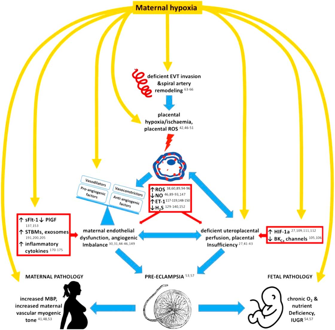
The suitability of gestational hypoxia induced by sustained reductions in the maternal inspired fraction of oxygen as a model of placental insufficiency has been questioned, because maternal compensatory cardiorespiratory responses may buffer impaired placental oxygenation. However, both human studies and animal models of maternal hypoxia now confirm that even exposure to chronic hypoxia for a third of gestation can lead to changes in the placental structure and function, indicative of increased uteroplacental vascular resistance and uteroplacental hypoxiaReference Zamudio, Wu and Ietta 27 , Reference Aljunaidy, Morton, Cooke and Davidge 41 – Reference Chang, Xiao, Huang, Longo and Zhang 43 (Fig. 2). For instance, in vivo and in vitro evidence shows that proliferation patterns of the uteroplacental vasculature are altered in response to chronic maternal hypoxia.Reference Rockwell, Dempsey and Moore 44 – Reference Mateev, Sillau and Mouser 46 These alterations in the placental vascular phenotype may underlie the diminished dilator and enhanced constrictor reactivity measured in the uteroplacental vascular bed of the hypoxic pregnant motherReference Rockwell, Dempsey and Moore 44 – Reference Mateev, Sillau and Mouser 46 (Fig. 2). Such changes will oppose the physiological increase in uteroplacental perfusion with advancing gestation, further compromising oxygen delivery to the fetoplacental unit and promoting placental oxidative stress, triggering a vicious cycleReference Turan, Aberdeen and Thompson 42 , Reference Mateev, Sillau and Mouser 46 – Reference Thaete, Dewey and Neerhof 51 (Fig. 2). Of interest, the placental response to the chronic hypobaric hypoxia of pregnancy at high altitude resembles many of these complications, not only in terms of symptoms and pregnancy outcome, but also in terms of global gene expression at the level of the placentaReference Zamudio, Wu and Ietta 27 (Fig. 2). This may, at least partly, explain the increased incidence of pregnancy complications at high altitude, such as the markedly increased prevalence of preeclampsiaReference Moore, Hershey, Jahnigen and Bowes 52 , Reference Reshetnikova, Burton, Milovanov and Fokin 53 (Fig. 2). Studies of human pregnancy at high altitude and in several animal models of maternal exposure to chronic hypoxia have confirmed that gestational hypoxia leads to significant FGRReference Kimball, Wayment and Merrill 54 – Reference Giussani and Davidge 57 (Fig. 2). In addition, there is evidence that placental oxidative stress may expose the fetus to potential oxidative injury, including protein or nucleic acid oxidation, translational inhibition or cell death, which in turn worsens pregnancy outcome and promotes IUGR.Reference Perrone, Tataranno and Negro 58 – Reference Yung, Cox, Tissot van Patot and Burton 60
Maternal hypoxia modulates placental, fetal and maternal pathways to mimic the pathogenesis of preeclampsia.
Exposure to maternal hypoxia during early gestation may have a profound influence on placentation by altering the characteristics of trophoblast proliferation.Reference Genbacev, Joslin, Damsky, Polliotti and Fisher 61 , Reference Thompson, Pence, Pinkas, Song and Telugu 62 Considering the importance of local oxygen tension in determining the onset of spiral artery conversion and development of the uteroplacental circulation at the end of the first trimester, it is not surprising that a suboptimal oxygen environment early in pregnancy will have significant adverse consequences on the morphological and functional maturation of the uteroplacental vascular bed, leading to reduced placental and fetal weights at termReference Thompson, Pence, Pinkas, Song and Telugu 62 – Reference Rodesch, Simon, Donner and Jauniaux 66 (Fig. 2). Gestational hypoxia and placental oxidative stress in the first trimester have also been linked to adverse effects on fetal brain development and are associated with several psychological disorders in later life, such as schizophrenia.Reference Phillips, Scott and Menassa 67 , Reference Fatemi and Folsom 68 Similarly, exposure of experimental animals to hypoxia during early gestation is associated with defects in cardiac development, impairing morphogenesis and ventricular function.Reference Sharma, Lucitti and Nordman 69 – Reference Ream, Ray, Chandra and Chikaraishi 72 On the other hand, many experimental studies on early-onset hypoxia during gestation show evidence of placental adaptation to adverse intrauterine conditions. These include an increase in placental weight, vascularization and capillary surface area for exchange, which depend on the severity and duration of the hypoxic insult and may not necessarily lead to significant effects on fetal body weight.Reference Rosario, Konno and Soares 73 – Reference Nuzzo, Camm and Sferruzzi-Perri 77 In this context, studies in rodent pregnancy by our own laboratory have shown that late-onset hypoxia for the last third of gestation leads to significant FGR.Reference Camm, Hansell and Kane 78 In contrast, early-onset hypoxic pregnancy increases placental weight, cushioning the adverse effects on fetal development, leading to maintained birth weight.Reference Nuzzo, Camm and Sferruzzi-Perri 77 It is also important to acknowledge that before 10 weeks of gestation in humans, intrauterine development occurs under relatively hypoxic conditions until the haemochorial placenta is fully established. Accordingly, at least in vitro, cytotrophoblast cells are insensitive to hypoxic conditions before 7 weeks of gestation, while between 10 and 12 weeks of gestation the same degree of hypoxia will significantly affect the invasion profile of the cytotrophoblast.Reference Genbacev, Joslin, Damsky, Polliotti and Fisher 61 , Reference Rodesch, Simon, Donner and Jauniaux 66 Thus, in human pregnancy, maternal hypoxia before 10 weeks of gestation may not significantly affect fetal organogenesis. On the contrary, premature onset of intervillous blood flow and oxygenation within the intrauterine environment may have adverse consequences on placental and fetal development through the development of placental oxidative stress.Reference Hutter, Kingdom and Jaeggi 79 – Reference Watson, Skepper, Jauniaux and Burton 82
Many molecular mechanisms have been proposed to mediate the adverse effects of maternal hypoxia on the fetoplacental unit, most of which impact on uteroplacental vascular function.
Nitric oxide
The important gasotransmitter nitric oxide (NO) has been identified as a key vasodilator in the uteroplacental circulation, in which pregnancy induces an increase in endothelial NO.Reference Aljunaidy, Morton, Cooke and Davidge 41 , Reference Xiao, Liu, Pearce and Zhang 83 , Reference Magness, Sullivan, Li, Phernetton and Bird 84 NO signalling may be one of the mechanisms underlying the pregnancy-induced uterine artery vasodilatation, allowing the crucial increase in uteroplacental blood flow to support the growing fetus.Reference Xiao, Liu, Pearce and Zhang 83 , Reference Bird, Zhang and Magness 85 – Reference Herrera, Kane and Hansell 87 Furthermore, eNOS knockout mice show significant FGR associated with a substantial increase in resistance in the uteroplacental vascular bed, impaired uteroplacental perfusion, placental hypoxia, oxidative stress and reduced transplacental nutrient transportReference Kulandavelu, Whiteley, Bainbridge, Qu and Adamson 25 , Reference Aljunaidy, Morton, Cooke and Davidge 41 , Reference Kusinski, Stanley and Dilworth 88 (Fig. 1). This was coupled with maternal proteinuria and alterations in maternal cardiovascular function, including a reduction in endothelium-dependent vasorelaxation, increased uterine artery vasoconstriction and hypertension.Reference Giussani and Davidge 57 , Reference Longini, Perrone and Vezzosi 59 Under gestational hypoxia the effects of NO on uterine artery vasodilatation are decreasedReference Mateev, Sillau and Mouser 46 , Reference Matsubara, Higaki, Matsubara and Nawa 89 (Fig. 2). This may occur due to decreased expression of NO synthesizing enzymes, or due to free radical scavenging of NO.Reference Matsubara, Higaki, Matsubara and Nawa 89 – Reference Fish, Yan and Matouk 91 Further, the sequestration of NO by superoxide forms peroxinitrite, which is thought to accumulate in placental tissues and itself has pro-oxidant effects capable of disrupting placental cellular proliferation and vascular function.Reference Matsubara, Higaki, Matsubara and Nawa 89 , Reference Kossenjans, Eis, Sahay, Brockman and Myatt 92 , Reference Miller, Voelker and Olister 93
Reactive oxygen species
This reduction in NO-induced increase in placental perfusion may be explained by the excess generation of reactive oxygen species (ROS) during chronic hypoxia; they act to sequester the available NO within the oxidatively stressed uteroplacental bedReference Vaziri, Liang and Ding 94 (Fig. 2). In an ovine model of gestational hypoxia, increased ROS production by NADPH oxidase 2 (NOX2) was responsible for increasing uterine artery myogenic tone, which was not observed when NOX2 was inhibited using apomycin.Reference Xiao, Hu and Huang 38 Hypoxia-induced oxidative stress has also been found to provide a strong stimulus for endoplasmic reticulum stress, which is associated with protein synthesis inhibition and impaired trophoblast survival and proliferation, further contributing to the increased prevalence of FGR and pregnancy complications at high altitude.Reference Yung, Cox, Tissot van Patot and Burton 60 , Reference Malhotra and Kaufman 95 , Reference Yung, Korolchuk, Tolkovsky, Charnock-Jones and Burton 96 Many studies in animal models have also reported antioxidant protection against FGR in pregnancy complicated by gestational hypoxia.Reference Aljunaidy, Morton, Cooke and Davidge 41 , Reference Giussani and Davidge 57 , Reference Giussani, Camm and Niu 97 , Reference Richter, Camm and Modi 98 Out of these agents, the mitochondrial antioxidant MitoQ has recently gained special interest due to its ability to specifically target mitochondrial oxidative stress, which is a major source of ROS in the placenta by nature of electron transport during oxidative phosphorylation.Reference Aljunaidy, Morton, Cooke and Davidge 99 , Reference Burton, Yung and Murray 100 This may explain the hypoxia-induced decrease in mitochondrial oxygen consumption and decrease in mitochondrial complex I activity in particular, which is the main site of electron leakage and mitochondrial ROS production.Reference Aljunaidy, Morton, Cooke and Davidge 99 , Reference Tissot van Patot, Murray and Beckey 101 – Reference Galkin, Abramov, Frakich, Duchen and Moncada 103 Maternal treatment with MitoQ in animal models of chronic hypoxia has led to the improvement of both placental mitochondrial stress and fetal outcomes, including birth weight and developmental programming of cardiovascular and psychiatric diseases, highlighting the importance of mitochondrial stress in mediating hypoxia-induced pathology.Reference Phillips, Scott and Menassa 67 , Reference Nuzzo, Camm and Sferruzzi-Perri 77 , Reference Aljunaidy, Morton, Cooke and Davidge 99
Calcium-activated potassium channels
During pregnancy, Ca2+-activated K+ (BKCa) channels in vascular smooth muscle cells have gained traction in being important mediators of uterine artery vasodilatation, and their inhibition reduces uteroplacental blood flow, contributing to IUGRReference Lorca, Wakle‐Prabagaran, Freeman, Pillai and England 104 (Fig. 1). Interestingly, hypoxia and oxidative stress suppress BKCa channel activity expression, which may explain the maladaptive myogenic response of the uteroplacental circulation in response to chronic hypoxiaReference Xiao, Hu and Huang 38 , Reference Liu, Liu and Shi 105 , Reference Hu, Xiao and Zhu 106 (Fig. 2). In addition, ROS have been identified as key inhibitors of BKCa channel activity, an effect not seen in the presence of the antioxidant N-acetylcysteine.Reference Zhu, Huang, Hu, Xiao and Zhang 107 Therefore, ROS may affect the vasculature of the uteroplacental bed in at least two ways, leading to dysfunction of both the endothelial layer by impairing NO-mediated mechanisms and of the smooth muscle cell layers by interfering with the function of Ca2+ channels.
Hypoxia-inducible factor 1-alpha
Some of the key regulators of cellular responses to hypoxia are the hypoxia-inducible factors (HIFs), which are rapidly stabilized upon the onset of oxygen deprivation, and interact with a variety of cellular enzymes and transcription factors to control cellular oxygen homeostasis.Reference Ziello, Jovin and Huang 108 HIF-1α expression, along with the expression of HIF-regulated proteins, such as transforming growth factor β-3 and vascular endothelial growth factor (VEGF), negatively correlates with fetal to placental weight ratio and positively relates with the adverse clinical outcome of chronic gestational hypoxia at high altitudeReference Zamudio, Wu and Ietta 27 (Fig. 2). HIF-1α and HIF-1α-regulated genes are similarly dysregulated in placentas during preeclampsia, with increased circulating levels of HIF-1α measured in preeclamptic mothersReference Iwagaki, Yokoyama and Tang 109 – Reference Rajakumar, Doty, Daftary, Harger and Conrad 111 (Fig. 1). Levels of HIF-1α only decline following delivery of the placenta, indicating that it may be useful as a predictive biomarker of failed placentation in preeclampsia, and further supporting the theory that the pathogenesis of preeclampsia is at least partly driven by hypoxia-mediated signalling.Reference Iwagaki, Yokoyama and Tang 109 , Reference Rajakumar, Doty, Daftary, Harger and Conrad 111 , Reference Caniggia and Winter 112
Endothelin-1 signalling
One of the many downstream effectors of HIF-1α is endothelin-1 (ET-1), which is an important antagonist of NO-mediated vasodilatation within a complex network of mediators acting on the vascular endothelium.Reference Bourque, Davidge and Adams 113 ET-1 interacts with NO by altering gene expression and ligand–receptor interactions, providing a close link between NO and ET-1 signalling, and generating a powerful vasoconstrictor effect.Reference Bourque, Davidge and Adams 113 – Reference Khimji and Rockey 115 Many investigators have proposed that an imbalance between these two essential endothelial agonists is implicated in various vascular pathologies, notably in several different forms of hypertension.Reference Bourque, Davidge and Adams 113 , Reference Alonso and Radomski 116 This interplay also seems to play an important role in the impairment of uteroplacental perfusion in hypoxia-induced FGR, which is one of the major complications of intrauterine exposure to hypoxiaReference Thaete, Dewey and Neerhof 51 , Reference Julian, Galan and Wilson 117 (Fig. 1). Although ET-1 shows little effect on uteroplacental vascular tone under physiological conditions, ET-1 and its receptors are markedly upregulated under conditions of chronic hypoxia through HIF-mediated signallingReference Takahashi, Soma, Muramatsu, Oka and Fukuchi 118 , Reference Moore, Shriver and Bemis 119 (Fig. 2). ET-1 binding to endothelin receptor A appears to be causative of impaired uteroplacental blood flow during chronic hypoxia and pregnancy at high altitude; a higher ET-1 to NO ratio shows a clear association with FGR at high altitude.Reference Thaete, Dewey and Neerhof 51 , Reference Julian, Galan and Wilson 117 The importance of the effects of ET-1 and of chronic maternal hypoxia on the uteroplacental vascular bed is further supported by the presence of different single nucleotide polymorphisms in the ET-1 gene in Andeans compared with Europeans.Reference Moore, Shriver and Bemis 119 While Andeans show a pregnancy-related fall in plasma ET-1 levels with advancing gestation, this does not occur in Europeans at high altitude. This may explain the relative protection against high altitude-induced FGR in highland native populations, such as the Andeans and Tibetans.Reference Moore, Shriver and Bemis 119 – Reference Soria, Julian, Vargas, Moore and Giussani 123
Placental hydrogen sulphide biology
Initially simply regarded as a toxic gas, H2S has come into physiological focus due to its role as an antioxidant, second messenger and regulator of vascular function.Reference Wang 124 – Reference Forgan, McNeill, DeLeon, Gao and Olson 126 Specifically, H2S is vasoactive and an important modulator of angiogenesis, thereby involved in the maintenance of optimal placental vascular function during healthy pregnancy.Reference Zhao, Zhang, Lu and Wang 127 – Reference Bryan, Yang, Wang and Khaper 130 H2S is also cytoprotective, involved in ischaemic preconditioning and in the enhancement of the mitochondrial redox balance.Reference Bryan, Yang, Wang and Khaper 130 The rate-limiting enzyme for H2S production, cystathionine γ-lyase (CSE), is localized in the smooth muscle cells of placental stem villi and its expression and activity are reduced in placentas under conditions of hypoxia and oxidative stress.Reference Yang, Pei, Cao and Wang 131 – Reference van Goor, van den Born, Hillebrands and Joles 134 Both women with hypertension and preeclampsia present with lower circulating levels of H2S, indicating that H2S has important antihypertensive propertiesReference Yang, Pei, Cao and Wang 131 – Reference van Goor, van den Born, Hillebrands and Joles 134 (Fig. 1). A growing body of evidence suggests that maternal chronic hypoxia suppresses placental levels of CSE via miR-21-mediated mechanisms, and that this is associated with mitochondrial depolarization, increased apoptosis and villous remodellingReference Lu, Kingdom, Burton and Cindrova-Davies 129 , Reference Yang, Pei, Cao and Wang 131 , Reference Cindrova-Davies, Herrera and Niu 132 , Reference Tsatsaris, Goffin and Munaut 135 – Reference Zhou, McMaster and Woo 138 (Fig. 2). These changes are further associated with evidence of impaired uteroplacental blood flow, uteroplacental hypoxia, IUGR and maternal vascular dysfunction, making H2S an agent of increasing interest in the inter-relationship between uteroplacental dysfunction and adverse fetal and maternal outcomes in complicated pregnancy.Reference Lu, Kingdom, Burton and Cindrova-Davies 129 , Reference Yang, Pei, Cao and Wang 131 , Reference Tsatsaris, Goffin and Munaut 135 – Reference Zhou, McMaster and Woo 138 Different mechanisms of action have been proposed for the protective effects of H2S in adverse pregnancy, including the sequestration of ROS, the regulation of potassium channels, the modulation of the renin-angiotensin system and the inhibition antiangiogenic factors.Reference Lu, Kingdom, Burton and Cindrova-Davies 129 , Reference Bryan, Yang, Wang and Khaper 130 , Reference Dongó, Beliczai-Marosi, Dybvig and Kiss 139 Therefore, decreased levels of placental CSE expression and activity following gestational hypoxia may have widespread adverse effects.Reference Dongó, Beliczai-Marosi, Dybvig and Kiss 139 , Reference Osmond and Kanagy 140 These may include direct adverse effects on systemic and uteroplacental vascular tone through impaired vasodilator actions on the endothelium and/or smooth muscle cells, or indirect adverse effects, for instance by exacerbating oxidative stress due to loss of its antioxidant properties.Reference Dongó, Beliczai-Marosi, Dybvig and Kiss 139 , Reference Osmond and Kanagy 140
Effects of maternal hypoxia on maternal health
In parallel with the plethora of evidence indicating adverse effects of gestational hypoxia on the fetoplacental unit, the same applies to the promotion of adverse effects of gestational hypoxia on the maternal circulationReference Aljunaidy, Morton, Cooke and Davidge 41 , Reference Zhou, Xiao and Hu 48 , Reference Thompson, Pence, Pinkas, Song and Telugu 62 (Fig. 2). Thus, dysregulation of many signalling pathways affecting the fetoplacental unit also seem to adversely affect the maternal cardiovascular system in gestational hypoxia, such as those involving ROS, NO, ET-1 and H2SReference Xiao, Hu and Huang 38 , Reference Zhou, Xiao and Hu 48 , Reference Thompson, Pence, Pinkas, Song and Telugu 62 , Reference Wang, Ahmad and Cai 133 , Reference Al-Magableh, Kemp-Harper and Hart 141 , Reference Vural 142 (Fig. 1). For example, in a human case–control study, treatment of women suffering gestational hypertension with antihypertensive drugs supplemented with NO donors and plasma volume expansion improved the uteroplacental resistance index and reduced both maternal hypertension and FGR compared with pregnant women treated with antihypertensive agents alone.Reference Valensise, Vasapollo and Novelli 143 Altered NO bioavailability is thought to be closely linked to oxidative stress and to ROS-mediated oxidative damage, which is increased in both the ischaemic placenta and the systemic vasculature of women suffering from preeclampsia for example by activated immunocytes in the maternal endothelium.Reference Matsubara, Higaki, Matsubara and Nawa 89 These immunocytes are also the source inflammatory cytokines, such as tumour necrosis factor-α (TNF-α), which are at least partly responsible for the increase in ET-1 concentrations in maternal serum in response to placental ischaemia.Reference LaMarca, Speed and Fournier 144 , Reference LaMarca, Cockrell, Sullivan, Bennett and Granger 145 ET-1 has potent vasoactive effects on the maternal endothelium and is an important mediator of maternal hypertension during preeclampsia, while endothelin receptor A antagonism has been shown to prevent the ET-1-mediated rise in blood pressure in many animal models.Reference LaMarca, Speed and Fournier 144 – Reference Jain 147 Women suffering from preeclampsia also show decreased plasma levels of the vasoactive agent H2S, which contributes to the maternal hypertension and renal damage of the preeclamptic phenotype.Reference Wang, Ahmad and Cai 133 , Reference Holwerda, Burke and Faas 148
In addition to the contribution of signalling pathways involving ROS, NO, ET-1 and H2S, the maternal vascular function also relies on the complex interaction between an exhaustive list of angiogenic and vasoactive factors, as well as cytokines and growth factorsReference Tissot van Patot, Ebensperger, Gassmann and Llanos 30 , Reference Gilbert, Ryan and LaMarca 31 , Reference Shah and Khalil 149 (Fig. 2). Any disruption of this intricate balance of circulating factors by the stressed placenta, as occurs in gestational hypoxia, may promote widespread endothelial dysfunction and vascular inflammation with detrimental effects on the maternal vasculature.Reference Tissot van Patot, Ebensperger, Gassmann and Llanos 30 , Reference Gilbert, Ryan and LaMarca 31 , Reference Shah and Khalil 149
Angiogenic imbalance
The placenta-derived placental growth factor (PlGF) and VEGF and are crucial for maternal endothelial health, promoting trophoblast survival and placental angiogenesis in the uteroplacental vascular bed.Reference Tsatsaris, Goffin and Munaut 135 – Reference Zhou, McMaster and Woo 138 The soluble fms-like tyrosine kinase-1 (sFlt-1) acts as a soluble receptor and antagonist against VEGF and PlGF.Reference Tanbe and Khalil 150 , Reference Shibuya 151 Chronic hypoxia, both in early and late gestation, placental oxidative stress and preeclampsia are all associated with an increased ratio of sFlt-1 to PlGF and VEGF in both the trophoblast and the maternal circulation, and administration of sFlt-1 itself has been found to further increase tissue ROSReference Li, Gu, Zhang, Lewis and Wang 137 , Reference Maynard, Min and Merchan 152 – Reference Nevo, Lee and Caniggia 157 (Figs. 1 and 2). While early in pregnancy the effect of sFlt-1 is mostly mediated via direct effects on placentation by inhibiting cytotrophoblast invasion and differentiation, the inhibitory effect of sFlt-1 on PlGF and VEGF has been suggested to be causative in the development of proteinuria and maternal endothelial dysfunction in preeclampsia in the later stages of gestation.Reference Zhou, McMaster and Woo 138 , Reference Maynard, Min and Merchan 152 , Reference Karumanchi and Bdolah 156 , Reference Sugimoto, Hamano and Charytan 158 , Reference Fisher 159 Interestingly, at least part of the suggested beneficial effects of H2S supplementation on maternal vascular function in pregnant women with preeclampsia is thought to occur via H2S-mediated upregulation of miR-133b, which in turn downregulates sFlt-1 release.Reference Hu, Guo and Wang 160 Such findings have sparked interest in the suitability of measuring the sFlt-1 to PlGF ratio as a biomarker for obstetric disorders related to placental insufficiency, such as preeclampsia.Reference Saleh, Vergouwe and Verdonk 161 – Reference Sovio, Gaccioli and Cook 163 This has led to the development of commercial bioassays to be used as additional diagnostic tools for preeclampsia, the efficacy of which has been validated.Reference Zeisler, Llurba and Chantraine 164 , Reference Verlohren, Galindo and Schlembach 165 An aspect of this story less well investigated is the potential of manipulating these angiogenic pathways in the treatment of preeclampsia, for example through the administration of VEGF or PlGF to increase their bioavailability. Although increased VEGF levels may be associated with adverse side effects, such as oedema, PlGF administration has been found to abolish the maternal hypertension in a rat model of preeclampsia with no adverse effects on the maternal extracellular water content.Reference Spradley, Tan and Joo 166 – Reference Chau, Hennessy and Makris 168 Reports on PlGF administration are limited and details on its mechanisms of protective action in adverse pregnancy are not completely understood. The beneficial effects of PlGF in compromised pregnancy have been attributed thus far to be partly mediated by a reduction in placental oxidative stress and an improvement in maternal endothelial function via NO and cyclic guanosine monophosphate-derived vasorelaxation.Reference Spradley, Tan and Joo 166 – Reference Suzuki, Ohkuchi and Matsubara 169
Inflammatory cytokine signalling
One of the downstream consequences of placental hypoxia, excess ROS availability and oxidative damage is cellular inflammation, triggering the release of inflammatory stress markersReference Mittal, Siddiqui, Tran, Reddy and Malik 170 – Reference Benyo, Miles and Conrad 172 (Fig. 2). These may be of either placental or endothelial origin and they act in synergy to generate a systemic endovascular inflammatory state, which contributes to the development of hypertension and kidney disease in preeclampsiaReference Schoots, Gordijn, Scherjon, van Goor and Hillebrands 28 , Reference Cuffe, Holland, Salomon, Rice and Perkins 173 – Reference Pantham, Aye and Powell 175 (Fig. 1). Of interest, the hypoxic placenta undergoes a clear shift in its inflammatory cytokine profile, showing reduced expression of anti-inflammatory cytokines, such as interleukin-10 (IL-10), and up-regulation of pro-inflammatory cytokines, such as TNF-α, IL-6, IL-8 and IL-1ß. However, the role of IL-6 in pregnancy is controversial and it may be involved in a variety of functions in the female reproductive tract.Reference Shah and Khalil 149 , Reference Royle, Lim and Xu 176 – Reference Prins, Gomez-Lopez and Robertson 182 The onset of this altered inflammatory cytokine profile in response to hypoxic exposure is thought to occur after 11 weeks of gestation in humans and becomes more prominent with advancing gestation, but is detectable in the maternal circulation and in the amniotic fluid prior to the manifestation of preeclampsia-related symptoms.Reference Benyo, Miles and Conrad 172 , Reference Conrad and Benyo 180 , Reference Pober and Cotran 183 , Reference Coussons-Read, Mazzeo and Whitford 184 In addition, TNF-α is a potent stimulus for ET-1 signalling, which may at least partly underlie TNF-α-mediated maternal hypertension as well as provide a link between placental, maternal and fetal pathology in inflammatory conditions during gestation.Reference LaMarca, Cockrell, Sullivan, Bennett and Granger 145 , Reference LaMarca, Speed and Fournier 185
Extracellular vesicles
Another consequence of hypoxic damage to the placenta may be the increased release of placental debris or so-called syncytiotrophoblast microparticles (STBM) alongside placental exosomes into the maternal circulation (Fig. 2). This process is exacerbated in pathological pregnancies, such as in preeclampsia and hypoxia as a result of poor placentation.Reference Burton and Jones 186 – Reference Germain, Sacks, Soorana, Sargent and Redman 193 This causes necrotic trophoblast damage following placental ischaemia and it has been proposed to trigger widespread maternal endothelial dysfunction, possibly due to the release of inflammatory cytokines and endothelial phagocytosis of microparticlesReference Burton and Jones 186 – Reference Germain, Sacks, Soorana, Sargent and Redman 193 (Fig. 1). STBMs negatively impact endothelial proliferation, while promoting the secretion of anti-angiogenic factors and inflammatory cytokines.Reference Smárason, Sargent, Starkey and Redman 187 This was confirmed in vitro in isolated perfused maternal resistance vessels, in which the presence of STBM vesicles in the perfusate reduced acetylcholine-mediated vasodilatation in subcutaneous fat arteries. This suggests that the mechanism of action of STBM-related maternal hypertension may be mediated via adverse effects on peripheral vasodilatation.Reference Cockell, Learmont and Smárason 194 The presence of increased necrotic trophoblast debris in the maternal circulation is found to be a characteristic of pregnancy-induced hypertension, but not IUGR in the absence of hypertension, thereby appearing to be central to the maternal component of the preeclamptic syndrome.Reference Goswami, Tannetta and Magee 192
Exosomes are, like STMBs, constituents of cell-derived extracellular vesicles released into the extracellular environment, containing agents destined for paracrine and endocrine signalling, such as miRs and growth factors.Reference Kowal, Tkach and Théry 195 , Reference Colombo, Raposo and Théry 196 The placenta is an active source of exosomes during pregnancy and maternal plasma concentration of exosomes increases with advancing gestation, potentially responsible for the maternal physiological adaptation to pregnancy and maternal-fetal immune regulation.Reference Sabapatha, Gercel-Taylor and Taylor 197 – Reference Mincheva-Nilsson and Baranov 199 This process occurs under physiological conditions during pregnancy, but is increased under conditions of hypoxia and during some complications of pregnancy, such as preeclampsia, as a result of placental apoptosis and necrosis, which also alters the exosomal content.Reference Salomon, Kobayashi and Ashman 200 – Reference Baig, Kothandaraman and Manikandan 203 These have been shown to affect maternal endothelial and vascular function, possibly through dysregulation of pro-inflammatory cytokines or endothelial-related miRs, such as miR-126, miR-17, miR-155 and miR-210, with potential roles in endothelial dysfunction in preeclampsia. 204 – Reference Atay, Gercel-Taylor, Suttles, Mor and Taylor 206
Maternal hypertension itself may further compound the formation of STBMs and propagate placental dysfunction and FGR. For example, an increased maternal myogenic tone and decreased diameter of maternal resistance vessels increases the velocity of blood entering the uteroplacental bed. This promotes turbulent flow in the intervillous blood spaces, which in turn may exacerbate mechanical and necrotic damage to the placental vasculature.Reference Burton and Jones 186 , Reference Roberts and Escudero 207 , Reference Toal, Chan and Fallah 208 This triggers the dislocation of microparticulate debris and necrotic trophoblast into the maternal circulation.Reference Burton and Jones 186 , Reference Roberts and Escudero 207 , Reference Toal, Chan and Fallah 208 Significant damage to the spiral arteries can cause occlusion of the villous blood spaces, resulting in placental infarction, further fuelling placental dysfunction.Reference Kliman 209 – Reference Soma, Yoshida, Mukaida and Tabuchi 211
Direct effects of chronic hypoxia on the fetus
A plethora of research interest has focussed on the combined effects of maternal hypoxia on the fetoplacental unit as a whole, most of which address effects on uteroplacental blood flow with indirect effects on fetal development. This is due to the fact that intrauterine hypoxia usually occurs as a result of preplacental or uteroplacental hypoxia. Occasionally, postplacental hypoxia can occur due to villous defects preventing sufficient oxygen uptake in the placental vascular bed or due to fetal cardiovascular dysfunction, such as that induced by umbilical cord occlusion or thrombosis.Reference Kingdom and Kaufmann 65 , Reference Stanek 212 This is associated with IUGR with absent or reversed end-diastolic blood flow in the umbilical artery, giving some insight into the direct effects of hypoxia on fetal development.Reference Kingdom and Kaufmann 65 , Reference Baergen 213 However, it is important to note that even postplacental hypoxia can be associated with significant alterations in placental and villous morphology, typical of nonbranching angiogenesis resulting in villous hypoplasia, which in itself is associated with an increase in uteroplacental vascular resistance. This makes the distinction between direct and indirect effects of hypoxia on the fetus less straightforward.Reference Baergen 213 Another line of research has focussed on incubation of the chick embryo under hypoxic conditions, in which developmental complications occur in the absence of any maternal or placental influences, such as maternal hypertension or placental insufficiency. Such studies have reported that both hypobaric and isobaric hypoxic incubation of the chick embryo lead to significant FGR with a similar ‘brainsparing’ redistribution of the cardiac output compared to the mammalian fetus.Reference Giussani, Salinas, Villena and Blanco 214 – Reference Mulder, Miedema, De Mey, Giussani and Blanco 218 Hypoxic incubation also resulted in embryonic cardiovascular dysfunction, which was absent in normoxic chick embryos from sea-level incubation or from high altitude incubation with oxygen supplementation.Reference Salinas, Blanco and Villena 219 In addition, development under hypoxic conditions had long-term adverse effects on systemic and pulmonary blood pressure regulation and presented with altered baroreflex sensitivity in the adult chicken.Reference Herrera, Salinas, Blanco, Villena and Giussani 220 , Reference Salinas, Blanco, Villena and Giussani 221 The development of cardiovascular dysfunction in the hypoxic chick embryo could be effectively prevented using antioxidant therapies, such as sildenafil or melatonin.Reference Itani, Skeffington, Beck and Giussani 222 , Reference Itani, Skeffington, Beck, Niu and Giussani 223 This provides evidence that fetal development can be influenced directly by both hypoxic conditions and antioxidant treatment, highlighting the importance of considering fetal and uteroplacental hypoxia as separate complications. However, it can be argued that the chorioallantoic membrane may be considered the avian homologue to the mammalian placenta, which may itself be influenced by hypoxic incubation.Reference Giussani, Salinas, Villena and Blanco 214 , Reference Mehta and Mehta 215 , Reference Metcalfe and Stock 224
Conclusions
In summary, uteroplacental hypoxia may link many of the effects of placental dysfunction with adverse effects on the mother and the fetus, which occur in many complications of pregnancy, such as in preeclampsia. Chronic hypoxia induces morphological, molecular and functional changes in the placenta that closely resemble those observed in placentae from women suffering from preeclampsia. In addition, chronic hypoxia recapitulates maternal and fetal adverse outcomes associated with the preeclamptic syndrome. Thus, pregnancy compromised by maternal exposure to hypoxia is not only a major risk factor for FGR, but it also promotes adverse changes in the placenta, with potential consequent adverse effects on the physiology of the mother and the offspring. Therefore, gestational hypoxia provides a useful model to study the inter-relationship between placental dysfunction and adverse outcomes in the mother and offspring in a wide array of examples of complicated pregnancy, such as in preeclampsia.
Acknowledgements
D.A.G. is the Professor of Cardiovascular Physiology & Medicine at the Department of Physiology, Development and Neuroscience at the University of Cambridge, Professorial Fellow and Director of Studies in Medicine at Gonville & Caius College, a Lister Institute Fellow and a Royal Society Wolfson Research Merit Award Holder.
Financial support
The study was supported by The British Heart Foundation, The Centre for Trophoblast Research and Trinity College, Cambridge.
Conflicts of interest
None