INTRODUCTION
The east Atlantic–Mediterranean prawn Melicertus kerathurus (Forskal, 1775) lives in coastal marine and brackish waters but it has nearly disappeared from Israel, southern Turkey and up to the Southern Aegean Sea (Galil & Kevrekidis, Reference Galil and Kevrekidis2002), its habitat being overrun mainly by Marsupenaeus japonicus (Kevrekidis & Thessalou-Legaki, Reference Kevrekidis and Thessalou-Legaki2006).
Reproductive pattern is a major factor determining the reproductive potential of a species. Therefore, it is very important to understand the reproductive biology (ovarian development) for future domestication of this species in local waters. Increased attention has been paid in recent years to the ovarian development of penaeid shrimp and macroscopic, histological characterization of penaeid shrimp maturation has been studied on various species such as Melicertus japonicus (Yano, Reference Yano1988), Melicertus vannamei (Arcos et al., Reference Arcos, Racotta and Palacios2005), Melicertus semisulcatus (Badawi, Reference Badawi1975), Melicertus kerathurus (Medina et al., Reference Medina, Vila, Mourente and Rodriguez1996), Melicertus monodon (Tan-Fermin, Reference Tan Fermin1991; Quinitio et al., Reference Quinitio, Caballero and Gustilo1993), Litopenaeus setiferus (King, Reference King1948), Penaues indicus (Subrahmanyam, Reference Subrahmanyam1965), Melicertus aztecus (Duronslet et al., Reference Duronslet, Yudin, Wheeler and Clark1975), Melicertus duorarum (Caillouet, Reference Caillouet1972), Fenneropenaeus merguiensis (Crocos & Kerr, Reference Crocos and Kerr1983), Lucifer orientalis (Oka & Sirahata, Reference Oka and Shirata1965) and Melicertus plebejus (Kelemec & Smith, Reference Kelemec and Smith1980) in the world. Medina et al. (Reference Medina, Vila, Mourente and Rodriguez1996) have also stated that the ovarian histology of M. kerathurus is typical of that described previously for other penaeids and based on their histological features, five distinct stages have been identified throughout ovarian development: previtellogenic, early vitellogenic, late vitellogenic, mature and spent or degenerating.
There are many studies on this species in the Mediterranean but very little is known about the seasonal cycle of gametogenic development, spawning periodicity and length at sexual maturity in Turkey. Until now, there have been many systematic studies of ecology (Geldiay, Reference Geldiay1969; Geldiay & Koçataş, Reference Geldiay and Koçataş1973; Alpbaz, Reference Alpbaz1978; Özden, Reference Özden1989) and cultivation (Köse et al., Reference Köse, Hindioğlu, Özden and Alpbaz1999; Kumlu et al., Reference Kumlu, Eroldogan and Saglamtimur2001; Aktaş et al., Reference Aktaş, Eroldogan and Kumlu2004). The present study represents the first documentation of the gametogenic cycle of shrimp in Turkey. Therefore, the current study aimed to refine the existing methods of staging by further characterizing reproduction in Melicertus kerathurus using: (1) qualitative techniques such as histology of the ovaries; and (2) quantitative parameters such as morphometry (carapace length, body length, gonad weight, body weight, gonadosomatic index and average/maximum oocyte diameter) and oocyte frequency as they occur in wild specimens.
MATERIALS AND METHODS
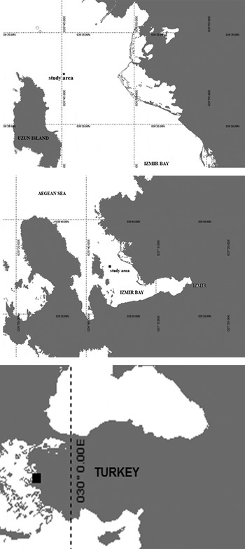
Wild specimens of Melicertus kerathurus (Forskal, 1775) were captured monthly (from May 2004 to April 2005) from Izmir Bay (032°32′00N 026°46′00E) (Figure 1). All individuals (N = 60) were weighed to the nearest 0.01 g and measured for their body length (BL, distance between the post orbital margin and the end of the telson extended in a straight line) and also carapace length (CL, from the posterior margin of the orbit to the posterior margin of the carapace) using Vernier callipers. The ovaries were dissected out and weighed. Since the different regions of the ovary exhibited similar oocyte stages and size–frequencies at all stages of development, only the mid-region of the abdominal lobe of the ovaries was analysed. The diameters of about 30 oocytes and follicles from each animal were measured using an ocular micrometer. Mean diameter was calculated from the long and short axes of the oocytes. The gonadosomatic index (GSI) was expressed as the percentage of the ovarian weight in relation to the total body weight (Medina et al., Reference Medina, Vila, Mourente and Rodriguez1996). Temperature (with a mercury thermometer that ranged –10 to 100 ± 0.5°C), salinity (with a hand refractometer, ‰) and pH (with pH meter) levels were monitored regularly throughout the study.
Shrimp (Melicertus kerathurus) sampling area in Izmir Bay, Turkey.

Histology
Following the removal and weighing of the ovaries, fragments of the middle region of the ovary were fixed for 24 hours in 10% formaldehyde. Thereafter, the ovarian tissues were dehydrated in increasing concentrations of alcohol. Ovarian tissues in paraffin wax were embedded at 6 µm and stained with haematoxylin and eosin. Oocyte diameters were measured from stained sections. The stained tissue glass slide was then examined, photographed (Olympus PM-C3566) under an optical microscope (Olympus CH-2) and qualitatively classified into a stage of development.
Statistics
Means of body weight, carapace length, GSI and oocyte diameter were compared by one-way analysis of variance (ANOVA) followed by the Duncan multiple comparison test at P = 0.05, in order to determine significant differences in each sample of shrimp separately. Statistical analyses were performed using SPSS 11.0 software.
RESULTS
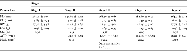
Data on the quantitative parameters from shrimps are presented in Table 1. All parameters showed significant differences over the five stages (P < 0.05). The mean body length and body weight of the shrimps in stages I to IV was significantly different from each other but stage IV individuals were significantly larger. Mean gonad weight was significantly increased through stages previtellogenic and mature and decreased at stage spent to a weight similar to stage previtellogenic. GSI mean values also increased significantly from stage previtellogenic through stage mature but decreased at stage spent to the previtellogenic. Average and maximum oocyte diameter values were significantly different at all stages (P < 0.05) and were largest for stage mature individuals (Table 1).
Summary of data on the quantitative parameters of wild Melicertus kerathurus. Data are presented as mean ± SEM.
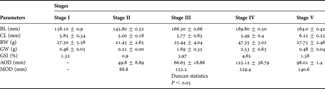
CL, carapace length; BL, body length; BW, body weight; GW, gonad weight; GSI, gonadosomatic index; AOD, average oocyte diameter; MOD, maximum oocyte diameter; SEM, standard error of the mean.
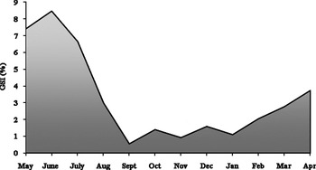
Changes in the mean GSI are shown in Figure 2. The GSI values of females started to increase in January and reached maximum level in June (8.45%). GSI values abruptly decreased in September (0.53%) which corresponds with the spawning season. After spawning, GSI started to increase again gradually.
Changes in the mean gonadosomatic index (GSI) of female Melicertus kerathurus during an annual reproductive cycle.

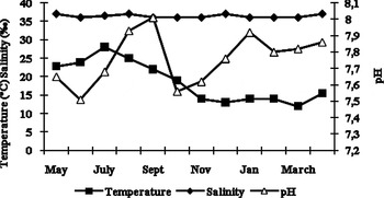
Temperature showed a clear seasonal pattern with maximal values observed in July (28°C), and minimal in March (12°C) and December (13°C). pH ranged from 7.51 to 8.01 with a maximum in September and minimum in June. Salinity was not changed so much and ranged from 36‰ to 37‰ without seasonal variability (Figure 3).
Variation of temperature, salinity and pH in sampling area 2004/2005.

Ovarian histology
Ovarian development of Melicertus kerathurus was classified into five stages based on the presence of the most advanced oocyte stage.
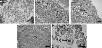
STAGE I (PREVITELLOGENIC): M. kerathurus captured from the wild in late December and January, the ovaries contained mainly previtellogenic oocytes (77.5%). The oogonia measured 34.53 SEM 1.88 indiameter (14–16 cm body length) µm. The GSI ranged from 1.57% to 1.08% (P < 0.05) (Figure 4A);
Sections of wild-caught Melicertus kerathurus ovaries: stage I (4A) (December–January), stage II (4B) (June), stage III (4C) (February–May), stage IV (4D) (June–October) and stage V (4E) (September). EVO, early vitellogenic oocytes; LVO, late vitellogenic oocytes; MO, mature ooctyes; DO, degenerating oocytes. Scale bars: 100 µm.

STAGE II (EARLY VITELLOGENIC): this stage consists mainly of previtellogenic oocytes (41.4%). Late vitellogenic oocytes were also present in this stage. The cytoplasm becomes more basophilic and undergoes a significant increase in size. These oocytes are gradually surrounded by follicle cells when they are relatively small. A basophilic nucleus lies centrally within the cytoplasm with one or two large, dark-staining nucleoli. Nucleoli appear around the periphery of the nucleus (perinucleus stage) and are stained slightly darker than the cytoplasm. This stage could be seen in June (Figure 4B);
STAGE III (LATE VITELLOGENIC): the ovaries contain large oocytes (90–150 µm in diameter) with cytoplasm filled with yolky substances. These yolky oocytes are preferentially located in the outer regions of the ovary, while previtellogenic is located in the cytoplasm of the ovary. The cytoplasm of previtellogenic oocytes is acidophilic, whereas the nucleolus is basophilic. This stage was observed from February to May (Figure 4C);
STAGE IV (MATURE): in this stage, from late June to October, ovaries are characterized by the presence of mostly mature oocytes that surpass 150–200 µm in diameter. As a typically distinctive feature, the cytoplasm of mature oocytes shows conspicuous cortical rods at the cell periphery (Figure 4D);
STAGE V (GVBD): in the middle of September, nucleus disappears (germinal vesicle breakdown, GVBD). Cortical rods elongate, extending radially inward. The GSI was very low, 0.53% (P < 0.05) (Figure 4E).
In September, ovaries are distinguished by the presence of atretic (degenerating) oocytes although in this month some animals will still have mature oocytes. It was observed that the ovaries were spent or had a resorbed appearance. Moreover, ovaries are characterized by the presence of empty oocytes when the shrimps are mating.
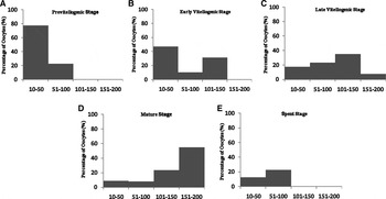
The oocyte size–frequencies for the posterior abdominal region of the ovary at the different stages of maturation are summarized in Figure 5. Oocytes at stage I (Figure 5A) were all in the size-range 10–100 µm and were mostly primary (previtellogenic) oocytes. At stage IV (Figure 5D), approximately 75% of the total number of oocytes per section had cortical rods with about 50% larger than 150 µm. The size-ranges of vitellogenic (yolky) oocytes with cortical rods overlap but their presence was mutually exclusive, that is, cortical rods were not observed at stage V (Figure 5E). Cortical rods occurred at stage V but were not counted since they were scattered singly and/or were without nuclei.
Oocytes size–frequency histogram showing the development of the oocytes in wild Melicertus kerathurus as represented by the posterior abdominal region during the different stages of maturation.

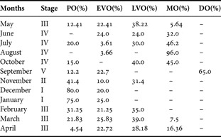
Percentages of vitellin in all stages are summarized in Table 2. In all M. kerathurus captured from wild in December and January the ovaries contained only previtellogenic and early vitellogenic oocytes (most ovaries were found at stage I). Stage III ovaries were predominant in shrimp sampled from February to May. Females samples collected between June and October showed stage IV ovaries with minimum percentages of previtellogenic oocytes (about 8%) and maximum percentages of mature (about 55%) oocytes.
Monthly changes of vitellin in Melicertus kerathurus during the study.

PO, previtellogenic oocyte; EVO, early vitellogenic oocyte; LVO, late vitellogenic oocyte; MO, mature oocyte; DO, degenerating oocyte; SEM, standard error of the mean.
Table 3 shows oocytes diameters of M. kerathurus. Changes in size of developing oocytes assist in differentiating between these stages. In stage II, diameter of oocytes ranged from 30.91 ± 1.19 µm to 68.69 ± 1.28 µm. In stage III, oocytes grew larger to 38–102 µm. In stage IV, cortical rods started to appear; oocytes varied from approximately 86–163 µm. In stage V, only late vitellogenic and degenerate oocytes could be seen (about 96–99 µm). The follicle size ranged from 468 to 858 µm (stages II–IV)
Oocyte diameters of grooved shrimp (Melicertus kerathurus) during developmental stages (the sample size was between 145.80 ± 0.50 and 189.8 ± 0.50 mm body length).

PO, previtellogenic oocyte; EVO, early vitellogenic oocyte; LVO, late vitellogenic oocyte; MO, mature oocyte; DO, degenerating oocyte; SE, standard error.
DISCUSSION
It has been believed that there is a close inverse relationship between reproduction (ovarian maturation and spawning) and growth (moulting) in penaeid prawns (Browdy, Reference Browdy and Wyban1992), therefore, the understanding of reproduction is important for the development of a complete culture technology (Primavera, Reference Primavera, Taki, Primavera and Llobrera1985). This study confirms the variability in Melicertus kerathurus reproductive performance at different times of the year. The results of the present study indicate that GSI peaked in June and after spawning decreased as pointed out by Medina et al. (Reference Medina, Vila, Mourente and Rodriguez1996) who observed that GSI and percentage of vitellogenic oocytes showed on upward trend from May to July. Türkmen & Yilmazyerli (Reference Türkmen and Yilmazyerli2006) also reported that a seasonal reproduction pattern for M. kerathurus was observed from April to September in Izmir Bay. Similarly, the GSI in Fenneropenaeus indicus also increased during vitellogenesis but decreased slightly after spawning to reach levels similar to immature females (Quinitio & Millamena, Reference Quinitio and Millamena1992). GSI values showed in Pandalus kessleri the same pattern with a rise during vitellogenesis and a sharp decrease after spawning (Quinitio et al., Reference Quinitio, Hara, Yamauchi, Mızushima and Fuji1989).
The significant differences in the mean values of gonad weight and GSI, but not in the mean values of body length and body weight, among the first three stages support the concept that energy is channelled to reproductive rather than somatic growth during the adult stage (Tan-Fermin & Pudadera, Reference Tan-Fermin and Pudadera1989). The current study shows also that significantly different GSI and gonad weight were accorded to the stages while there was not so much difference for the other criteria. The mean carapace length of stage spent (62.20 mm) was relatively higher, but not significantly different from the average of the other stages. Our results are in agreement with Tan-Fermin & Pudadera (Reference Tan-Fermin and Pudadera1989) who found that the highest carapace length was in spent stage (73.0 mm).
While histology can precisely characterize oocyte development stages, classification schemes vary from one species to another. Based on light and electron microcopy, 5 stages of oogenesis were identified in Farfantepenaeus. aztecus and Liptopenaeus setiferus (Duronslet et al., Reference Duronslet, Yudin, Wheeler and Clark1975), as demonstrated in a previous study with M. indicus (Subramanyam, 1965). Oka & Sirahata (1965) classified ovarian maturity in M. orientalis into 8 stages. Yano (Reference Yano1988) further demonstrated 10 stages of oocyte development in M. japonicus. Oocyte development was classified into 4 stages in M. monodon (Tan-Fermin & Pudadera, Reference Tan-Fermin and Pudadera1989) and M. chinensis (Matsuyama & Matsuura, Reference Matsuyama and Matsuura1983).
Medina et al. (Reference Medina, Vila, Mourente and Rodriguez1996) have stated that the ovarian histology of M. kerathurus is typical of that described previously for other penaeids. Based on histological features, five distinct stages have been identified throughout ovarian development: previtellogenic, early vitellogenic, late vitellogenic, mature and spent or degenerating. Similarly, the present results showed that 5 maturation stages were identified.
Cortical rods are supposedly the precursors of the jelly layer around penaeid eggs which have an important role in their activation (Clark et al., Reference Clark, Yudin, Lynn, Griffin and Pillai1990; Kruevoisayawan et al., Reference Kruevaisayawan, Vanichviriyakit, Weerachatyanukul, Magerd, Witnyachumnarnkul and Sabhon2007). Subrahmanyam (Reference Subrahmanyam1965) indicated that yolk started to appear in stage II oocytes, marginal bodies (cortical rods in this paper) were observed at stage IV and stage V, and ovaries were spent in Penaeus indicus. Results from the present study indicate that five maturation stages were followed starting from stages II and III, as categorized by Subrahmanyam (Reference Subrahmanyam1965) these were similar in histological features except for the degree of yolk accumulation. Quinitio et al. (Reference Quinitio, Caballero and Gustilo1993) have shown that M. monodon with stage V ovaries either spawned or resorbed their eggs before moulting, as was noted in P. indicus, like in Melicertus kerathurus in this study.
Medina et al. (Reference Medina, Vila, Mourente and Rodriguez1996) reported that in Melicertus kerathurus ovarian, as a typically distinctive feature, the cytoplasm of mature oocytes show conspicuous cortical rods at the cell periphery. Our results agree with this where cortical rods were seen in stage IV as indicated by the presence of mature oocytes with CO in their ovaries (Peixoto et al., Reference Peixoto, Cavalli, D'Incao, Milach and Wasielesky2003).
According to Tan-Fermin & Pudadera (Reference Tan-Fermin and Pudadera1989), investigation of the reproductive cycles of female M. monodon showed that almost 40% of the primary oocyte was recorded in vitellogenic stages (I–II), approximately 50% of the total number of oocytes per section had cortical rods in cortical rod stages (III–IV). Peixoto et al. (Reference Peixoto, Wasielesky, Martino, Milach, Soares and Cavalli2008) found that PO frequency was 74 ± 2.6% and CO frequency was 25.9 ±3.5% in the ovary. The present study found that females were reaching stages II, III and IV–V in November, February to May and June to September at a rate of 10%, 22%, and 12%, respectively. Our results agree with those of Medina et al. (Reference Medina, Vila, Mourente and Rodriguez1996), who also report over 10% (stage II in April), 20% (stage III in June) and 25% (stage IV or stageV in July).
The oocyte size–frequency and oocyte diameter are thought to be good indicators of the stage of ovarian maturity in wild M. monodon (Tan-Fermin & Pudadera, Reference Tan-Fermin and Pudadera1989). Undeveloped ovaries of Melicertus semisulcatus had 29–41 µm, vitellogenic stages had 110–281 µm and cortical rod stages had 356–380 µm oocyte diameters (Browdy et al., Reference Browdy, Fainzilber, Tom, Loya and Lubzens1990). Similar results have been reported for M. monodon. Tan-Fermin & Pudadera (Reference Tan-Fermin and Pudadera1989) gave an average diameter of 250 µm for yolky and 340 µm for cortical rod oocytes. This trend was also reported by Vogt et al. (Reference Vogt, Quinitio, Pascual, De Pauw, Jaspers, Ackefors and Wilkins1989)—in the same species that the diameter of vitellogenic and cortical rod oocytes ranged from 150 to 300 µm and from 300 to 400 µm, respectively. It was measured in Farfantepenaeus paulensis that PO diameter was 69.7 ± 2.8 µm; CO diameter was 247.2 ± 3.5 µm (Peixoto et al., Reference Peixoto, Wasielesky, Martino, Milach, Soares and Cavalli2008). In the current study, oocytes diameter ranged from 30.91 ± 1.19 to 38.15 ± 1.57 µm in previtellogenic stages, 102.32 ± 7.25 µm in late vitellogenic stages and 163.92 ± 2.25 µm in cortical rods stages.
In conclusion, in tropical countries, supply of mature broodstock may be possible throughout the year, but gravid females can only be obtained during certain seasons in sub-tropical Mediterranean countries (Aktaş et al., Reference Aktaş, Kumlu and Eroldogan2003). Reproductive pattern is a major factor determining the reproductive potential of a species. Therefore, according to the GSI and ovarian development results in this study, the best period of capturing shrimp adults for breeding purposes from Izmir Bay is between April and June. At other times, although full ovary is seen in image, this time is not convenient for breeding. Ovarian histology has not been studied on the grooved shrimp M. kerathurus in Turkey until now. The results presented in this paper are additional information to help understand the reproductive biology of M. kerathurus important for the culture of shrimp which is captured at the spawning time from Izmir Bay, Turkey.
ACKNOWLEDGEMENTS
The authors of this paper thank Erol Hindioğlu, Selim Köseoğlu and Gülnur Metin for the supply of shrimps and assistance in the field.