I. INTRODUCTION
Biosensor technology has truly revolutionized the quality of human life by providing selective, sensitive, and rapid (point-of-care) detection tools for disease diagnosis and monitoring. Whether it is detection of disease specific, small molecule metabolites like glucose, protein biomarkers, DNA or pathogenic microorganisms, biosensors are being increasingly used in developing advanced detection techniques in healthcare and medicine.
In general, a biosensor can be broadly defined as a device which can detect presence of chemical or biological molecules like enzymes or proteins or nucleic acid, microorganisms, using specific biorecognition elements like receptors, antibodies, enzymes, substrate etc. The information about specific binding of analyte to biorecognition element is converted into a measurable output as a signal using the transducer. Depending upon the type of transducer used, biosensors can be categorized as electrochemical, optical, thermal, piezoelectric etc. To limit the scope of this review, we will be specifically focusing on the electrical/electrochemical biosensors used in healthcare. In electrical/electrochemical biosensors, transducer element converts the biological event such as affinity capture or biocatalytical conversion corresponding to concentration of analyte into an electrical signal such as current or voltage, based on either amperometric, potentiometric, conductometric, or field effect transistor principles. Figure 1 shows the basic design of electrochemical/electrical biosensor.
Schematic of electrochemical/electrical biosensor design.

With the discovery of carbon nanomaterials and advancement in nanofabrication methods and tools, carbon nanotubes (CNTs) and graphene have been extensively explored as transducer elements in the development of biosensor devices. Graphene, in particular has become a popular choice in the development of electrochemical biosensor devices due to its excellent electrical, chemical, and material properties which include large surface area, charge carrier concentrations, and exceptional electron mobility with ballistic electron transport. Moreover the reactivity of graphene edges and functional groups introduced in the basal plane due to its synthesis procedure for e.g., oxygen containing groups introduced during graphene oxide synthesis by Hummer’s method are crucial in electrochemical reactions and its functionalization with biorecognition elements.
In this paper, we specifically review electrical/electrochemical biosensors developed using graphene oxide, reduced graphene oxide, CVD graphene, and various graphene nanostructures including nanoribbons and nanowalls and their application in healthcare for clinical diagnosis. Various aspects of graphene based biosensors development such as material synthesis, properties, electrode fabrication, and biofunctionlization have been comprehensively discussed. Finally we have focused on graphene biosensor applications in detection of DNA, metabolites/small molecules, protein biomarkers and pathogens in the form of in vitro POC devices as well as wearable and implantable biosensors.
II. MATERIAL SYNTHESIS AND PROPERTIES
A. Graphene
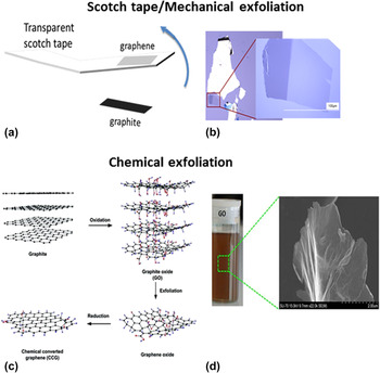
Graphene is a sp 2 hybridized carbon, arranged perfectly in a honeycomb structure to form a 2D monolayer of graphitic structure analogous to a polycyclic aromatic hydrocarbon of quasi infinite size. Reference Fitzer, Kochling, Boehm and Marsh1 Though theoretically graphene was known many decades ago, Geim and Novoselov first isolated the graphene by repeatedly peeling the bulk graphite using simple scotch tape in 2004. Reference Novoselov, Geim, Morozov, Jiang, Zhang, Dubonos, Grigorieva and Firsov2 This mechanical exfoliation method resulted in pristine monolayer to few-layer graphene material [Fig. 2(a)]. The exceptionally superior properties of graphene have been demonstrated using this mechanically exfoliated graphene. Reference Novoselov, Geim, Morozov, Jiang, Zhang, Dubonos, Grigorieva and Firsov2 Though this method resulted in superior quality graphene, it has shortcomings such as large scale synthesis, poor transfer methods which limits its use in fabricating devices at a commercial scale. Another class of method, used in 19th century which is also based on the exfoliation of graphite using strong acids and oxidizers, is named as chemical exfoliation. Brodie, Staudenmaier, and Hummer methods are the examples of chemical exfoliation methods. Reference Dreyer, Park, Bielawski and Ruoff3 Hummer’s method is one of the most commonly used methods. This method does not produce pristine graphene, rather results in graphene oxide (GO) sheets [Fig. 2(b)]. These oxygenated functional groups generate defects in sp 2 plane of graphene, rendering it electronically less conductive. But the conductivity can be restored to greater extent by subsequent reduction methods (thermal, chemical, or electrochemical) to produce reduced graphene oxide (RGO). Low cost, ease of processing, aqueous solubility/dispersibility, the presence of reactive edges due to abundance of oxygen functional groups makes GO a popular material in electrochemical biosensors development.
Mechanically exfoliated graphene (a) schematic of scotch tape method (b) optical image of monolayer graphene on SiO2/Si produced by scotch tape. Adapted from Ref. Reference Zan, Ramasse, Jalil and Bangert9 chemically exfoliated graphene (c) Schematic of Hummer’s method of exfoliating graphene layers from graphite. Adapted from Ref. Reference Bai, Li and Shi10. Reprinted with permission from John Wiley and Sons (2011) (d) optical and SEM image of GO. Adapted from Ref. Reference Marques, Gonçalves, Cruz, Almeida, Singh, Grácio, Sousa and Hashim11.
Other synthesis methods reported include, epitaxial growth of high-crystalline graphene using single crystalline silicon carbide (SiC) substrate (4H or 6H-SiC). Reference Vanbommel, Crombeen and Vantooren4,Reference de Heer, Berger, Ruan, Sprinkle, Li, Hu, Zhang, Hankinson and Conrad5 In this process, Si undergoes thermal desorption in ultra-high vacuum with subsequent rearrangement of carbon atoms to produce mono Reference Berger, Song, Li, Wu, Brown, Naud, Mayou, Li, Hass, Marchenkov, Conrad, First and de Heer6 to multilayer graphene. Reference de Heer, Berger, Ruan, Sprinkle, Li, Hu, Zhang, Hankinson and Conrad5,Reference Hass, de Heer and Conrad7 The advantage of epitaxial graphene (EG) is that it can be grown on large area semi-insulating SiC wafer without need of any graphene layer transfer process. Reference First, de Heer, Seyller, Berger, Stroscio and Moon8 This makes it amenable for device fabrication for semiconductor based miniaturized electrical biosensors. Obtaining uniform thickness with large domains, cost of SiC and transfer of graphene on to another substrate due to its interfacial bonding to SiC, possess some challenges in their application. Epitaxial growth of graphene using hydrocarbon decomposition on various other single crystal metal surfaces like Pt, Ru, TiC, Cu has been reported in the past. Reference Sutter, Flege and Sutter12
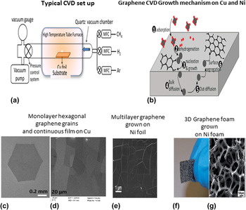
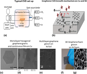
Chemical vapor deposition (CVD) method is currently being extensively studied in synthesis of wafer-scale, large area graphene for large scale device fabrication. Efforts are ongoing in the development of CVD growth protocol with controlled number of graphene layers with desired grain morphology and dimensions. Most commonly, in a typical CVD synthesis of graphene, a metallic planar foil (e.g., Cu or Ni) or 3D foam (e.g., Ni foam) is used as catalytic substrate for the growth. Carrier gas (argon), reducing gas (hydrogen), and carbon precursor gas (mostly methane) are passed at a desired flow rates ratio through a tube loaded with a growth substrate in a temperature controlled furnace. Figures 3(a) and 3(b), respectively, show schematics of typical CVD set up and growth mechanism of graphene at Cu and Ni surfaces. At pyrolytic temperature (about 1000 °C) of carbon precursor gas, single layer graphene (SLG) can be grown on copper surface [Fig. 3(c)] based on a self-limiting process whereas few layer (FLG) to multilayer graphene (MLG) can be grown on the Ni substrate [Fig. 3(d)] due to higher solubility of C in Ni. Use of Ni foam as substrate in CVD growth process has been reported to synthesize the 3D graphene foam [Fig. 3(e)].
(a) Schematic of typical CVD set up. Adapted from Ref. Reference Kumar, Lee and Aliofkhazraei13. (b) Sketch of graphene growth by both direct chemisorptions/deposition on Cu (step 1 to 3) and precipitation/segregation on Ni (step 1, 2, 4, 5, and 6). Adapted from Ref. Reference Losurdo, Giangregorio, Capezzuto and Bruno14. SEM image of graphene grown on Cu foil (c) monolayer graphene grains. Reprinted by permission from Macmillan Publishers Ltd: [Nature communications] Ref. Reference Zhou, Yu, Liu, Cheng, Chen, Huang, Liu, Wang, Huang and Duan15, copyright (2013) (d) continuous graphene film. (e) Multilayer graphene film grown on Ni foil and transferred on SiO2/Si on Ni foam. Adapted from Ref. Reference Huang, Chang, Guo, Liu, Xie, Wang, Ling and Yang16. Reprinted with permission from Elsevier (2012) (f) optical image of 3D graphene foam. (g) SEM image of graphene foam. Adapted from Ref. Reference Zhang, Ma, Yang, Huang and Dong17 with permission of The Royal Society of Chemistry (2014).
Though graphene can be grown at large scale using CVD process, its typical transfer processes includes polymer coating, etching of metallic substrate using iron chloride solution which can introduce ion or polymer impurities, affecting its electrical characteristics. With ongoing development in improving transfer/processing technologies, Reference Yang, Oh, Jung, Choi, Yu, Shin, Choi, Cho and Choi18–Reference Zaretski, Moetazedi, Kong, Sawyer, Savagatrup, Valle, O'Connor, Printz and Lipomi20 CVD graphene holds great promise for electronics and sensor device fabrication.
As mentioned earlier, the defect rich, water soluble/dispersible GO based material is a popular material in electrochemical biosensing as reactive defects/edges are crucial for heterogeneous electron transfer (HET) at the electrode surface for redox reactions and for biofunctionlization. In addition, solution processibility of GO based materials is advantageous in incorporating electroactive molecules for electrochemical sensing. But at the same time GO as well as RGO lacks the ideal electrical properties of pristine graphene. Large area CVD graphene film is almost defect free material with intact sp 2 structure shows poor HET therefore more widely used in FET based biosensors than electrochemical based. Raman spectra and cyclic voltammetry measurements of these materials characterize the structural defects and corresponding electrochemical activity. Reference Dreyer, Park, Bielawski and Ruoff3,Reference Brownson, Kampouris and Banks21 To further enhance their performance in biosensing advanced graphene nanoarchitectures and graphene based hybrids/composites have been synthesized. To limit the scope of this article we haven’t discussed graphene hybrids in this review as our group have already comprehensively reviewed the synthesis and applications of graphene hybrids. Reference Badhulika, Terse-Thakoor, Chaves Villarreal and Mulchandani22
B. Advanced graphene nanostructures
Apart from planar graphene structures, various advanced graphene nanostructures have been synthesized to enhance their electrocatalytic activity, surface area, and conductivity for use in electrochemical biosensors. These nanostructures with improved properties over planar graphene, are quite newer to the field, and hence have very few literature reports of biosensor applications.
1. Graphene nanoribbons
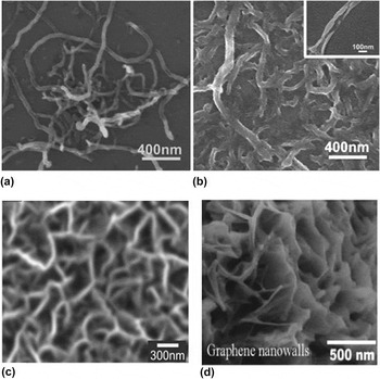
Graphene nanoribbons (GNRs), which are thin strips of graphene of width 100 nm or less, can be simply visualized as single or multiwall CNTs cut along its parallel axis and unfolded to form planar structure of width corresponding to diameter of the CNTs. GNRs can be synthesized by (i) bottom up organic synthetic approach, Reference Yang, Dou, Rouhanipour, Zhi, Raeder and Muellen23 (ii) e-beam/ion beam lithographic methods to pattern the exfoliated graphene Reference Han, Oezyilmaz, Zhang and Kim24,Reference Cai, Ruffieux, Jaafar, Bieri, Braun, Blankenburg, Muoth, Seitsonen, Saleh, Feng, Muellen and Fasel25 followed by oxygen plasma etching and (iii) unzipping CNTs through chemical oxidation Reference Kosynkin, Higginbotham, Sinitskii, Lomeda, Dimiev, Price and Tour26 or Ar plasma etching. Reference Jiao, Zhang, Wang, Diankov and Dai27 GNRs produced through chemical oxidation methods are referred as graphene oxide nanoribbons (GONRs) [Figs. 4(a) and 4(b)]. Similar to GO, these GONRs are further reduced to restore its electronic conductivity depending on the extent of oxidation. GNRs produced through lithographic methods have oxidized/rough edges due to oxygen plasma etching used in their fabrication. Compared to graphene and CNTs, high surface area, edge to basal plane ratio, electroactive defects, and edge chemistry/functional groups of GNRs are advantageous in electrochemical biosensing of analyte.
SEM images (a) multiwalled carbon nanotubes (MWCNTs) before chemical unzipping. Adapted from Ref. Reference Dong, Long, Wang, Chan-Park, Huang, Huang and Chen46. (b) Graphene oxide nanoribbons (GONRs) prepared by chemical unzipping of MWCNTs. Adapted from Ref. Reference Dong, Long, Wang, Chan-Park, Huang, Huang and Chen46 with permission of The Royal Society of Chemistry (2011). (c) MGNFs grown by microwave plasma enhanced CVD (MPCVD). Adapted from Ref. Reference Shang, Papakonstantinou, McMullan, Chu, Stamboulis, Potenza, Dhesi and Marchetto35. Reprinted with permission from John Wiley and Sons (2008) and (d) graphene nanowalls synthesized by electrophoretic deposition (EPD). Adapted from Ref. Reference Akhavan, Ghaderi and Rahighi43. Reprinted with permission from American Chemical Society (2012).

GNRs are also promising structures as a semiconductor gate/channel material in field effect transistor (FET) based biosensors. The graphene has a zero band gap which limits their sensitivity and limit of detection in FET biosensing Reference Sarkar, Liu, Xie, Anselmo, Mitragotri and Banerjee28 In GNRs, the band gap opening is possible by the electronic confinement of the charge carriers in quasi-one dimension due to their narrower widths. Extent of band gap opening largely depends upon the width of the GNRs as well as on presence of defects/edges. Attempts are being made to fabricate GNRs with width down to 10 nm. Still achieving GNRs with smooth edges without any structural disorders or functional groups, induced during their fabrication at high yields is a challenge. The method to produce high quality graphene nanoribbons from CNTs reported by the Dai group at Standford University Reference Jiao, Wang, Diankov, Wang and Dai29 has been widely used for studying their physical and electronic properties. Reference Wang, Ouyang, Jiao, Wang, Xie, Wu, Guo and Dai30 This method involves first calcination of MWCNTs in air to etch/oxidize the defect sites followed by sonication in 1,2-dichloroethane (DCE) organic solution of poly(m-phenylenevinylene-co-2,5-dioctoxy-p-phenylenevinylene) (PmPV) to unzip them. Other recent methods for high quality GNRs include electrochemical unzipping of MWCNTs. Reference Shinde, Debgupta, Kushwaha, Aslam and Pillai31
2. Graphene nanowalls
Graphene nanowalls, which are also called as carbon nanowalls, are mainly multiple graphene sheets assembled or in situ grown in vertical or parallel fashion with exposed sharp edges. These graphene nanowalls have a large specific area with higher conductivity and electroactivity, due to its increased edge plane sites, are suitable for development of electrochemical biosensors. The vertical graphene nanowalls structures are grown in situ by various PECVD methods using microwave plasma, Reference Wu, Qiao, Chong and Shen32–Reference Shang, Papakonstantinou, McMullan, Chu, Stamboulis, Potenza, Dhesi and Marchetto35 radio frequency (rF), Reference Davami, Shaygan, Kheirabi, Zhao, Kovalenko, Ruemmeli, Opitz, Cuniberti, Lee and Meyyappan36 inductively coupled plasma (ICP), Reference Jain, Karacuban, Krix, Becker, Nienhaus and Buck37 radical injection, Reference Kondo, Hori, Yamakawa, Den, Kano and Hiramatsu38 or e-beam excited, conductively coupled, Reference Hiramatsu, Shiji, Amano and Hori39 on different substrates such as glass, Reference Kim, Joung and Choi40 SiO2, Reference Tanaka, Yoshimura, Okamoto and Ueda34 sapphire, Al2O3, Reference Fang, Levchenko, Kumar, Seo and Ostrikov41 NiFe, Reference Wu, Qiao, Chong and Shen32 epitaxial growth on SiC Reference Kumar Roy, Ganguly, Yang, Wu, Hwang, Tai, Chen, Chen and Chattopadhyay42 etc., without need of additional catalyst. Whereas parallel graphene nanowalls or layered GO sheets can be synthesized by electrophoretic deposition (EPD) of GO from an electrolyte solution on graphite electrode Reference Akhavan, Ghaderi and Rahighi43,Reference Akhavan, Ghaderi, Hashemi and Rahighi44 or by simple ultrasonication assisted intercalation methods. Reference Yang, Guan, Meng, Yang, Li and Jiao45 Figs. 4(c) and 4(d) shows the SEM images of graphene nanowalls structures grown by microwave plasma enhanced CVD (MPCVD) and EPD method.
III. FABRICATIONS AND BIOFUNCTIONALIZATION OF GRAPHENE ELECTRODES
A. Fabrication of graphene electrodes
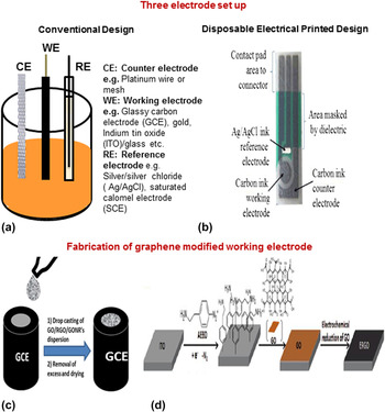
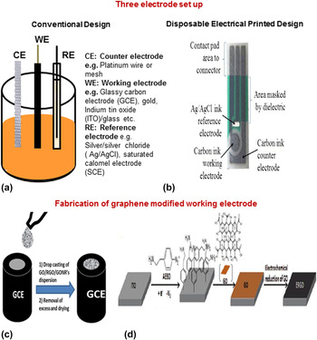
Graphene biosensors involving electrochemical measurements using the conventional three-electrode set up employ gold (Au) or glass carbon (GC) Reference Hu, Li, Bai, Li, Hua, Wang and Niu47,Reference Hu, Li, Han, Wu, Zhang, Niu and Bao48 disk electrodes, indium tin oxide (ITO) coated glass plates, Reference Singh, Sinsinbar, Choudhary, Kumar, Pasricha, Verma, Singh and Arora49 or pyrolytic graphite electrode (PGE) Reference Qiu, Qu, Dong, Ai and Han50 as working electrodes or use disposable electrical printed (DEP) chips with printed carbon electrodes Reference Giovanni, Bonanni and Pumera51 [Fig. 5(a)]. For modification of electrodes with GO, RGO, and GNRs (oxidative unzipped SWCNTs), the materials are first dispersed uniformly in water or ethanol using sonication and the resulting suspension is then drop casted or screen printed on the electrode surface. The solvent is allowed to evaporate and electrode is washed to remove the excess loosely bound material. In another method, negatively charged GO was deposited on the electrode surface through formation of positively charged self-assembled monolayers (SAM) of amine terminated benzenediazonium (ATBD) cation. Reference Haque, Park, Sung, Jon, Choi and Kim52 Fig. 5(b) shows fabrication of graphene electrode using drop casting method as well as through SAM of ATBD on ITO surface.
Three electrode set up for electrochemical measurements using (a) conventional and (b) screen printed disposable designs. Adapted from Ref. Reference Truong53. Fabrication of graphene working electrodes by (c) simple drop casting and (d) self-assembled monolayer (SAM) of ATBD cation and negatively charged GO. Adapted from Ref. Reference Haque, Park, Sung, Jon, Choi and Kim52. Reprinted with permission from American Chemical Society (2012).
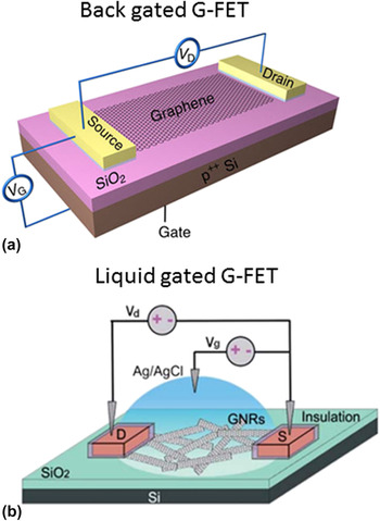
FET based devices reported in the literature mostly have used CVD grown graphene with few reports of RGO and RGONR based devices. Reference Dong, Long, Wang, Chan-Park, Huang, Huang and Chen46,Reference Kim, Sohn, Jung, Yoon, Lee and Park54 CVD graphene grown on Cu or Ni is first coated with polymethylmethacrylate (PMMA) polymer for flexibility in transfer process and then its metal substrate is etched in ferric chloride or ferric nitrate solution. PMMA/graphene films floating in water are then transferred by scooping it on suitable substrates to be used as a working electrode and PMMA is removed using solvents such as acetone. For FET device fabrication, the device has either photolithography or EBL patterned source (S) or drain (D) electrodes made of Cr/Au or Ti/Au on graphene/SiO2/Si substrate Reference Zhang, Liu, Meng, Li, Liang, Hu and Wang55 or silver paint is used as the S and D electrode on the graphene coated glass slide Reference Dong, Shi, Huang, Chen and Li56 or SiO2/Si substrate. Reference Lin, Phan Thi Kim, Chen, Liu, Chen, Wei and Li57 Further, for liquid-gated (LG) FET, a liquid chamber is created by insulating electrodes either by using silicon rubber or polydimethylsiloxane (PDMS). In LG-FET, gate voltage is applied through silver wire immersed in the liquid drop placed on the device. Figure 6 shows the design of graphene FET based biosensor including back gate and liquid gated FETs.
FET-based graphene biosensor design: back gated G-FET (a) device structure and the circuit diagram corresponding to measurement of I SD (V G) Adapted from Ref. Reference Zhang, Liu, Meng, Li, Liang, Hu and Wang55. Reprinted by permission from Macmillan Publishers Ltd: [Nature communications], copyright (2013). (b) Liquid gated G-FET using RGONR thin film as a conducting channel with Ag/AgCl electrode immersed in phosphate buffer saline for applying the gate voltage and electrodes are insulated using silicon rubber. Adapted from Ref. Reference Dong, Long, Wang, Chan-Park, Huang, Huang and Chen46 with permission of The Royal Society of Chemistry (2011).

Advanced structures like graphene nanowalls are grown directly in situ by PECVD or EPD on the desired electrode substrates as mentioned above. Epitaxial graphene also do not need any transfer process as they are grown directly on the SiC substrate.
B. Biofunctionalization of graphene electrodes
Functionalization of graphene electrode with biorecognition elements such as proteins, aptamer, or DNA can be done using simple physical adsorption through hydrophobic interactions, using electrostatic or covalent binding through oxygenated functional groups of GO or RGO or noncovalent binding through chemical bi-linker molecules.
For covalent functionalization of GO based electrode, widely used approach is to link its carboxylic groups to amine groups of biorecognition elements through amide bond formation, using crosslinker chemistry. For example, probe DNA with 5′-NH2 group was immobilized on GO/GCE electrode by covalent linkage to carboxylic groups on GO using EDC [1-ethyl 3-(3-dimethylaminopropyl)carbodiimide·HCl] crosslinker and N-hydroxysuccinimide (NHS) chemistry. Reference Gong, Yang, Dong and Zhang58
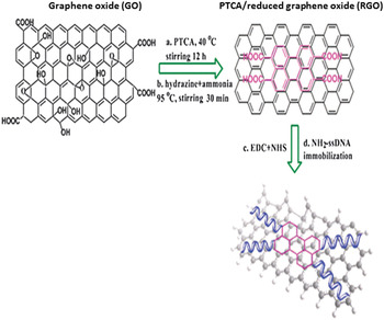
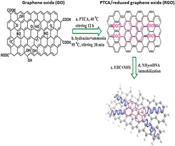
Biomolecules functionalization through π–π interaction using sp 2 basal plane of GO, RGO, or CVD graphene or hydrophobic interactions by bi-linker molecules is a common approach used in graphene based devices. In this method one end of the bi-linker molecule such as 3,4,9,10-perylene tetracarboxylic acid (PTCA), 1-pyrenebutanoic acid succinimide ester (pyrene-NHS) and poly(BMA-r-PEGMA-r-N-acryloxysuccinimide), binds to the graphene surface via π–π bond or hydrophobic interaction while the other end reacts with the –NH2 group of the biomolecule. Reference Hu, Li, Bai, Li, Hua, Wang and Niu47,Reference Feng, Chen, Ren and Qu59,Reference Huang, Dong, Liu, Li and Chen60 Fig. 7 shows the NH2-ssDNA functionalization of graphene through PTCA linker and EDC/NHS chemistry.
Schematic representation of noncovalent functionalization of graphene using 3,4,9,10-perylene tetracarboxylic acid (PTCA) linker to introduce carboxylic groups and subsequent functionalization with probe ssDNA. Adapted from Ref. Reference Hu, Li, Bai, Li, Hua, Wang and Niu47 with permission of The Royal Society of Chemistry (2010).
IV. GRAPHENE ELECTRICAL/ELECTROCHEMICAL BIOSENSOR APPLICATIONS
A. DNA biosensing
DNA biosensing in healthcare is crucial in screening and diagnosis of human genetic diseases by detecting single nucleotide polymorphisms (SNPs), human pathogenic diseases to identify the pathogens based on their specific DNA sequences, Reference Liao, Mastali, Li, Gau, Suchard, Babbitt, Gornbein, Landaw, McCabe, Churchill and Haake61–Reference Drummond, Hill and Barton64 DNA damage caused by chemical agents e.g., acrylamide etc., anticancer drugs, nucleases, ionizing radiations, and studying the interaction. Reference Qiu, Qu, Dong, Ai and Han50,Reference Oliveira and Oliveira-Brett65,Reference Palecek, Fojta, Tomschik and Wang66 Development of DNA diagnostics from biosensor to gene chips to miniaturized ‘Lab-on-Chip’ device for rapid, simple, massively parallel yet accurate and highly sensitive analysis of DNA is intense. Reference Drummond, Hill and Barton64,Reference Wang67
Graphene has been successfully used as a platform in the development of DNA biosensor due to difference in the binding affinity of ssDNA and dsDNA to graphene. It has been reported that ssDNA binds strongly to graphene than dsDNA due to its structural flexibility and maximally exposed nucleobases for interaction with the graphene plane. These nucleobases mainly bind through their aromatic groups to sp 2 basal plane of graphene by π–π interaction. Also, they are shown to bind to graphene in different morphological shapes (spherical particles or elongated networks) depending upon the magnitude of the interaction energies between nucleobases and between graphene and nucleobases. Reference Akca, Foroughi, Frochtzwajg and Postma68
Graphene based electrochemical DNA sensing is based on two major principles: (i) detection of hybridization event between the probe DNA (pDNA) and target DNA (tDNA) (ii) direct detection of bases of DNA by monitoring their oxidation.
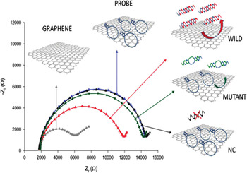
Electrochemical impedance spectroscopy (EIS) is a popular method in hybridization based DNA biosensing due to its sensitivity in translating the binding events at the electrode and electrolyte interface. “Signal-off” and “signal-on” mode are the two formats for the EIS-based graphene DNA biosensor. In the “signal-off” mode format, ssDNA is immobilized through physical adsorption as a pDNA which upon hybridization with tDNA forms dsDNA duplex and detaches from the graphene surface resulting in the decrease in the impedance signal (“signal-off” mode). Reference Bonanni and Pumera69 Figure 8 shows the schematics of protocol and Nyquist plots for “signal-off” mode impedimetric DNA biosensing. Different chemically modified graphene platforms such as GO, electrochemically RGO (ERGO), and thermally RGO (TRGO), were studied in “signal-off” mode EIS based SNP detection for Alzheimer disease. Reference Giovanni, Bonanni and Pumera51 GO was found to be the best platform amongst them to detect the SNP. In another study, few layer graphene was found to be the most sensitive platform with a LOD of 6.6 pM (linear detection range from 3 × 10⁻¹³ to 3 × 10⁻¹⁰ M) when compared to single (LOD 50 nM) and multilayer graphene (LOD 66 μM) for detection of wild type target and was chosen for detecting SNP correlated to the Alzheimer disease. Reference Bonanni and Pumera69 The unique morphology of large area, wavy GO nanowalls providing large specific area with rich conjugated structure for efficient pDNA immobilization and sharp edges for faster electron transfer have resulted in more sensitive “signal-off” based EIS detection of PML/RARA [fusion gene sequence formed from promyelocytic leukemia (PML) and retinoic acid receptor alpha (RARA)] with LOD of 5.20 fM and detection range of 1.0 × 10⁻⁷–1.0 × 10⁻¹⁴ mol/L. Reference Yang, Guan, Meng, Yang, Li and Jiao45
Schematic of the protocol and Nyquist plots for “signal-off” impedimetric DNA biosensing of the graphene surface (gray), hairpin pDNA (blue), complementary target (red), 1-mismatch target (green), and negative control with a noncomplementary sequence (black). All measurements were performed in PBS buffer solution containing K3[Fe(CN)6]/K4[Fe(CN)6]. Adapted from Ref. Reference Bonanni and Pumera69. Reprinted with permission from American Chemical Society (2011).

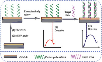
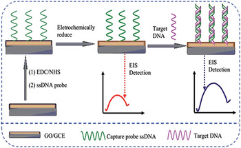
DNA immobilized on GO or RGO by covalent modification through oxygenated groups or by π–π interaction through bifunctional linkers like perylene derivatives anchoring functional groups for DNA attachments results “signal-on” mode EIS detection due to change in the conformation and negative charge accumulation after hybridization with target DNA instead of the detachment in case of “signal-off” EIS. Figure 9 shows the fabrication and “signal-on” impedimetric DNA detection of specific HIV gene sequence. Reference Gong, Yang, Dong and Zhang58 GO was used for immobilization of pDNA through carbodiimide chemistry and then converted the GO electrochemically to ERGO to improve the electron transfer characteristics. The resulting electrode had a LOD of 3 × 10⁻¹³ M with linear detection range from 1 × 10⁻¹² to 1 × 10⁻⁹ M.
Schematic diagram of the fabrication of the impedimetric DNA biosensor and the detection of target DNA for “signal-on” mode impedimetric DNA biosensing. Adapted from Ref. Reference Gong, Yang, Dong and Zhang58 with permission of The Royal Society of Chemistry (2015).
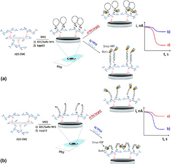
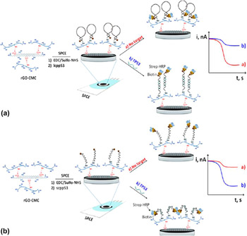
A disposable amperometric DNA biosensor was reported for the direct detection of tDNA sequence on p53 tumor suppressor (TP53) gene and discriminating between wild and SNP containing sequences using RGO-carboxymethyl cellulose (CMC) nanomaterial. Reference Esteban-Fernandez de Avila, Araque, Campuzano, Pedrero, Dalkiran, Barderas, Villalonga, Kilic and Pingarron70 Two different DNA sensing platforms were evaluated by immobilizing biotinylated hairpin capture probe of two different lengths (33 versus 20 nucleotides) on the RGO/CMC modified screen printed carbon electrode (SPCE). Streptavidin-Horseradish peroxidase (Strep-HRP) conjugate was captured on the hybridized duplex and amperometric detection was carried out using 3,3′,5,5′-tetramethylbenzidine (TMB)–H2O2 system (Fig. 10). In the case of long pDNA, due to increased distance between Strep-HRP conjugate and electrode, the current response was decreased due to hindrance of enzyme catalyzed electron transfer. On the other hand, in the case of short pDNA amperometric response increased due to decreased hindrance to electron transfer rate.
Schematic illustration of the steps involved in the preparation of the electrochemical DNA biosensor for the detection of the TP53 gene using rGO−CMC hybrid nanomaterial as a scaffold [(a) the long TP53 capture probe (lcpp53) or (b) the short (spp53) TP53 capture probe] and labeling with Strep-HRP (relative sizes of the components are not drawn on a real scale). Adapted from Ref. Reference Esteban-Fernandez de Avila, Araque, Campuzano, Pedrero, Dalkiran, Barderas, Villalonga, Kilic and Pingarron70. Reprinted with permission from American Chemical Society (2015).
Anodized EG with high edge plane defects was found to be a superior platform than pristine EG, boron doped diamond, CNTs, and glassy carbon in simultaneous detection of all four nucleic acids (A, T, G, C) based on their oxidation. Reference Lim, Hoh, Ang and Loh71 All nucleic acids were resolved as individual oxidation peaks using differential pulse voltammetry (DPV) from sample containing free bases, ssDNA and dsDNA.
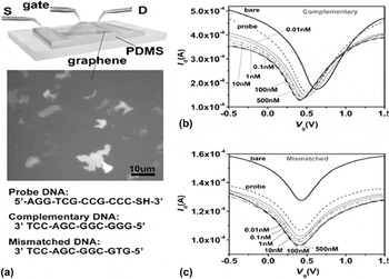
FET-based devices are of great choice in developing miniaturized chip based POC biosensors for label free detection. CVD graphene has been used to fabricate G-FET (graphene-FET) devices due to ease and reproducibility of device fabrication. Reference Zhang, Liu, Meng, Li, Liang, Hu and Wang55,Reference Lin, Phan Thi Kim, Chen, Liu, Chen, Wei and Li57,Reference Mohanty and Berry72 Liquid gated G-FETs have been reported in DNA sensing with single base mismatch specificity. Reference Dong, Shi, Huang, Chen and Li56,Reference Lin, Phan Thi Kim, Chen, Liu, Chen, Wei and Li57 Synthetic oligonucleotide (pDNA) was immobilized on the graphene patterned with source and drain electrodes. A drop of buffer containing fully matched tDNA or one-base mismatched ssDNAs was then added. After hybridization reaction, chip is rinsed and fresh drop of buffer was added. Liquid gating was established by using silver wire immersed into the top of the drop. Figure 11(a) shows the schematic of liquid gated G-FET device with DNA sequences used in sensing. Detection was based on the n-doping of graphene with DNA as evident by left shifting of V g,min in transfer curve [drain current (I d) versus gate voltage (V g) curve] as shown in Fig. 11(b). Liquid gated G-FET biosensor was developed and Hall effect measurements were performed based on the Van der Pauw method for DNA hybridization detection. Reference Lin, Phan Thi Kim, Chen, Liu, Chen, Wei and Li57 It was found that sheet resistance increases and hole carrier mobility of graphene FET devices decreases with increasing concentration of complementary DNA or single base mismatch DNA. Whereas hole carrier concentration increases with added complementary DNA but no significant effect with added one base mismatch DNA.
(a) Schematic illustration of the graphene device operated by liquid gating. Middle is an optical microscopy image of the graphene films. Bottom shows the DNA sequences used in the experiments. Transfer characteristics for the graphene transistors before adding DNA, after immobilization with probe DNA, and after reaction with (b) complementary or (c) one-base mismatched DNA molecules with the concentration ranging from 0.01 to 500 nM. Adapted from Ref. Reference Dong, Shi, Huang, Chen and Li56. Reprinted with permission from John Wiley and Sons (2010).
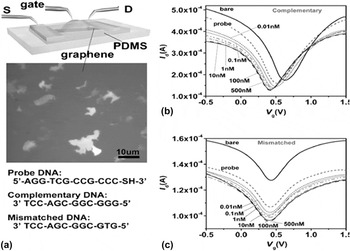
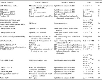
Table I summarizes the recently reported DNA biosensor using graphene based electrodes.
Summary of recently reported DNA biosensors using graphene based electrodes.
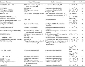
PDI: perylene Reference Kim, Sohn, Jung, Yoon, Lee and Park54 derivative, N,N-bis-(1-aminopropyl-3-propylimidazol salt)-3,4,9,10-perylene tetracarboxylic acid diimide, CMC: carboxymethylcellulose, PEG: pyrolytic graphite electrode, HRP: horseradish peroxidase, PAN: polyaniline, CHI: chitosan, Ph-NH2: phenyl-amino.
Modified nucleoside and nucleobases are the selective markers for the wholebody turnover of RNA including tRNA, mRNA, and rRNA. Increased levels of modified nucleoside are found in the urine of the patients with different types of malignancies Reference Seidel, Brunner, Seidel, Fritz and Herbarth81 and AIDS. Reference Borek, Sharma, Buschman, Cohn, Penley, Judson, Dobozin, Horsburgh and Kirkpatrick82 Therefore detection and quantification of bases of nucleic acids is important in the diagnosis and therapy control of these diseases. Individual and simultaneous detection of adenine and guanine has been reported using carboxylic acid functionalized graphene electrode. Reference Huang, Niu, Sun, Han, Wu, Li and Xiong83 The direct electroxidation of adenine and guanine was studied using cyclic voltammetry and differential pulse voltammetry methods.
B. Protein biomarkers biosensing/immunosensing
Diseases biomarkers are mainly proteins whose presence or abnormal levels in the body fluids can be used as indicator for diagnosis, progression of a disease or to assess pathophysiological response to a therapeutic treatment. Rapid, selective, and sensitive point of care detection of single or array of biomarkers from body fluids, i.e., blood or urine, on a single biosensor device has opened a promising avenue for early and accurate detection of life threatening diseases like cardiovascular diseases, cancer etc. The promising progress in the biomarkers discovery research through advanced genomic and proteomic methods have facilitated the development of biosensors including POC devices in clinical diagnosis based on biomarkers detection.
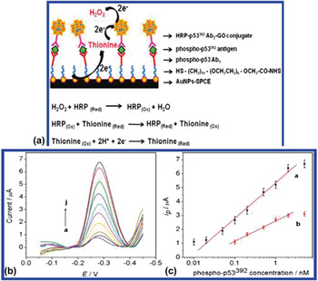
Graphene has been explored mostly in the detection of cancer protein biomarkers such as phosphorylated p53 protein (ovarian cancer), Reference Xie, Chen, Du and Lin84,Reference Du, Wang, Shao, Wang, Engelhard and Lin85 carcinoembryonic antigen (CEA) (colorectal cancer), Reference Chen, Jia, Han, Ma and Ma86 alpha-fetoprotein (AFP) (liver cancer), Reference Chen, Jia, Han, Ma and Ma86 and cardiac biomarkers such as cardiac troponin I, Reference Tuteja, Kukkar, Suri, Paul and Deep87 C-reactive protein, Reference Yuan, Yu, Xia, Gao, Xu, Li and He88 soluble CD40 ligand, Reference Yuan, Yu, Xia, Gao, Xu, Li and He88 etc. Graphene, mainly ERGO has been used as the platform for the antibody/probe immobilization through its functional groups in various sandwich type immunosensors Reference Haque, Park, Sung, Jon, Choi and Kim52 resulting in higher detection sensitivity due to its high electronic conductivity. Apart from using as a platform, graphene has also been used as a nanocarrier in linking enzyme label and signal antibody in signal amplification strategy due to its high loading ratio than gold nanoparticle (AuNP), high surface area, and biocompatibility. Reference Du, Wang, Shao, Wang, Engelhard and Lin85 Enhanced sensitivity was reported when bioconjugate featuring HRP and p53392 (phosphorylated p53 at Ser392) signal antibody linked to functionalized GO was used in sandwich immunoreaction [Fig. 12(c)]. For immunosensing, AuNP modified SPCE is first functionalized with primary antibodies.
(a) Schematic illustration of the multienzyme labeling amplification strategy using HRP-p53392Ab2-GO conjugate. (b) Square wave voltammetry curves acquired at HRP-p53392Ab2-GO/phosphop53392/Ab1/NHS/AuNPs-SPCE after incubation with (a) 0–5 ng mL-1 phospho-p53392 antigen in pH 7.4 PBS containing 25 μM thionine and 2 mM H2O2. (c) Calibration plots of the (a) proposed HRP-p53392Ab2-GO/phospho-p53392/Ab1/NHS/AuNPs-SPCE and (b) traditional HRP-streptavidin-biotin-p53392Ab2/phospho-p53392/Ab1/NHS/AuNPs-SPCE for detecting phospho-p53392 antigen. Adapted from Ref. Reference Du, Wang, Shao, Wang, Engelhard and Lin85. Reprinted with permission from American Chemical Society (2011).

Upon sandwich immunoreaction, HRP-p53392Ab2-GO conjugate is captured on the electrode. Enzymatic products of HRP in presence of H2O2 and thionine are then electrochemically detected. Figures 12(a) and 12(b) shows the schematic of multienzyme labeling amplification strategy and square wave voltammetry measurements at different concentrations of phospho-p53392 antigen. This amplification strategy has dynamic range from 0.02 to 2 nM and LOD of 0.01 nM for phosphorylated p53-S392 (phospho-p53392). Reference Du, Wang, Shao, Wang, Engelhard and Lin85
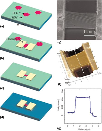
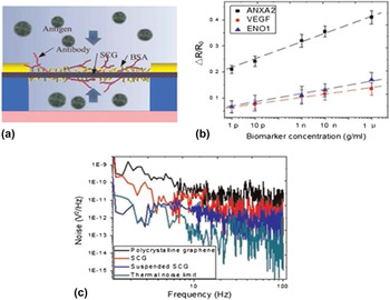
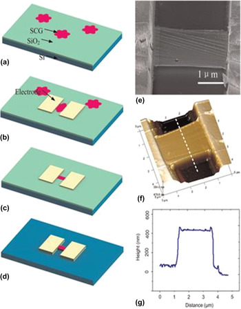
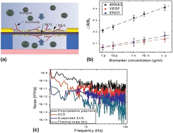
Recently, CVD grown, 3D monolithic and macroporous graphene foam has been reported for sensitive detection of CEA. Reference Liu, Wang, Wang, Li, Xi, Wang and Wang89 Macroporous scaffold with large electroactive area, superior substrate diffusion along with high conductivity and stability makes 3D graphene foam (3D G foam) superior electrode over conventional planar electrodes. During sensor fabrication, concanvaline A (ConA) lectin was first immobilized on the graphene foam through polydopamine as the linker. HRP labeled anti-CEA antibody was further immobilized on the 3D G foam via biospecific “lectin-sugar protein” interaction between ConA and HRP. Electrochemical detection of CEA was based on the measuring the inhibited current response of redox probe using DPV with increasing concentrations of CEA. Without any complex amplification strategy, the 3D electrode showed good dynamic detection range of 0.1–750 ng/mL with LOD of ∼90 pg/mL for CEA detection. In case of CVD grown polycrystalline graphene (PCG), the grain boundaries and carriers scattering from substrate such as SiO2, largely affects the intrinsic properties of graphene and thus the performance of the sensor. Biosensing performance of LPCVD grown, free standing single crystal graphene (SCG) in multiplex detection of lung cancer biomarkers was studied and compared to the device made with PCG. Reference Li, Zhang and Cui90 Fig. 13 shows the SCG sensor fabrication and characterization. The immunoreaction between three tumor biomarkers namely ANXA2, VEGF, and ENO1 in increasing concentration with their corresponding antibodies were detected based on the change in the resistance due to change in the carrier concentration and therefore the mobility of the graphene [Figs. 14(a) and 14(b)]. The SCG biosensor can measure the concentrations as low as 0.1 pg/mL with low frequency of noise over PCG [Fig. 14(c)]. SCG based biosensor showed superior uniformity over PCG based ones. It also exhibited specificity and large linear detection range from 1 pg/mL to 1 µg/mL.
Suspended, single crystalline graphene (SCG) sensor fabrication. (a–d) process. (a) Transfer SCGs from copper foil onto SiO2/Si substrate by PMMA assisted process. (b) Choose a SCG and fabricate metal electrodes on it. (c) Etch SCG into rectangular beam shape. (d) Etch SiO2 beneath to release the suspended SCG beam. (e) SEM image of a suspended SCG beam. (f) AFM topographic image of a SCG beam. (g) The AFM lines scan of SCG beam marked with white dot line confirming SCG is suspended and the distance between the beam and substrate is about 300 nm. Adapted from Ref. Reference Li, Zhang and Cui90. Reprinted with permission from Elsevier (2015).
(a) Schematic illustration of immunoreaction of suspended SCG biosensor. (b) The SCG sensor’s response to 3 different types of lung cancer tumor markers. Tumor markers with higher concentration result in larger resistance shift of the sensor. (c) Comparison of low-frequency noise in polycrystalline graphene, nonsuspended SCG and suspended SCG. Suspended SCG has the smallest low-frequency noise. Adapted from Ref. Reference Li, Zhang and Cui90. Reprinted with permission from Elsevier (2015).
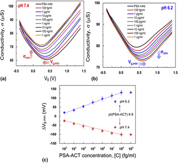
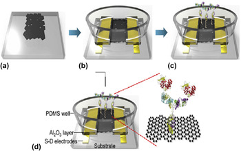
Liquid gated RGO-FET biosensors is reported for label free ultrasensitive detection of prostate cancer biomarker, prostate specific antigen/α1-antichymotrypsin (PSA-ACT) complex. Reference Kim, Sohn, Jung, Yoon, Lee and Park54 Fig. 15 shows the schematic of fabrication and detection of PSA-ACT complex. The channel conductivity σ (V g) is obtained at different analyte concentration using PBS solution of pH of 7.4 and 6.2.
Schematic of RGO-FET fabrication and detection of PSA-ACT complex. (a) Self-assembly of GO nanosheets on amine SAM substrate and reduction to RGO. (b) Formation of Ti/Au source and drain electrodes, capping of the electrodes by Al2O3 and PDMS layers for minimization of leakage currents. (c) Functionalization of RGO channel by linker molecules, immobilization of PSA mAb to the linker molecules on RGO channel, and treatment of ethanolamine (ETA) for nonspecific binding. (d) Illustration of RGO-FET immunosensor with Pt reference electrode in the analyte solution. The immunoreactions between antibody (PSA mAb) and the antigen (PSA-ACT complex) caused changes in the carrier density in the RGO channel due to the doping effects. Adapted from Ref. Reference Kim, Sohn, Jung, Yoon, Lee and Park54. Reprinted with permission from Elsevier (2013).
The isoelectric point of PSA-ACT is 6.8 so it is negatively and positively charged at pH 7.4 and 6.2, respectively. Using ΔV g,min as sensing parameter, RGO-FET could detect PSA-ACT complex down to LOD of 100 fg/mL (∼1.1 fM). Figure 16 shows the detection characteristic of RGO-FET sensor.
Detection characteristics of RGO-FET immunosensors. (a–b) σ-V g plot of RGO-FET at a V sd of 0.6 V with various concentrations of PSA-ACT complex in the analyte solutions at (a) pH 7.4 and (b) pH 6.2. (c) Shift in the minimum conductivity point (ΔV g,min) with the concentration of PSA-ACT complex in the pH 7.4 and pH 6.2 analyte solutions. The ΔV g,min value was obtained by calculating the difference in V g,min as a reference for the device with no binding of PSA-ACT complex. Adapted from Ref. Reference Kim, Sohn, Jung, Yoon, Lee and Park54. Reprinted with permission from Elsevier (2013).
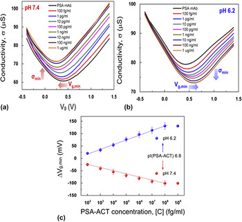
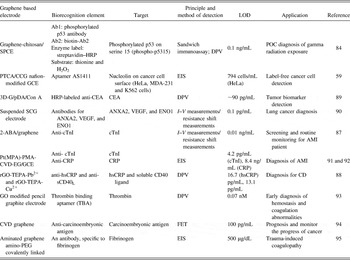
Table II summarizes the reports of graphene based biosensors in protein biomarkers detection and their performance.
Summary of reports of graphene based biosensors in protein biomarkers detection and their performance.
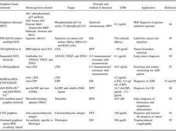
SPCE: screen printed carbon electrode, PTCA: 3,4,9,10-perylene tetracarboxylic acid, CCG: chemically converted graphene, 3D G: three dimensional graphene foam, pDA: polydopamine, ConA: concanvaline A, SCG: single crystal graphene, cTnI: cardiac troponin I, 2-ABA: 2-aminobenzyl amine, AMI: acute myocardial infarction, hsCRP: high sensitivity C-reactive protein, TEPA: tetraethylenepentamine, CD: cardiovascular disease, SNAP-25: synaptosomal-associated protein 25, BoNT-LcA: botulinum neurotoxin serotype A light chain, MPA: mercaptopropionic acid, EG: electroactive monolayer graphene.
C. Metabolites/small molecules biosensing
Low molecular weight compounds such as glucose, dopamine (DA), ascorbic acid (AA), uric acid (UA) which are normally present in the body fluids play important role in the normal functioning of some of the biological processes such as DA in central nervous system or AA in tissue and immune system. They can also be present in the system, being the metabolic products for e.g., UA is the oxidation product in urine metabolism. Their quantitative detection is important in diagnostics as abnormal levels indicates certain metabolic disorders like diabetics, Reference Harris96 gout/kidney dysfunctioning, Reference Mohandas and Johnson97 neurological disorders like schizophrenia and Parkinson. Reference Wightman, May and Michael98
Electrochemical glucose biosensors have played a key role in the development of accurate and reliable glucose detection for the diagnosis and management of diabetes. Among them enzyme based amperometric biosensors have been the focus of advanced research and development. Most of the enzyme based glucose biosensors, use glucose oxidase (GOx) enzyme to catalyze the oxidation of glucose and indirectly detect glucose by measurement of its oxidation product i.e., hydrogen peroxide (H2O2). Direct electrochemical quantification of glucose oxidation can be achieved either using redox mediators or more recently through direct electron transfer (DET) of redox factor of GOx using advanced materials and electrode geometry.
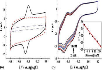
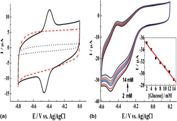
Graphene has been studied for DET and in amperometric glucose biosensing based on the reduction of oxygen. Reference Wu, Shao, Hu, Jin, Yin, Zhang and Cai99–Reference Unnikrishnan, Palanisamy and Chen105 GOx/polyethylenimine-functionlized ionic liquid (PFIL)/RGO electrode has been reported to exhibit DET of GOx and electrocatalytic activity for glucose detection based on oxygen reduction. Reference Shan, Yang, Song, Han, Ivaska and Niu104 Negatively charged GOx was immobilized through counter anion exchange from ionic liquid.
DET redox peak at the electrode and its electrocatalytic activity toward O2 and H2O2 were studied using cyclic voltammetric analysis. Based on the high electrocatalytic/reduction activity of the electrode toward O2 and H2O2, an amperometric glucose biosensor was developed. The biosensor showed a linear response in the clinically relevant glucose concentration ranging from 2 to 14 mM. Figure 17 shows the DET peaks at GOx/PFIL/RGO electrode and glucose response based on decrease in oxygen reduction current in response to glucose.
(a) Cyclic voltammograms of graphene-PFIL (dashed), graphite-GOx-PFIL (dotted), and graphene-GOx-PFIL (solid) modified electrodes in 0.05 M PBS solution (pH = 7.4) saturated with N2 at a scan rate of 0.05 V/s. (b) Cyclic voltammetric measurements at the graphene-GOx-PFIL modified GC electrode in various concentrations of glucose PBS solution saturated with O2: 2–14 mM from outer to inner. The inset is the calibration curve (R = 0.994) corresponding to amperometric responses at −0.49 V. Scan rate: 0.05 V/s. Adapted from Ref. Reference Shan, Yang, Song, Han, Ivaska and Niu104. Reprinted with permission from American Chemical Society (2009).
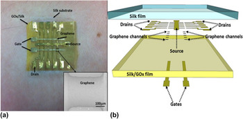
Implantable or wearable glucose biosensors are becoming more popular over strip-based tests in patients where frequent monitoring of glucose is crucial and also strip tests are inconvenient as it involves multiple finger pricking. Atomic thick graphene with unique sensing properties in FET based devices can be easily integrated into miniaturized devices with flexible substrate to develop sensitive and reliable wearable or implanted sensors. Silk fibroin-encapsulated G-FET based enzymatic flexible biosensor has been reported in detection of glucose (Fig. 18). Reference Xueqiu and Pak106
(a) Optical image of silk-based graphene FET glucose biosensors attached onto the wrist. Each patch contains 10 graphene FET biosensor channels. The inset is an SEM image of a typical graphene FET channel between source and drain electrodes. (b) Exploded-view schematic illustration of the silk-based graphene FET biosensors. Adapted from Ref. Reference Xueqiu and Pak106. Reprinted with permission from Elsevier (2014).
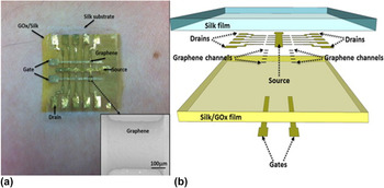
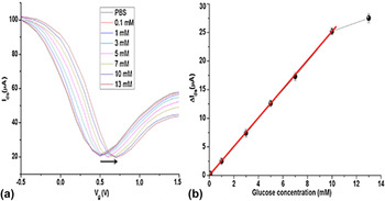
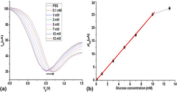
In this device, silk protein acts as both device substrate and GOx enzyme immobilization material. The biosensing was based upon measuring the differential drain–source current and shift in the dirac point of the graphene in response to doping caused by H2O2, the oxidation product of glucose [Fig. 19(a)]. The biosensor showed a linear detection range of 0.01–10 mM of glucose with LOD of 0.1 mM and average sensitivity of 2.5 μA/mM [Fig. 19(b)]. This silk based glucose sensor can be used as a wearable biosensor to measure the glucose from the sweat due to its mechanical flexibility and stability. It can also be implanted in the body for continuous monitoring of glucose as silk is the biocompatible and biodegradable natural polymer. Another similar flexible G-FET glucose biosensor was developed using polyethylene terephthalate (PET) substrate. Reference Kwak, Choi, Kim, Kim, Yoon, Ahn, Yang, Yang and Seo107
(a) Charge transfer curves of silk/GOx functionalized graphene FET in response to the addition of glucose to various concentrations at V ds = 100 mV. (b) The response curve of the silk/GOx functionalized graphene FET to glucose at V g = 0 V. Adapted from Ref. Reference Xueqiu and Pak106. Reprinted with permission from Elsevier (2014).
Compounds like DA, AA, and UA generally coexist in the biological matrices. Since their oxidation potentials are very close to each other, their voltammetric peaks cannot be resolved at traditional bare electrodes. Graphene modified electrodes have been reported in the selective detection of these compounds. Reference Shang, Papakonstantinou, McMullan, Chu, Stamboulis, Potenza, Dhesi and Marchetto35,Reference Lian, Sun, Sun and Liu108–Reference Chao, Ma and Li114 Vertically oriented multilayer graphene nanoflake (MGNF) film with 2–3 nm thick sharp edges possess high electron transfer kinetics and demonstrated well-resolved voltammetric peaks from oxidation of AA, DA, and UA. Reference Shang, Papakonstantinou, McMullan, Chu, Stamboulis, Potenza, Dhesi and Marchetto35 The biosensing performance of the electrode was measured using DPV as a function of DA concentration and found to be superior to untreated and unmodified carbon electrodes with LOD of 0.17 μM.
Oxidative stress at cellular level has been shown to be an crucial factor in pathogenesis of various diseases such as atherosclerosis, Reference Heinecke115 cancer, Reference Sosa, Moline, Somoza, Paciucci, Kondoh and Lleonart116 male infertility Reference Sharma, Pasqualotto, Nelson, Thomas and Agarwal117 etc. Hydrogen peroxide (H2O2) is one of the reactive oxygen species (ROS) produced as a by-product of oxidative metabolism in vivo. Therefore detection of H2O2 is important in clinical diagnosis. Graphene electrodes including RGO, Reference Xu, Dai and Chen118,Reference Zhou, Liu, Jiang, Yang and Chen119 single layer graphene nanoplatelets Reference Lu, Dong, Li and Hu120 have been reported for DET of hemoglobin, HRP and microperoxidase-11 and H2O2 biosensing. To further improve the sensitivity of H2O2 biosensing, graphene hybrids have been used with improved surface area for e.g., recently heme peptide-graphene–CNT was shown to have ∼15 times more catalytic current for H2O2 reduction compared to heme peptide-graphene electrode. Reference Komori, Terse-Thakoor and Mulchandani121
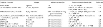
Table III present examples of graphene based electrodes in other metabolites/small molecules sensing.
Examples of graphene based electrodes in other metabolites/small molecules sensing.
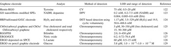
ChOx: cholesterol oxidase, ChEt: cholesterol esterase, PSS: polystyrene sulfonic acid.
D. Pathogen biosensing
Detection of pathogens in biological tissues and food samples is of utmost importance in the field of forensics, medical diagnostics, and clinical research as it helps in the timely identification and prevention of problems related to health and safety. The conventional techniques to detect microbial pathogens often rely upon clinical microbiology monitoring approaches that are time consuming, expensive and involve labor intensive standard cultures and tests. Hence these methods are inadequate to meet the rapidly growing demands of the industry and cannot be used in situations where managing large numbers of environmental or clinical samples is essential.
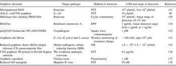
TRGO, Reference Liu, Choi, Park, Lee and Seo130 GO Reference Mohanty and Berry72,Reference Liu, Kim, Cheon and Seo131,Reference Wang, Su, Xu, Xiang, Yuan and Chai132 and CVD graphene Reference Huang, Dong, Liu, Li and Chen60,Reference Mannoor, Tao, Clayton, Sengupta, Kaplan, Naik, Verma, Omenetto and McAlpine133 have been explored in the detection of pathogenic bacteria Reference Huang, Dong, Liu, Li and Chen60 as well as viruses. Reference Liu, Choi, Park, Lee and Seo130,Reference Liu, Kim, Cheon and Seo131 These bacteria and viruses are specifically captured on the graphene based sensor platform using antibody–antigen interactions. This biospecific binding is then quantified based on the electrical measurements such as current, resistance, or impedance change.
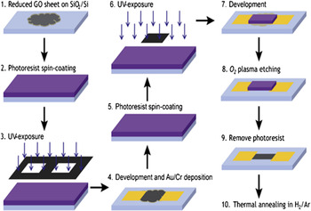
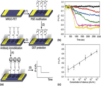
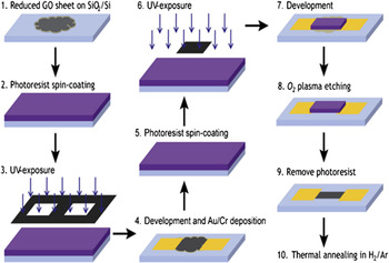
GO produced using chemical exfoliation method produces graphene with irregular edge and shape along with size of GO. These morphological features of graphene greatly affect the electronic properties of graphene therefore the random GO based FET are expected to suffer from low reproducibility. To overcome this issue, micropatterned RGO (MRGO) on the SiO2/Si substrate was fabricated and integrated in the polydimethylsiloxane (PDMS) microchannel to be applied for label-free, real-time, and ultrasensitive detection of rotavirus. Reference Liu, Kim, Cheon and Seo131 Fig. 20 shows the schematic of an MRGO-FET fabrication.
Schematic illustration for fabricating an MRGO-FET. Adapted from Ref. Reference Liu, Kim, Cheon and Seo131. Reprinted with permission from Elsevier (2013).
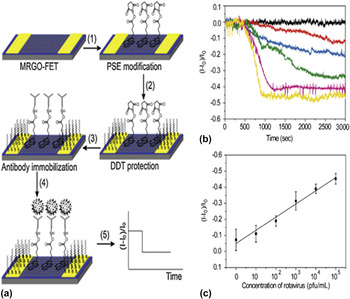
The rotavirus specific antibody was immobilized on the MRGO-FET through covalent chemistry modifications and then the current (I 0) was stabilized with a PBS buffer. Once the target virus was injected, the relative current change (I) was monitored in real time. Figure 21(a) shows the step by step procedure for detection of rotavirus using MRGO-FET. Concentration of the virus sample was varied from 0 to 105 pfu/mL and current drop was measured as shown in Fig. 21(b). As the concentration increased from 0 to 105 pfu/mL, the normalized I d [(I − I 0)/I 0] of the MRGO-FET gradually decreased by 7.23%, 11.06%, 19.0%, 31.45%, 39.0%, and 45.33%, respectively. The current drop was approximately proportional to the virus concentration. The LOD for rotavirus was found to be as 102 pfu which is superior to the conventional ELISA method. These results suggest that the MRGO-FET could be used for highly sensitive pathogen detection with improved reproducibility.
(a) Schematics of the MRGO-FET based rotavirus detection. (1) 1-Pyrenebutyric acid N-hydroxysuccinimide ester (PSE) modification, (2) blocking of the gold electrode by 1-dodecanethiol (DDT), (3) antibody immobilization and blocking the MRGO surface by ethanolamine, (4) rotavirus capture and (5) electrical signal detection. (b) Real-time and label-free virus detection on the MRGO-FET device at V ds = 0.1 V. (b) Current response of the MRGO-FET device depending on the concentration of a rotavirus sample [blank (black), 10 (red), 102 (blue), 103 (green), 104 (pink), and 105 pfu/mL (yellow)], (c) calibration plot of the detection. Adapted from Ref. Reference Liu, Kim, Cheon and Seo131. Reprinted with permission from Elsevier (2013).
In another report, rotavirus was captured on antibody functionalized multilayer free standing TRGO film. Reference Liu, Choi, Park, Lee and Seo130 The input cell number dependent binding event on the graphene surface was monitored electrochemically using cyclic voltammetry measurements in presence of redox probe. 105 pfu/mL and 103 pfu/mL of input cells were detected with approximately 30.7% and 1.3% sensitivity respectively demonstrating application of TRGO for electrochemical biosensing of rotavirus.
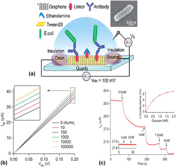
CVD grown G-based biosensor for a fast, label-free, and selective detection of bacteria E. coli with high sensitivity (10 cfu/mL) and specificity was reported. Reference Huang, Dong, Liu, Li and Chen60 Fig. 22(a) shows the schematics of anti-E. coli antibody functionalized graphene-FET for detection of E. coli biosensor. The binding of E. coli to its specific antibodies immobilized on the graphene electrode and subsequent increase in the conductance was recorded [Fig. 22(b)]. The anti-E. coli functionalized G-FET was found to be pH sensitive and also studied for real time detection of metabolic activities of E. coli triggered by glucose [Fig. 22(c)]. This nanoelectronic biosensor could serve as a high throughput platform for fast and reliable detection of any pathogenic bacteria, and for functional studies or screening of antibacterial drugs.
(a) Illustration of anti-E. coli antibody functionalized graphene-FET for detection of E. coli. Inset: scanning electron microscopy (SEM) image of an E. coli on antibody functionalized graphene. (b) I ds versus V ds curves of anti-E. coli antibody functionalized graphene device after incubated with E. coli of different concentrations. V g = 0 V. (c) Real-time current recording (V ds = 100 mV and V g = 0 V) of a graphene device immobilized with E. coli bacteria with addition of glucose to the PBS recording buffer at the indicated time points to reach the final concentrations of 0.5, 1, 1.5, and 2 mM. Adapted from Ref. Reference Huang, Dong, Liu, Li and Chen60 with permission of The Royal Society of Chemistry (2011).
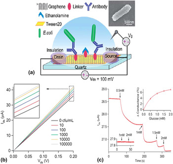
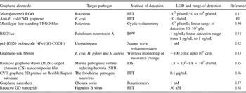
Table IV is a summary of the recently reported pathogen biosensors using graphene based electrodes.
Summary of the recently reported pathogen biosensors using graphene based electrodes.
V. SUMMARY
In summary, exceptional electrical and chemical properties of graphene and its derivatives provide an ideal platform for the development of reliable and highly sensitive sensors for healthcare. This chapter comprehensively discusses the synthesis of such structures and their functionalization for fabricating electrochemical sensors that help detect a wide array of biological entities such as DNA, proteins, and pathogens with enhanced sensitivity and specificity. Research on the interface of graphene and a variety of materials such as metals, metal oxides, polymers, quantum dots, and small molecules is emerging as one of the most dynamic areas in healthcare research as it establishes excellent coupling with bio-entities that forms the basis of developing efficient, long lasting sensors for detection of a multitude of diseases, toxins, and biomarkers. It is indeed without any doubt that given the unique selling point of graphene, such research will proliferate into related areas of medicine, drug/gene delivery, cancer therapies, and other biomedical applications.
ACKNOWLEDGMENT
We acknowledge the financial support of the National Science Foundation (1307671), U.S. Department of Agriculture (2014-67021-21589), and W. Ruel Johnson Chair in Environmental Engineering.