Introduction
Opsins are members of the G protein-coupled receptor (GPCR) superfamily that is distinguished by a Schiff base linkage between a conserved lysine residue and a retinaldehyde chromophore (Ebrey & Koutalos, Reference Ebrey and Koutalos2001). The photosensitive chromophore absorbs light to activate the opsin protein, initiating a signaling cascade in the photoreceptor cell (Burns & Baylor, Reference Burns and Baylor2001; Shichida & Morizumi, Reference Shichida and Morizumi2007). Gene duplication events throughout vertebrate evolution have given rise to several opsin families. There are five groups of visual opsins, including rhodopsin (rh1) and four cone opsin groups: rhodopsin-like (rh2), short-wavelength sensitive 1 (sws1), short-wavelength sensitive 2 (sws2), and long-wavelength sensitive (lws). Visual opsins are expressed in photoreceptor cells of the retina and, while being primarily responsible for initiating the visual transduction cascade (Menon et al., Reference Menon, Han and Sakmar2001), are also involved in nonvisual processes (Altimus et al., Reference Altimus, Güler, Villa, McNeill, Legates and Hattar2008; Lall et al., Reference Lall, Revell, Momiji, Al Enezi, Altimus, Güler, Cameron, Allender, Hankins and Lucas2010). Nonvisual opsins, such as pinopsin, melanopsin, and exorhodopsin, are expressed in a wide variety of tissues and thought to only be involved in nonvisual light-dependant biological processes, such as the circadian system (Newman et al., Reference Newman, Walker, Brown, Cronin and Robinson2003; Pierce et al., Reference Pierce, Noche, Ponomareva, Chang and Liang2008; Peirson et al., Reference Peirson, Halford and Foster2009).
GPCRs comprise one of the largest known families of integral membrane proteins. Rhodopsin (rh1), a highly characterized GPCR, is the visual opsin that initiates dim-light vision in vertebrates (Okawa & Sampath, Reference Okawa and Sampath2007). Because of its stability and abundance in rod photoreceptor outer segments, rhodopsin is amenable to detailed studies of structure and function, often through site-directed mutagenesis and in vitro expression (Imai et al., Reference Imai, Kefalov, Sakurai, Chisaka, Ueda, Onishi, Morizumi, Fu, Ichikawa, Nakatani, Honda, Chen, Yau and Shichida2007; Knierim et al., Reference Knierim, Hofmann, Ernst and Hubbell2007; Nickle & Robinson, Reference Nickle and Robinson2007). Rhodopsin was also the first opsin with a high-resolution crystal structure of its dark state (Palczewski et al., Reference Palczewski, Kumasaka, Hori, Behnke, Motoshima, Fox, Le Trong, Teller, Okada, Stenkamp, Yamamoto and Miyano2000), with crystal structures of both its activated (Scheerer et al., Reference Scheerer, Park, Hildebrand, Kim, Krauss, Choe, Hofmann and Ernst2008) and chromophore-free (Park et al., Reference Park, Scheerer, Hofmann, Choe and Ernst2008) states also having been resolved. Due to high levels of sequence and structural conservation among GPCRs (Rosenbaum et al., Reference Rosenbaum, Rasmussen and Kobilka2009), data collected and methods developed on rhodopsin have also facilitated further studies on other visual and nonvisual opsin families (Teller et al., Reference Teller, Stenkamp and Palczewski2003; Ramon et al., Reference Ramon, Mao and Ridge2009).
It is rare for a vertebrate to have more than one rh1 gene. Despite two ancestral genome duplication events that are thought to predate the vertebrate radiation (Dehal & Boore, Reference Dehal and Boore2005), the majority of vertebrates maintain only a single copy of rh1. In contrast, gene numbers of some cone opsin groups can vary greatly in vertebrates. No mammalian rh2 opsins have been identified (Trezise & Collin, Reference Trezise and Collin2005; Bowmaker, Reference Bowmaker2008), while many other species have up to four rh2 genes (Chinen et al., Reference Chinen, Hamaoka, Yamada and Kawamura2003; Parry et al., Reference Parry, Carleton, Spady, Carboo, Hunt and Bowmaker2005; Matsumoto et al., Reference Matsumoto, Fukamachi, Mitani and Kawamura2006). Derived primates have experienced lws opsin duplication (Dulai et al., Reference Dulai, von Dornum, Mollon and Hunt1999), as have teleost fish, where it is not uncommon to have at least four lws genes (Weadick & Chang, Reference Weadick and Chang2007; Owens et al., Reference Owens, Windsor, Mui and Taylor2009). However, having multiple rh1 genes is a very unique trait. Comprising 96% of living fish species due to an adaptive radiation after a genome duplication event 150 million years ago (Taylor et al., Reference Taylor, Braasch, Frickey, Meyer and Van de Peer2003), teleosts are the only known group of vertebrates containing species expressing multiple rh1 genes, although most are still believed to have only one. One example is the eel, having multiple species that express a second rh1 gene during maturation, coupled with its migration from freshwater to seawater (Beatty, Reference Beatty1975; Hope et al., Reference Hope, Partridge and Hayes1998; Zhang et al., Reference Zhang, Futami, Horie, Okamura, Utoh, Mikawa, Yamada, Tanaka and Okamoto2000, Reference Zhang, Futami, Yamada, Horie, Okamura, Utoh, Mikawa, Tanaka, Okamoto and Oka2002). Another example is the short fin pearleye (Scopelarchus analis), a deep-sea teleost that expresses up to two rh1 genes depending on its lifecycle (Pointer et al., Reference Pointer, Carvalho, Cowing, Bowmaker and Hunt2007). Our investigation of the genome of zebrafish (Danio rerio), a well-studied teleost and model vertebrate, revealed an open reading frame for a previously uncharacterized rh1-like gene, suggesting yet another case of multiple rh1 genes in a species of teleost fish.
With version Zv9 of the zebrafish genome project having recently been made available, researchers are increasingly identifying and characterizing zebrafish genes and gene families of interest, including opsins. So far, nine visual opsins have been identified in zebrafish, including rh1, four rh2, sws2, sws1, and two lws (Chinen et al., Reference Chinen, Hamaoka, Yamada and Kawamura2003), a large complement even among teleosts. Additionally, many nonvisual opsins have been discovered in zebrafish, including exorhodopsin (Mano et al., Reference Mano, Kojima and Fukada1999), two melanopsins (Bellingham et al., Reference Bellingham, Whitmore, Philp, Wells and Foster2002, Reference Bellingham, Chaurasia, Melyan, Liu, Cameron, Tarttelin, Luvone, Hankins, Tosini and Lucas2006), teleost multiple tissue (tmt) opsin (Moutsaki et al., Reference Moutsaki, Whitmore, Bellingham, Sakamoto, David-Gray and Foster2003), and two vertebrate ancient long (VAL) opsins (Kojima et al., Reference Kojima, Torii, Fukada and Dowling2008); a nonannotated sequence for encephalopsin has also been highlighted in the zebrafish genome. The advantage of studying opsin genes in the zebrafish is not only due to the availability of a genome sequence, but also to the considerable body of work assembled on the zebrafish visual system (Bilotta & Saszik, Reference Bilotta and Saszik2001; Neuhauss, Reference Neuhauss2003; Fleisch & Neuhauss, Reference Fleisch and Neuhauss2006; Fadool & Dowling, Reference Fadool and Dowling2008), and to the variety of existing and emerging tools available to study genes of interest in zebrafish (Amsterdam & Becker, Reference Amsterdam and Becker2005; Amacher, Reference Amacher2008; Halpern et al., Reference Halpern, Rhee, Goll, Akitake, Parsons and Leach2008).
In this study, we used zebrafish as a model to identify and characterize a novel rh1 gene. Data mining of the zebrafish genome revealed an open reading frame encoding a novel rhodopsin gene, herein referred to as rh1-2. The amino acid sequence of rh1-2 contains several unique amino acid substitutions compared to normally conserved rh1 residues and some substitutions more characteristic of cone opsins. Despite these substitutions, the rhodopsin-like protein was successfully expressed in vitro, and not only bound chromophore, yielding an absorption spectra in the visible range (λ max ≈ 500 nm), but also activated in response to light indicative of a functional opsin. In contrast to rh1, rh1-2 is not expressed during the first 4 days of embryonic development but is expressed by day 21 and is expressed in the adult retina but not in adult brain or muscle tissue. While rh1-2 sequences have only been identified in cyprinid fish, phylogenetic analyses suggest a much older origin. The novel gene is also intronless, similar to other rh1 sequences of teleosts. The discovery of a new previously uncharacterized opsin gene in zebrafish retina is surprising, given its status as a model system for studies of vertebrate vision and visual development.
Materials and methods
Opsin sequences
The zebrafish genome assembly, currently in version Zv9 (http://www.sanger.ac.uk/Projects/D_rerio/) was explored through a BLAST search, using the full-length sequence of zebrafish rh1 (GenBank: AB087811) to identify putative rh1 homologs; various BLAST homology algorithms were employed to ensure the best possible coverage, including blastn, blastp, and tblastn. Genomic DNA was extracted from fish tissues using the DNeasy Blood & Tissue Kit (QIAGEN). Primers were designed to amplify fragments of the coding region of the rh1-2 gene from cyprinid fish, including zebrafish, using genomic DNA as a template (Table S1). RNA was extracted from fish eyes using the TRIzol reagent (Invitrogen), and complementary DNA (cDNA) libraries were generated using the SMART cDNA Library Construction Kit (BD Biosciences). Teleost rh1 gene fragments were amplified from these cDNA libraries using either custom rh1 primers (Table S1) or previously designed acanthomorph rh1 primers (Chen et al., Reference Chen, Bonillo and Lecointre2003). PCR was performed using PfuTurbo (Stratagene) or FastStartTaq (Roche), with resulting bands being cloned into the pJET1.2 cloning vector (Fermentas) and sequenced using a 3730 DNA Analyzer (Applied Biosystems).
Protein expression
Complete coding sequences of zebrafish rh1 and rh1-2 were cloned into the p1D4-hrGFP II expression vector (Morrow & Chang, Reference Morrow and Chang2010). This vector was used to transiently transfect cultured HEK293T cells using Lipofectamine 2000 (Invitrogen; 12 μg of DNA per 10-cm plate). Cells were harvested 48 h posttransfection and regenerated using 11-cis-retinal, generously provided by Dr. Rosalie Crouch (Medical University of South Carolina). The pigments were then solubilized in 1% dodecyl maltoside and immunoaffinity purified with the 1D4 monoclonal antibody (Molday & MacKenzie, Reference Molday and MacKenzie1983), as previously described (Chang et al., Reference Chang, Jönsson, Kazmi, Donoghue and Sakmar2002; Morrow & Chang, Reference Morrow and Chang2010). The ultraviolet-visible absorption spectra of purified zebrafish rh1 and rh1-2 were recorded at 25°C using the Cary4000 double-beam spectrophotometer (Varian). For zebrafish rh1-2, a difference spectrum was calculated as the difference between dark- and light-bleached spectra after bleaching for 60 s.
Localizing expression of zebrafish rh1-2
RNA was extracted from zebrafish embryos (1, 2, 3, 4 dpf), juvenile heads (21 dpf), and adult tissues (retina, eye, brain, and muscle) using TRIzol Reagent (Invitrogen), and cDNA libraries were generated using the SMART cDNA Library Construction Kit (BD Biosciences), with identical amounts of RNA used as template in all reactions. Primers were designed to amplify ∼300 bp fragments of zebrafish rh1 and rh1-2; primers for rh1-2 were designed specifically to avoid amplification of rh1 (Table S1). Primers for β-actin, used as a positive control for RT-PCR experiments, were obtained from a previous study (Wang et al., Reference Wang, Wong and Ge2003). PCR was performed using PfuTurbo (Stratagene) under standard cycling conditions, with resulting bands being cloned into the pJET1.2 cloning vector using the CloneJET PCR cloning kit (Fermentas) and sequenced using a 3730 DNA Analyzer (Applied Biosystems).
Phylogenetic analyses
All rh1 and rh1-2 nucleotide sequences isolated from zebrafish, Danio roseus, Danio albolineatus, and E. bicolor in this study were aligned with other teleost rh1 sequences obtained from NCBI (Table S2) using ClustalW (Larkin et al., Reference Larkin, Blackshields, Brown, Chenna, McGettigan, McWilliam, Valentin, Wallace, Wilm, Lopez, Thompson, Gibson and Higgins2007). Teleost exorhodopsin sequences were included as outgroups, in order to root the tree. Due to the short length of some of the teleost rh1 sequences obtained from NCBI, this aligned data set was trimmed to include only nucleotides 94–912 and subjected to phylogenetic analyses using both maximum likelihood and Bayesian methods (Guindon & Gascuel, Reference Guindon and Gascuel2003; Ronquist & Huelsenbeck, Reference Ronquist and Huelsenbeck2003). Maximum likelihood phylogenetic methods were implemented in the program PHYML 3.0 (Guindon & Gascuel, Reference Guindon and Gascuel2003; Guindon et al., Reference Guindon, Lethiec, Duroux and Gascuel2005), under the HKY+I+G model. For the likelihood analyses, bootstrapping methods were used to assess the degree of confidence in nodes of the phylogeny (Felsenstein, Reference Felsenstein1985). Bayesian inference was performed in MrBayes 3.1.2 (Ronquist & Huelsenbeck, Reference Ronquist and Huelsenbeck2003), under the HKY+I+G model. To identify the most appropriate models for the molecular analyses, we used MrModeltest 2.3 (Nylander, Reference Nylander2004) and selected the best models favored under the Akaike Information Criterion (AIC). Two independent analyses were performed, each composed of four Markov chains with default heating values. Markov chains were run for 10 million generations, sampling trees (and parameters) every 1000 generations. Convergence was assessed using a number of methods. The average s.d. of split frequencies, as calculated in MrBayes, were all well below 0.01 at stationarity. Also implemented in MrBayes, a convergence diagnostic for branch length posterior probabilities, the potential scale reduction factor, roughly approached 1 as the runs converged (Gelman & Rubin, Reference Gelman and Rubin1992). Convergence to stationarity was also assessed by plotting log-likelihood scores and other parameter values in the program Tracer 1.4.1 to ensure that there were no trends in the data post burn-in (Rambaut & Drummond, Reference Rambaut and Drummond2007). Finally, adequacy of mixing was assessed by examining acceptance rates for parameters in MrBayes, and by calculating in Tracer the effective sample sizes, the number of independent samples from the marginal posterior distribution for each parameter; higher values being indicative of better sampling from the posterior distribution. These values were all well above 100. By these measures, convergence was achieved within the first 25% of trees sampled, which were discarded as burn in, and remaining trees were taken as representative of the posterior probability distribution.
Results
Sequence analysis of rh1-2
Zebrafish rh1-2 shares 78% amino acid sequence similarity to zebrafish rh1, and, like other teleost rh1 genes, is intronless (Fitzgibbon et al., Reference Fitzgibbon, Hope, Slobodyanyuk, Bellingham, Bowmaker and Hunt1995). In general, the novel opsin gene shares much greater amino acid sequence similarity with the other visual opsins of zebrafish (38–78%), as opposed to its nonvisual opsins (25–32%); the only exception is the nonvisual exorhodopsin, paralog to mammalian Rh1 genes (Mano et al., Reference Mano, Kojima and Fukada1999), which shares 70% sequence similarity. Zebrafish rh1-2 is not a tandem duplicate of rh1, as the two genes are located on chromosomes 11 and 8, respectively. Full-length rh1-2 coding sequences were amplified from two other species from the Danio genus: D. roseus and D. albolineatus, while partial coding sequence was amplified from the cyprinid E. bicolor; partial rh1 coding sequences were also isolated for these three fish, with all rh1 and rh1-2 sequences amplified in this study being deposited in GenBank under accession numbers HQ286326–32.
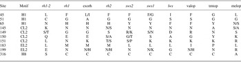
The rh1-2 gene family contains a series of amino acid substitutions that suggest it might have properties different from other rhodopsins. The most unique substitutions are sites C51, K145, L151, E199, and S316, all of which are conserved in all four rh1-2 sequences and not found in any other opsin gene family. There is also a group of residues, L45, N65, S/T149, Q150, and L183, whose identities are usually found in cone opsins or nonvisual opsins but rarely in rhodopsins (Table 1). All of these residues group into one of three distinct opsin motifs: helix 1/helix 8, cytoplasmic loop 2, or extracellular loop 2.
Dominant identities of key sites of rh1-2 in other opsin families

The following opsin gene families were sampled: rh1-2, rh1 (rhodopsin), exorh (exorhodopsin), rh2 (rod-like opsin), sws2 (short-wavelength sensitive 2), sws1 (short-wavelength sensitive 1), lws (long-wavelength sensitive), valop (vertebrate ancient opsin), tmtop (teleost multiple tissue opsin), and melop (melanopsin).
In addition to unique and cone-like substitutions, rh1-2 contains many of the sites and motifs required for visual opsin function. Both K296, the lysine residue that forms a Schiff base linkage with the chromophore (Zhukovsky et al., Reference Zhukovsky, Robinson and Oprian1991), and E113, the glutamate counter-ion to the Schiff base linkage (Sakmar et al., Reference Sakmar, Franke and Khorana1989) are present. Both E134 and R135 of the D/ERY motif are present, being highly conserved in opsins and involved in light-dependant proton uptake (Arnis et al., Reference Arnis, Fahmy, Hofmann and Sakmar1994) and transducin activation (Fanelli & Dell’Orco, Reference Fanelli and Dell’Orco2008). Cysteine residues, C110 and C187, required for the formation of a critical disulfide bond (Davidson et al., Reference Davidson, Loewen and Khorana1994; Hwa et al., Reference Hwa, Reeves, Klein-Seetharaman, Davidson and Khorana1999), are present as well as conserved glycosylation sites N2 and N15 (Kaushal et al., Reference Kaushal, Ridge and Khorana1994; Murray et al., Reference Murray, Fliesler and Al-Ubaidi2009) and palmitoylation sites C322 and C323 (Traxler & Dewey, Reference Traxler and Dewey1994).
In vitro expression
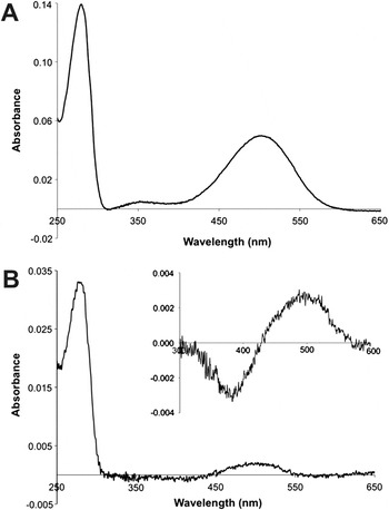
Complete coding sequences of zebrafish rh1 and rh1-2 were cloned into the p1D4-hrGFP II expression vector. These genes were expressed in mammalian cell culture, regenerated with 11-cis-retinal, solubilized with dodecyl maltoside, and purified with the 1D4 monoclonal antibody, as previously described (Chang et al., Reference Chang, Jönsson, Kazmi, Donoghue and Sakmar2002; Morrow & Chang, Reference Morrow and Chang2010). Zebrafish rh1 was expressed and purified with relative ease (Fig. 1A) and produced spectra similar to previous studies (Chinen et al., Reference Chinen, Hamaoka, Yamada and Kawamura2003). Zebrafish rh1-2 was more difficult to express but still bound 11-cis-retinal and produced a stable photopigment with an absorption maximum (λ max) of approximately 500 nm (Fig. 1B), a typical λ max value among fish rhodopsins (Johnson et al., Reference Johnson, Grant, Zankel, Boehm, Merbs, Nathans and Nakanishi1993; Yokoyama et al., Reference Yokoyama, Knox and Yokoyama1995; Chang et al., Reference Chang, Chiao and Yan2009). When bleached with light, the absorption peak of zebrafish rh1-2 shifted to approximately 380 nm, characteristic of the biologically active meta II state of visual opsins. Both expression level and absorbance ratio (A 280/A MAX) of zebrafish rh1-2 were significantly lower than those of zebrafish rh1, suggesting that the former is less stable and more likely to misfold than the latter. These results imply that, while unique substitutions in zebrafish rh1-2 might reduce its stability, it can be expressed, has an appropriate λ max for a teleost rhodopsin, and properly activates when bleached with light.
Absorbance spectra of zebrafish rh1 and rh1-2 after in vitro expression and purification. (A) Dark spectrum of zebrafish rh1 comparable to previous expressions (Chinen et al., Reference Chinen, Hamaoka, Yamada and Kawamura2003). (B) Dark spectrum of zebrafish rh1-2, whose expression level and absorbance ratio (A 280/A MAX) are significantly lower than zebrafish rh1. The absorption maximum of zebrafish rh1-2 is approximately 500 nm, which is highlighted in the dark-light difference spectrum (inset).

Zebrafish rh1-2 expression pattern
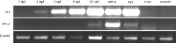
The temporal and spatial expression pattern of zebrafish rh1-2 was investigated using RT-PCR. Expression of zebrafish rh1-2 was not detected during the first 4 days of embryonic development but was detected in the heads of juveniles by 21 days postfertilization (dpf). Expression of rh1-2 was also detected in the adult retina but not in the adult brain or muscle (Fig. 2). Zebrafish rh1 expression was detected as early as 2 dpf and strictly in the adult eye and retina, as previously reported (Raymond et al., Reference Raymond, Barthel and Curran1995; Takechi & Kawamura, Reference Takechi and Kawamura2005). These results suggest that zebrafish rh1-2 is not expressed as early as rh1 in zebrafish development.
RT-PCR analysis of zebrafish rh1-2 expression. Zebrafish rh1-2 is not expressed during the first 4 days of embryonic development but is expressed by the 21-day mark. In the adult, zebrafish rh1-2 is expressed in the retina but not in brain or muscle tissue. Zebrafish rh1 expression was similar to previous studies (Raymond et al., Reference Raymond, Barthel and Curran1995; Takechi & Kawamura, Reference Takechi and Kawamura2005). β-actin serves as a positive control.

Phylogenetic analyses
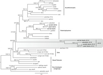
Maximum likelihood and Bayesian phylogenetic analyses were performed on a dataset of aligned rh1 and rh1-2 sequences (Fig. 3A) in order to investigate the origins of the rh1-2 gene family and its relationship to rh1 visual pigments. These analyses indicate that all four rh1-2 sequences isolated from cypriniform fish form a well-supported monophyletic group and that this novel gene was the result of a duplication that most likely occurred within the rh1 gene family during teleost evolution (Fig. 4). Although the rh1-2 genes form a well-defined group, due to the low support for many of the early teleost rh1 lineages, it remains difficult to pinpoint exactly when in teleost evolution this novel gene family may have emerged. With respect to teleost rh1 sequences, this phylogeny recovers many of the commonly accepted systematic relationships among ray-finned fish (Forey et al., Reference Forey, Littlewood, Ritchie, Meyer, Greenwood, Miles and Patterson1996; Hurley et al., Reference Hurley, Mueller, Dunn, Schmidt, Friedman, Ho, Prince, Yang, Thomas and Coates2007; Wang et al., Reference Wang, Li and He2007). This includes support for both acanthopterygian and ostariophysian groups of fishes, although positions of the dory, cod, and pearl eye rh1a sequences are somewhat unresolved. With respect to duplications within teleost rh1 genes, it is interesting that our analyses do not show strong support for a grouping of the rh1-2 sequences with the duplicate pearl eye and eels sequences. This might suggest multiple gene duplications within teleost rh1 genes, although due to the low support of many of these divergences, at the moment, this is difficult to conclude with certainty. Interestingly, in our analyses the pearl eye rh1b sequence does appear to group, albeit with variable support, with the duplicate copies of the eel rh1 sequences.
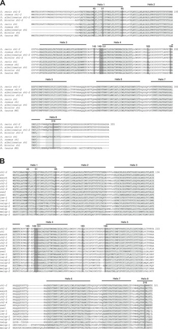
Amino acid alignments of zebrafish rh1-2. (A) The rh1-2 gene family aligned with corresponding rh1 sequences and bovine rhodopsin. The seven transmembrane helices of rhodopsin, along with the eighth cytoplasmic helix, are labeled and shaded in light gray. Unique substitutions of the rh1-2 gene family are numbered and shaded in dark gray. Sequences obtained from GenBank are listed in Table S2. (B) Zebrafish rh1-2 aligned with other visual and nonvisual opsin genes from zebrafish. N- and C-termini were excluded as they were too variable to align.

Phylogeny of teleost rh1 nucleotide sequences, including the rh1-2 gene family, generated using both maximum likelihood and Bayesian methods; rh1-2 sequences form a monophyletic group within the rh1 gene family. Sequences amplified in this study are indicated in bold. Numbers above nodes indicate ML bootstraps (100 replications) followed by posterior probabilities. Branches are proportional to ML estimated branch lengths under HKY+I+G model, the best model as determined by Modeltest AIC. Sequences obtained from GenBank are listed in Table S2.

Discussion
In this study, we investigate rh1-2, a new family of opsin genes discovered in cyprinid fish. All four rh1-2 sequences form a monophyletic group within the rh1 clade of actinopterygian fish. While being most closely related to the rh1 family of visual opsins, rh1-2 has multiple substitutions that are either cone opsin like in nature or unique to the rh1-2 gene family. Despite these unique substitutions at otherwise highly conserved sites, zebrafish rh1-2 was successfully expressed in vitro and found to bind 11-cis-retinal with an absorption maximum around 500 nm; when bleached with light, it activated and changed conformation to its meta II intermediate. This activity coincides with the presence of conserved amino acids required for opsin function in the rh1-2 gene sequence, including lysine at site 296 that forms a Schiff base linkage with 11-cis-retinal (Zhukovsky et al., Reference Zhukovsky, Robinson and Oprian1991) and glutamate at site 113 that acts as a counterion to the protonated Schiff base linkage (Sakmar et al., Reference Sakmar, Franke and Khorana1989). Unlike rh1, rh1-2 is not expressed during the first 4 days of embryonic development but is expressed by 21 days post fertilization. In the adult, rh1-2 is expressed in the retina but not in the brain or muscle.
Although rare, zebrafish is not the only teleost to express a second rh1 gene. The rod photoreceptors of eels start expressing an alternate rh1 gene, during the transition from a freshwater habitat as a juvenile to a deep-sea habitat as an adult (Beatty, Reference Beatty1975; Archer et al., Reference Archer, Hope and Partridge1995; Zhang et al., Reference Zhang, Futami, Horie, Okamura, Utoh, Mikawa, Yamada, Tanaka and Okamoto2000). This switch in rh1 gene expression also results in a blueshift of λ max of approximately 40 nm (Beatty, Reference Beatty1975; Hope et al., Reference Hope, Partridge and Hayes1998). The short fin pearleye expresses an rh1a gene throughout its lifespan but only expresses the rh1b gene as an adult; the λ max of rh1b is also blueshifted by 6 nm compared to rh1a (Pointer et al., Reference Pointer, Carvalho, Cowing, Bowmaker and Hunt2007). Zebrafish rh1 is expressed as early as 2 dpf and is expressed at high levels throughout adulthood (Raymond et al., Reference Raymond, Barthel and Curran1995). Meanwhile, rh1-2 expression initiates sometime between 4 and 21 dpf and also remains at more modest levels throughout adulthood. The λ max values of zebrafish rh1 (501 nm) (Chinen et al., Reference Chinen, Hamaoka, Yamada and Kawamura2003) and rh1-2 (∼500 nm) are similar. Ultimately, the rh1 gene pair in zebrafish is similar to that of the pearleye, which has one gene expressed earlier in development at high levels, and a second gene expressed later in development at lower levels. However, unlike the pearleye, the two rh1 genes of zebrafish do not seem to differ significantly in their λ max values.
Like most protein families, there are key residues and motifs present in opsins that are required for proper structure and function (Iannaccone et al., Reference Iannaccone, Man, Waseem, Jennings, Ganapathiraju, Gallaher, Reese, Bhattacharya and Klein-Seetharaman2006). Alternatively, some sites in opsins act as determinants of rh1 and cone opsin function (Imai et al., Reference Imai, Kojima, Oura, Tachibanaki, Terakita and Shichida1997; Carleton et al., Reference Carleton, Spady and Cote2005; Kuwayama et al., Reference Kuwayama, Imai, Morizumi and Shichida2005) or differentiate between visual and nonvisual opsins (Bellingham et al., Reference Bellingham, Whitmore, Philp, Wells and Foster2002; Murakami & Kouyama, Reference Murakami and Kouyama2008). The rh1-2 gene family contains a number of substitutions at sites known to vary between rod, cone, and nonvisual opsins, as well as some that are entirely unique at otherwise highly conserved sites (Fig. 3B). Because opsins have been examined so extensively, many of these substitutions have been implicated in experimental opsin structure and function studies.
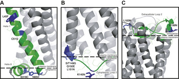
There are three motifs in rh1-2 that have substitutions suggesting altered function compared to rhodopsin: helix 1/helix 8, cytoplasmic loop 2, and extracellular loop 2 (Fig. 5). All three of these motifs are involved in meta II stability, transducin activation, or both, suggesting that rh1-2 does not behave as a traditional rhodopsin. Residues L45 and C51 are found in the first transmembrane helix of rh1-2. Helix 1 plays a role in assuring proper transmembrane packing of helices 2 and 7, with site 51 being of particular importance (Bosch et al., Reference Bosch, Ramon, del Valle and Garriga2003). It is possible that interfering with helix 7 specifically could lead to disruption of helix 8, a key motif in both meta II stability and transducin activation (Ernst et al., Reference Ernst, Meyer, Marin, Henklein, Fu, Sakmar and Hofmann2000; Marin et al., Reference Marin, Krishna, Zvyaga, Isele, Siebert and Sakmar2000). Additionally, N65, a cone-like substitution at the cytoplasmic surface of helix 1, is in close proximity to S316, an rh1-2-specific substitution in helix 8, and a residue involved in the meta I/II equilibrium (Weitz & Nathans, Reference Weitz and Nathans1992).
Structures of three key motifs of rh1 containing cone-like and unique substitutions in the rh1-2 gene family. (A) The interaction between helix 1 and 8. (B) The second cytoplasmic loop. (C) The second extracellular loop. Unique substitutions of the rh1-2 gene family are highlighted in blue, with key motifs being colored green, and other residues colored gray. Approximate plasma membrane boundaries with either the extracellular or cytoplasmic environment are emphasized with a dashed line. The crystal structure of dark state bovine rhodopsin (1U19) and MacPyMOL (DeLano, Reference DeLano2008) were used for all images.

There are also a number of substitutions in the second cytoplasmic loop (K145, S/T149, Q150), as well as L151, the first residue of helix 4, which are either unique to rh1-2 or cone-like in nature. Cytoplasmic loop 2 is a key motif in transducin interaction and activation (König et al., Reference König, Arendt, McDowell, Kahlert, Hargrave and Hofmann1989; Franke et al., Reference Franke, Sakmar, Graham and Khorana1992; Ernst et al., Reference Ernst, Hofmann and Sakmar1995; Natochin et al., Reference Natochin, Gasimov, Moussalf and Artemyev2003), with some residues of the loop also being implicated in phosphorylation and glycosylation patterning (Shi et al., Reference Shi, Osawa, Dickerson and Weiss1995; Zhu et al., Reference Zhu, Imanishi, Filipek, Alekseev, Jastrzebska, Sun, Saperstein and Palczewski2006). Finally, L183 and E199 are part of extracellular loop 2, which folds into two short β-strands that form a shield over the chromophore-binding site (Palczewski et al., Reference Palczewski, Kumasaka, Hori, Behnke, Motoshima, Fox, Le Trong, Teller, Okada, Stenkamp, Yamamoto and Miyano2000; Okada et al., Reference Okada, Sugihara, Bondar, Elstner, Entel and Buss2004); the movement of this motif is key to rh1 activation (Ahuja et al., Reference Ahuja, Hornak, Yan, Syrett, Goncalves, Hirshfeld, Ziliox, Sakmar, Sheves, Reeves, Smith and Eilers2009). Since other residues in extracellular loop 2 have been previously identified as key determinants of differences between rhodopsins and cone opsins, including meta II decay rate (Kuwayama et al., Reference Kuwayama, Imai, Hirano, Terakita and Shichida2002), these substitutions could also be involved in altering rh1-2 function.
The λ max of an opsin gene is a property often used as an indicator of biological function, with each group of visual opsins having a characteristic λ max range: rh1 ∼500 nm, rh2 = 470–510 nm, sws2 = 440–460 nm, sws1 = 360–430 nm, and lws = 510–560 nm (Trezise & Collin, Reference Trezise and Collin2005). The λ max values of nonvisual opsins have not been analyzed to the same extent as those from visual opsins, often because they are more difficult to express. The only nonvisual opsins in fish that have been successfully expressed in vitro are the two isoforms of zebrafish VAL-opsin, both of which absorb maximally around 500 nm (Kojima et al., Reference Kojima, Torii, Fukada and Dowling2008). Melanopsin, a nonvisual opsin, has been investigated in mammals and reported to have a λ max around either 480 nm (Dacey et al., Reference Dacey, Liao, Peterson, Robinson, Smith, Pokorny, Yau and Gamlin2005; Panda et al., Reference Panda, Nayak, Campo, Walker, Hogenesch and Jegla2005; Torii et al., Reference Torii, Kojima, Okano, Nakamura, Terakita, Shichida, Wada and Fukada2007; Terakita et al., Reference Terakita, Tsukamoto, Koyanagi, Sugahara, Yamashita and Shichida2008) or 420 nm (Newman et al., Reference Newman, Walker, Brown, Cronin and Robinson2003; Melyan et al., Reference Melyan, Tarttelin, Bellingham, Lucas and Hankins2005). The λ max of zebrafish rh1-2, at approximately 500 nm, falls within the range of both rh1 and rh2 visual opsin groups, but is also similar to some nonvisual opsins, such as VAL-opsin. While the λ max value of rh1-2 might not help in distinguishing its functional role, the fact that it is much more difficult to express relative to zebrafish rh1 is indicative of a protein less stable than rhodopsin.
The spatial and temporal expression pattern of an opsin is an integral functional feature. The onset of expression of the visual opsin repertoire in zebrafish varies from 2 dpf for rh1 and lws-2 to 7 dpf for rh2-2 (Takechi & Kawamura, Reference Takechi and Kawamura2005). Meanwhile, the expression of both isoforms of VAL-opsin can be seen as early as 1 dpf (Kojima et al., Reference Kojima, Torii, Fukada and Dowling2008). In adult vertebrates, visual opsins are expressed in the outer segments of photoreceptor cells found in the retina (Lamb et al., Reference Lamb, Collin and Pugh2007) and, in rare circumstances, in the brain (Koyanagi et al., Reference Koyanagi, Kawano, Kinugawa, Oishi, Shichida, Tamotsu and Terakita2004). Nonvisual opsins can be expressed in a wide variety of tissues, often simultaneously, including the retina (Hannibal et al., Reference Hannibal, Hindersson, Nevo and Fahrenkrug2002; Grone et al., Reference Grone, Zhao, Chen and Fernald2007), brain (Mano et al., Reference Mano, Kojima and Fukada1999; Philp et al., Reference Philp, Bellingham, Garcia-Fernandez and Foster2000), and even the testis (Tarttelin et al., Reference Tarttelin, Bellingham, Hankins, Foster and Lucas2003) and ovaries (Kojima et al., Reference Kojima, Torii, Fukada and Dowling2008). Zebrafish rh1-2 expression initiates between 4 and 21 dpf and is localized in the retina. These results suggest that rh1-2 is more likely to serve as a visual opsin, as few nonvisual opsins are expressed strictly in the retina. Furthermore, since the development of photoreceptors in vertebrates is dependent on rh1 expression (Lem et al., Reference Lem, Krasnoperova, Calvert, Kosaras, Cameron, Nicolò, Makino and Sidman1999), rh1-2 likely lacks a similar role in photoreceptor development.
The evolutionary origins of the rh1-2 gene family were investigated via phylogenetic analyses of rh1 and rh1-2 sequences. Both maximum likelihood and Bayesian methods recovered a phylogeny in which all rh1-2 sequences formed a well-supported monophyletic group within the rh1 gene family. So far, we have only isolated rh1-2 sequences from cyprinids, a family of fish including zebrafish, goldfish, and barbs that originated between 32 and 39 million years ago (Zardoya & Doadrio, Reference Zardoya and Doadrio1999; Wang et al., Reference Wang, Li and He2007). However, our phylogenetic analyses suggest a much older origin for this novel gene. As with rh1 genes of all actinopterygians, rh1-2 does not contain introns (Fitzgibbon et al., Reference Fitzgibbon, Hope, Slobodyanyuk, Bellingham, Bowmaker and Hunt1995), suggesting that the duplication event leading to the birth of the rh1-2 family likely occurred after the retrotransposition event that split rh1 and exorhodopsin in this group of fish (Bellingham et al., Reference Bellingham, Tarttelin, Foster and Wells2003). Although the exact time of actinopterygian divergence is a contentious issue, this would mean that the origin of the rh1-2 gene family is certainly no older than 350 million years ago (Hurley et al., Reference Hurley, Mueller, Dunn, Schmidt, Friedman, Ho, Prince, Yang, Thomas and Coates2007). According to our analyses, there is some evidence that the origins of the rh1-2 group might have occurred around the divergence of the eel rh1 sequences. The divergence of the elopomorphs (eels) from more derived teleosts occurred approximately 140 million years ago (Forey et al., Reference Forey, Littlewood, Ritchie, Meyer, Greenwood, Miles and Patterson1996).
Such origins of the rh1-2 gene family suggest that its distribution among teleost fish may be much wider than our current experiments would indicate, particularly as we have not yet attempted to find rh1-2 genes outside of cyprinid fishes. This raises the possibility that rh1 gene duplicates in the pearl eye and eels might in fact be part of the same gene duplication that gave rise to the rh1-2 gene family. This would be rather surprising, as these rh1 duplicates are not generally thought to be widespread among fish. However, this possibility cannot be ruled out, as our analyses show low support at key divergences deep within the teleost rh1 phylogeny. Better sampling of basal teleost rh1 sequences, as well as a wider sampling of rh1-2 sequences would be needed to resolve this issue.
While rh1-2 is most closely related to the rh1 visual opsin group, this alone cannot classify it as a visual opsin, as the same can be said about the nonvisual opsin, exorhodopsin (Mano et al., Reference Mano, Kojima and Fukada1999). Despite expressing zebrafish rh1-2 in cell culture and characterizing its expression pattern via RT-PCR, it is still difficult to ascertain its function, whether within the visual system or elsewhere. Regardless, the discovery of a new previously uncharacterized opsin gene in the zebrafish retina is an important and surprising discovery, given its status as a model system for studying vertebrate vision and visual development.
Additional insights into the function of this novel gene could help to further elucidate how the vertebrate visual system functions. Localizing the specific cell types within the retina that express rh1-2 would help to determine its functional role. This could be done through extensive in situ hybridizations of adult zebrafish retina or by generating a transgenic zebrafish with expression of a GFP reporter driven by elements found upstream of the zebrafish rh1-2 ORF. Additional biochemical characterization of rh1-2 expressed in vitro would also help to determine the functional repercussions of its many unique amino acid substitutions. Finally, obtaining additional rh1-2 nucleotide sequences from other teleosts would help to resolve the evolutionary origins of this novel gene family.
Acknowledgments
We would like to acknowledge Ashley Bruce, Ian Scott, and Vince Tropepe for advice, suggestions, and zebrafish tissue samples. This research was supported by an NSERC Discovery Grant (B.S.W.C.), an Early Researcher Award (B.S.W.C.), and a University Health Network (University of Toronto) Vision Science Research Program fellowship (J.M.M.).