Trisomy 18 is the second most common numerical chromosomal disorder with poor mortality and morbidity. Of the affected individuals, 90 percent die within the first year of life, and median survival time varies from 3 to 14.5 days.Reference Embleton, Wyllie, Wright, Burn and Hunter1–Reference Brewer, Holloway, Stone, Carothers and FitzPatrick5 However, trisomy 18 is not an absolutely lethal chromosomal disorder as 50% of patients survive longer than a week and 5–10% of patients survive over 1 year.Reference Root and Carey2, Reference Rasmussen, Wong, Yang, May and Friedman3 The most recent American Heart Association neonatal resuscitation guideline omitted trisomy 18 from the list of conditions for which cardiopulmonary resuscitation is withheld,6 and thus several neonatologists and paediatricians have provided aggressive management for trisomy 18 patients.
In trisomy 18 patients, congenital cardiac defect is the most common complication and occurs in 90% of patients.Reference Carey7, Reference Matsuoka, Misugi, Goto, Gilbert and Ando8 Cardiac surgery has been rarely performed in trisomy 18 patients, as they die within several days and have severe neurological disabilities even if they survive. Recently, several reports encourage aggressive cardiac surgery in trisomy 18 patients.Reference Graham, Bradley, Shirali, Hills and Atz9, Reference Kaneko, Kobayashi and Achiwa10 However, it remains unknown whether surgical interventions reduce the morbidity and mortality in trisomy 18 patients with cardiac complications. To clarify an efficacy of cardiac surgery in trisomy 18 patients, we analysed clinical data including the survival rate and the rate of discharge alive between patients who underwent cardiac surgery and those who were treated with conservative management.
Patients and methods
Data collection
We analysed 34 consecutive trisomy 18 patients, including 21 males, 62%, with cardiac complications who were admitted to Kyushu Koseinenkin Hospital from 1985 to 2009. Definitive diagnosis of trisomy 18 was based on chromosome analysis from peripheral blood by G-banding or fluorescence in situ hybridisation with written informed consents from their parents. We did not perform further karyotyping such as analysis of many peripheral blood lymphocytes or analysis of cells from other tissues in these patients. The present study was approved by our institutional ethical committee. Patient demographic data including gestational age, birthweight, gender, chromosomal mosaicism, cardiac anomalies, and extracardiac anomalies were recorded. As far as major extracardiac anomalies were concerned, we highlighted digestive and central nervous anomalies. Diagnosis of congenital cardiac defects was determined by echocardiography. Cardiac complications were classified as single or more complex cardiac lesions. Single cardiac lesion refers to a simple defect such as ventricular septal defect or patent ductus arteriosus, whereas complex lesion means concomitant lesions such as ventricular septal defect and atrial septal defect or more complex conotruncal anomalies. We also collected operative data including age, weight, pre-operative assisted ventilation, palliative or corrective cardiac surgery, and time to discharge alive. It depended on weight at the time of surgery, that is, whether or not weight is over 2.5 kilograms, respiratory stability and surgeon's preference as to whether palliative or primary intracardiac repair should be performed.
Patients were divided into two groups: patients who underwent cardiac surgery and those who were conservatively treated. In our institution, cardiac surgery in trisomy 18 is carried out when patients survived beyond 14 days of age; symptoms related to cardiac disease are hazardous in achieving home-care; and their parents preferred aggressive treatments. We provided conservative management to patients who did not undergo cardiac surgery. When oral or enteral tube feeding was established, diuretics and/or digoxin were administrated. When this was not established, intravenous diuretics or cardiac inotropic agents were administrated. All the patients were followed up in our hospital or affiliated hospitals. Discharge alive was defined as achieving home-care.
Statistical analysis
Results are expressed as median values. Comparisons between the surgery group and the conservative group were made using the chi-square test or Wilcoxon rank sum test, as appropriate. Kaplan–Meier survival and Cox proportional hazard regression analyses were performed to clarify the efficacy of cardiac surgery. The rate of discharge alive was also estimated using the Kaplan–Meier method. Two-sided p values were calculated with the use of log-rank tests. p-value less than 0.05 was considered statistically significant.
Results
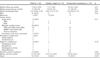
Patients’ profiles are summarised in Table 1. Median follow-up time was 74 months ranging from 0.2 to 242 months. Median gestational age and birthweight were 37 weeks ranging from 28 to 41 weeks and 1.67 kilograms ranging from 0.82 to 2.70 kilograms, respectively. There were six patients with single cardiac lesion, while 28 patients had two or more complex cardiac lesions including ventricular septal defect with atrial septal defect and/or patent ductus arteriosus in 19 patients, double outlet right ventricle with/without mitral valvular stenosis in eight patients, patent truncus arteriosus in one patient. Of the patients, five, that is, 15%, also had complications involving aortic coarctation. All of them developed respiratory distress or cardiomegaly on chest X-ray owing to left-to-right shunt. Polyvalvular mxyomatous thickening and regurgitation was observed in 24, that is, 75%, of 32 patients who obtained echocardiographic findings regarding valves.Reference Matsuoka, Misugi, Goto, Gilbert and Ando8 With respect to major extracardiac anomalies, 23 patients had neurological anomalies including cerebellar hypoplasia in 21 patients, defect of corpus in 2 patients, intracranial haemorrhage in 2 patients, arachnoid cyst in 2 patients, and meningocele with Chiari malformation in one patient. There were four patients with digestive anomalies including one patient with omphalocele, one with diaphragmatic hernia, and two patients with oesophageal atresia. There was one patient with meningocele and another with omphalocele who underwent surgical corrections soon after birth. There were 13 patients, that is 38%, who needed endotracheal intubation and assisted ventilation because of unstable respiration.
Data of patients who underwent cardiac surgery and those who were conservatively treated.

VSD = ventricular septal defect; PDA = patent ductus arteriosus; ASD = atrial septal defect; DORV = double outlet rigth ventricle; MS = mitral stenosis
Nine patients who underwent cardiac surgery
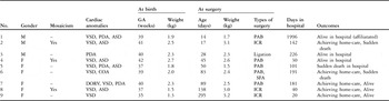
Table 2 shows the list of nine patients who underwent cardiac surgery, of whom six patients were male. Median age and weight at surgery were 50 days, ranging from 14 to 295 days, and 2.40 kilograms, ranging from 1.50 to 3.20, respectively. We performed intracardiac total repair in three patients, pulmonary arterial banding in five patients, and simple ligation of the ductus in one patient. There were three out of nine patients who had a single defect such as ventricular septal defect or patent ductus arteriosus, while the other patients had complex cardiac lesions. There was one patient, that is, patient 6 in Table 2, with ventricular septal defect and aortic coarctation who underwent subclavian flap aortoplasty and pulmonary arterial banding. Pre-operative assisted ventilation was performed in three patients. There was no statistical difference in follow-up duration, birthweight, gestational age, gender, chromosomal mosaicism, cardiac anomalies, extracardiac anomalies, and pre-operative assisted ventilation between patients who underwent cardiac surgery and those who were conservatively treated (Table 1). Despite the fact that three, that is, 33%, of the nine patients who underwent cardiac surgery carried chromosomal mosaicism, the Cox proportional hazardous model revealed no relationship between mosaicism and survival (p = 0.08).
The list of 9 patients who underwent cardiac surgery.

M = male; F = female; GA = gestational age; VSD = ventricular septal defect; ASD = atrial septal defect; PDA = patent ductus arteriosus; ICR = intracardiac repair; PAB = pulmonary arterial banding; SFA = subclavian flap aortoplasty
Median hospitalisation time was 142 days, ranging from 20 to 226 days, in nine patients who underwent cardiac surgery. As post-operative course, all the patients except one, that is, patient 5, were extubated within several days and did not need assisted ventilation subsequently. Patient 5 was re-intubated because of persistent apnoea. She needed tracheotomy and assisted ventilation.
Outcomes of the 34 patients with trisomy 18
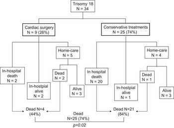
Clinical outcomes of 34 patients with trisomy 18 were summarised in Figure 1. Of the 34 patients, 25, that is, 74%, died at median age of 44 days, ranging from 1 to 1517 days. Median survival duration was 3.5 months, ranging from 0 to 241 months. In the patients who underwent cardiac surgery, median survival duration was 22.4 months, ranging from 3.3 to 241 months. There were two in-hospital deaths including one early hospital death. He died for a sudden arrhythmic event at 3 months of age (21st post-operative day). Of the nine patients who underwent cardiac surgery, five were discharged alive to achieve home-care, but two patients died after discharge at 18.5 and 49.5 months of age, respectively. No autopsy was obtained in the series of cases.
Clinical outcomes of 34 patients with trisomy 18 in patients who underwent cardiac surgery and those who were conservatively treated.

In patients who were conservatively treated, there were 20 in-hospital deaths and one death after discharge. Of the 25 patients who underwent conservative treatments, four, that is 16%, were discharged alive, but three patients died because of infection or acute respiratory failure probably owing to apnoea. There was a significant difference in the number of deaths between patients who underwent cardiac surgery and those with conservative treatments (p = 0.02). Median survival duration in patients who underwent cardiac surgery was 0.9 (0–36.6) months, which was longer than that in patients who were conservatively treated (p = 0.015; Table 1).
Survival rates
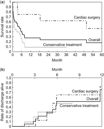
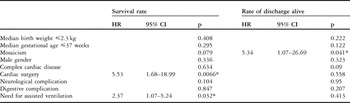
In Kaplan–Meier analysis, overall survival rates at 1 month, 6 months, 12 months, and 24 months were 63%, 38%, 25%, and 22%, respectively (Fig 2). Cox proportional hazard regression analysis revealed that the independent hazardous variable for survival rate was cardiac surgery, with a hazard ratio of 5.53, 95% confident interval between 1.68 and 18.99, p = 0.006, and pre-operative assisted ventilation, with a hazard ratio of 2.27, 95% confident interval between 1.07 and 5.24, p = 0.03 (Table 3). The other factors such as gender, gestational age, birthweight, types of cardiac anomalies and extracardiac anomalies, however, were not hazardous in survival rates. In patients who underwent cardiac surgery, the cumulative survival rates at 6 months, 12 months, and 24 months were 38%, 25%, and 22%, respectively, compared with 25%, 9%, and 9% in patients who were conservatively treated (Fig 2a). There was a significant difference in the survival rates between patients who underwent cardiac surgery and those who were conservatively treated (p = 0.002). These findings suggested that cardiac surgery contributed to increased survival rate in patients with trisomy 18. Of the 25 patients who were conservatively treated, two, however, died while waiting for cardiac surgery. When these two patients were sorted in the surgery group, the cumulative survival rate did not differ between patients who underwent cardiac surgery and those who were conservatively treated (p = 0.112).
(a) Kaplan–Meier curve shows the survival rate between patients who underwent cardiac surgery and those who were conservatively treated. The survival rate in patients who underwent cardiac surgery was significantly higher than those who were conservatively treated (p = 0.035). (b) The rate of discharge alive between two groups. There is no statistical significance (p = 0.35).

Cox proportional hazard regression analysis for death in 34 patients with trisomy 18.

HR = hazard ratio; CI = confident interval
*Means statistically significance (p value less than 0.05)
Rates of discharge alive
Of the nine patients who underwent cardiac surgery, five were discharged alive, while four of the 25 patients who underwent conservative treatments were discharged alive. Of the patients who underwent cardiac surgery, three stayed alive in hospital because of parents’ refusal to raise them, that is, patient 4 in Table 2, and assisted ventilation with tracheotomy owing to persistent apnoea, that is, patients 1 and 3 in Table 2. In patients who were conservatively treated, one patient who had complications involving oesophageal atresia and double outlet right ventricle did not achieve home-care because of the management of gastrostomy. The rates of discharge alive at 1 month, 3 months, and 6 months were 0%, 12%, and 65% in patients who underwent cardiac surgery, compared with 0%, 20%, and 47% in patients who were conservatively treated (Fig 2b). There was no statistical significance in rates of discharge alive between the two groups (p = 0.80). Cox proportional hazard regression analysis revealed that independent hazardous variable was carrying mosaicism, with a hazard ratio of 5.34, 95% confident interval between 1.07 and 26.69, p = 0.041. Median in-hospital duration in patients who underwent cardiac surgery was longer than that in patients who were conservatively treated for 5 months, ranging from 1 to 12 months, versus 3 months, ranging from 0.5 to 6 months (p = 0.009). There was no significance between median numbers of re-hospitalisation between patients who underwent cardiac surgery and those who were conservatively treated (4 versus 5, p = 0.949).
Discussion
We analysed 34 consecutive trisomy 18 patients including nine patients who underwent cardiac surgery. We found that cardiac surgery contributes to increased survival rate in trisomy 18 patients. Of the patients who underwent cardiac surgery, 44% died in spite of effective cardiac surgery. The issue about the indication of cardiac surgery seems to be equivocal in trisomy 18 patients. There could be significant bias in the present study, because we performed cardiac surgery for less severe cardiac lesions, less severe extracardiac anomalies, and less severe general status, as demonstrated in Table 1. This setting, however, was practical in the management of trisomy 18 patients.
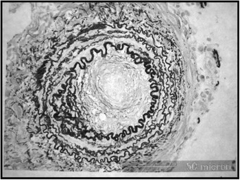
It has been considered that the majority of trisomy 18 patients die of central apnoea rather than congenital cardiac disease, while several authors advocated the causes of death in trisomy 18 patients to be cardiac failure and pulmonary haemorrhage resulting from congenital cardiac disease.Reference Kosho, Nakamura, Kawame, Baba, Tamura and Fukushima11, Reference Kinoshita, Nakamura and Nakano12 When a trisomy 18 patient with left-to-right shunt survives beyond several months, pulmonary vascular obstructive disease develops earlier and symptoms related to cardiac failure may disappear.Reference Carey7 In the present case series, one patient with large ventricular septal defect and patent ductus arteriosus underwent lung biopsy to evaluate the degree of pulmonary vasculature obstructive disease at 4 months of age (Fig 3). The histological finding showed organised peripheral small pulmonary arteries below 150 micrometres in diameter with fibrous and obstructive changes of the media, which made the patient refuse the operation. She has been surviving for 36 months with stable haemodynamics and respiration. The present case indicated that the development of pulmonary vascular obstructive disease was not always responsible for death in trisomy 18 patients.
Histological finding shows organised peripheral small pulmonary arteries <150 μm in diameter with fibrous and obstructive changes of the media (Elastica–Goldner stain, original × 400).

The issue of the management of trisomy 18 has been ethically discussed. Chervenak and McCulloughReference Chervenak and McCullough14 found that a choice between an aggressive and a non-aggressive management should be made owing to probable lethality or absent cognitive developmental capacity if the patient survived. Previous authors found that aggressive management, including cardiac surgery, could contribute to increased early survival rates in trisomy 18 patients.Reference Graham, Bradley, Shirali, Hills and Atz9, Reference Kosho, Nakamura, Kawame, Baba, Tamura and Fukushima11 There were, however, few reports that described the efficacy of cardiac surgery in the long-term follow-up of trisomy 18. Kaneko et alReference Kaneko, Kobayashi and Achiwa10 described that 64% of trisomy 18 patients who underwent cardiac surgery died from various causes with median survival of 6 months. We also demonstrated that 44% of patients died after cardiac surgery. These findings suggested that cardiac surgery could not always prevent death in all the trisomy 18 patients.
We also demonstrated that chromosomal mosaicism was a hazardous factor not for survival rates but for rates of discharge alive. As Baty et alReference Baty, Blackburn and Carey13 reported that long-term survival did not appear to be due to mosaicism, we assumed that mosaicism did not influence the overall survival time but the rate of discharge alive.
We consider that the management for cardiac complications should be individualised in trisomy 18 patients. We must inform the family that cardiac surgery may contribute to improving the survival rate to some extent, but that other problems including sudden death remain even after cardiac surgery. We emphasise that it is necessary to establish a trusting relationship and partnership between parents and health-care providers.
The study is limited by the small number of case series. In addition, the two groups were not identical in number of patients. Nationwide survey should be necessary to evaluate the true efficacy of cardiac surgery. Unfortunately, home-care programmes have not been systematically established in our hospital. The establishment of home-care programme by cardiologists, neonatologists, nurses, and other co-workers may contribute to achieving home-care and shortening of admission time.
In conclusion, we analysed the clinical outcomes of 34 patients with trisomy 18 and compared the rates of survival and discharge alive between patients who underwent cardiac surgery and those who were conservatively treated. Cardiac surgery contributed to increased survival rate but not the rate of discharge alive. The indication of cardiac surgery should be carefully individualised to improve the quality of life in trisomy 18 individuals and concerned surrounding people. We must inform their family that cardiac surgery contributes to improve the survival time but other problems including sudden death remain even after cardiac surgery.
Acknowledgement
We are grateful to Professor Shoichi Ohga, Kyushu University, Satochi Honjo, Fukuoka National Hospital, for the contribution of statistical analysis and helpful discussion and Toshiro Yamaki, Japanese Research Institute of Pulmonary Vasculature, for the contribution of histological analysis.