Introduction
Semiconductor photocatalysis technology has promising applications in environmental protection, solar energy utilization, and the development of novel functional materials (Fukina et al., Reference Fukina, Koryagin, Koroleva, Zhizhin, Suleimanov and Kirillova2021; Shi et al., Reference Shi, Li, Xia, Lu, Zuo, Luo and Yao2017; Tun et al., Reference Tun, Wang, Naing, Wang and Zhang2019). It refers to a new technology with major economic and social benefits. Among numerous photocatalytic materials, perovskite oxides (ABO3) are recognized as p-type photocatalytic semiconductor materials with stable structures, narrow band positions, and prominent photocatalytic activity (Gong et al., Reference Gong, Xiang, Zhang, Sun, Ye and Lin2019; Ismael & Wu, Reference Ismael and Wu2019; Li et al., Reference Li, Yan, Zuo, Lu, Luo, Li, Yao and Ni2017b). However, the disadvantages of pure perovskite oxide catalytic materials (e.g. uncontrollable particle size, easy agglomeration, and poor recyclability) limit their practical application to a significant extent (Maeda et al., Reference Maeda, Eguchi and Oshima2014; Peng et al., Reference Peng, Fu, Yang and Ouyang2016; Purohit et al., Reference Purohit, Yadav and Satapathi2021).
Over the past few years, a wide range of techniques has been developed to adjust and improve the performance of perovskite oxide photocatalytic materials (e.g. ion doping (Kim et al., Reference Kim, Jang, Ra, Kim, Kim and Lee2019), surface loading (Li et al., Reference Li, Tang, Zheng, Shao and Li2017a), morphology control (Maridevaru et al., Reference Maridevaru, Wu, Viswanathan Mangalaraja and Anandan2020), and heterostructure construction (Khaledian et al., Reference Khaledian, Zolfaghari, Nezhad, Niaei, Khorram and Salari2021)). Loading photocatalytic nanoparticles on the surfaces of matrix materials is effective in controlling the size of nanoparticles and increasing the specific surface area and the number of active sites, while also overcoming the inherent lack of stability, agglomeration, and the problematic reuse of unsupported nanoparticles (Belver et al., Reference Belver, Bedia, Álvarez-Montero and Rodriguez2016; Chen et al., Reference Chen, Wu, Liu, Wang and Song2019b; Khan et al., Reference Khan, Khan, Usman, Imran and Saeed2020; Landge et al., Reference Landge, Sonawane, Sivakumar, Sonawane, Uday Bhaskar Babu and Boczkaj2021; Yang et al., Reference Yang, Ke, Yang, Liu, Guo, Frost, Su and Zhu2010). It is necessary, therefore, to attach perovskite oxide particles to a suitable matrix.
Muscovite is a layered silicate mineral with many advantages, such as good light transmittance, large specific surface area, strong adsorption capacity, good heat resistance, and stable chemical properties (Barakat et al., Reference Barakat, Kumar, Lima and Seliem2021; Salam et al., Reference Salam, Abukhadra and Mostafa2020; Shao et al., Reference Shao, Siao, Lai, Hsieh, Tsao, Lu, Chen, Hsu and Chu2021; Touaa et al., Reference Touaa, Bouberka, Gherdaoui, Supiot, Roussel, Pierlot and Maschke2020); an ideal natural catalyst carrier. A nano-zero-valent iron (NZVI)-loaded muscovite composite was synthesized by Bao et al. (Reference Bao, Damtie, Hosseinzadeh, Frost, Yu, Jin and Wu2020) using a liquid-phase reduction method. This process prevented agglomeration, improved dispersibility, increased the number of catalytically active sites, and enhanced catalytic performance. A TiO2/muscovite nanocomposite was synthesized by Li et al. (Reference Li, Sun, Peng, You, Qin and Zeng2019) using a liquid-phase precipitation method and those authors found that the muscovite matrix delayed growth and phase transformation of TiO2 and enhanced photocatalytic activity. When perovskite oxide is loaded on the surface of muscovite, the structure and interfacial properties of the material will be changed, which is expected to improve the photocatalytic activity of the material.
In view of this, the purpose of the current study was to prepare photocatalytic LaNi0.95Fe0.05O3/muscovite composite materials by a sol-gel method to obtain materials with high photocatalytic degradation performance and recyclability, while reducing their amount and preparation costs, which are critical criteria for the industrial application of such photocatalysts.
Materials and Methods
Materials
The initial components used for the production of photocatalytic LaNi0.95Fe0.05O3/muscovite composite material were as follows: lanthanum nitrate (La(NO3)3·6H2O) (analytically pure, produced by Shanghai Aladdin Biochemical Technology Co., Ltd, Shanghai, China); nickel nitrate (Ni(NO3)2·6H2O), and ferric nitrate (Fe(NO3)3·9H2O) (analytically pure, produced by Chengdu Kelong Chemical Reagent Factory, Chengdu, China). Citric acid (analytically pure, produced by Chengdu Kelong Chemical Reagent Factory, Chengdu, China) served as a complexing agent, and muscovite (K{Al2[AlSi3O10](OH)2}) (600 mesh, obtained from Shijiazhuang Chenxing Industrial Co., Ltd, Shijiazhuang, China) was the supporting matrix. Other materials used were: methyl orange, sodium sulfite (Na2SO3), ammonium oxalate ((NH4)2C2O4), isopropanol (IPA), and anhydrous ethanol (analytically pure, produced by Chengdu Kelong Chemical Reagent Factory, Chengdu, China); deionized water (resistivity of 18.2 MΩ·cm, prepared in the laboratory).
Preparation of LaNi0.95Fe0.05O3/muscovite Composite Photocatalytic Material
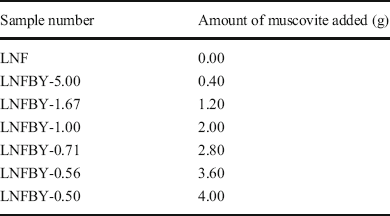
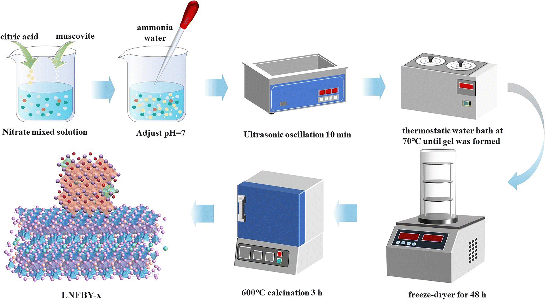
In a beaker, 4.3301 g of La(NO3)3·6H2O, 2.7625 g of Ni(NO3)2·6H2O, and 0.2020 g of Fe(NO3)3·9H2O were dissolved in 60 mL of a mixture of anhydrous ethanol and deionized water with a volume ratio of 2:1. Citric acid was added to the beaker in a molar ratio of 1.5:1 to the total amount of metal ions, ammonia water was added dropwise to regulate the pH to 7, and muscovite, calcined at 600°C for 3 h, was added in various mass ratios. The solution was stirred for 10 min at ambient temperature and shaken in an ultrasonic water bath for 10 min. The sample beaker was placed in a thermostatic water bath and stirred at 70°C until a gel was formed. The gel was frozen in a freezer and then dried in a freeze-dryer for 48 h to obtain a dry gel sample. The prepared dry gel was placed in a muffle furnace and heated to 600°C at a rate of 5°C·min–1. After natural cooling, the sample was ground into a powder for later use. The materials prepared with various LaNi0.95Fe0.05O3 to muscovite ratios (x) were named LNFBY-x, where LNF denotes LaNi0.95Fe0.05O3, BY represents the calcined muscovite, and x is the mass ratio of LNF to muscovite. The amounts of muscovite added in the various samples are listed in Table 1 and the preparation process is illustrated in Fig. 1.
Muscovite added to LaNi0.95Fe0.05O3/muscovite composite photocatalytic material

Process of preparing the LNFBY-1 sample

Characterization
The phase composition of the samples was studied using an Ultima IV X-ray diffractometer (XRD, Rigaku Co., Tokyo, Japan) with CuKα radiation (λ = 1.5406 nm) over an angular range of 10–65°2θ, and continuous scanning with X’Clelerator. The morphology of each composite was examined using an Ultra 55 field emission scanning electron microscope (SEM, Zeiss, Oberkochen, Germany) with a working voltage of 15 kV, for which the samples were spread onto aluminum sheets and coated with a thin layer of gold by sputter coating. Transmission electron microscopy (TEM) and high-resolution TEM (HRTEM) were performed using an FEI Tecnai G20 instrument (Thermo Fisher Scientific, Inc., Waltham, Massachusetts, USA) with a LaB6 electron gun filament, operated at 200 kV. Surface potentials were measured using a Zetasizer Nano ZS90 instrument (Malvern Panalytical Co., Ltd., Malvern, UK). Specific surface area and porosity were determined by N2 adsorption and desorption using an Autosorb IQ instrument (Anton Paar QuantaTec Inc., Boynton Beach, Florida, USA). The elemental analysis was performed on a K-Alpha+-type X-ray photoelectron spectrometer (XPS, Thermo Fisher Scientific, Inc., Waltham, Massachusetts, USA), using a monochromatic AlKα source with an energy of 1486.6 eV. The IR absorption characteristics of the composites were investigated as KBr disks using a Nicolet-5700 FTIR spectrometer (Nicolet Instrument Co., Ltd., Fitchburg, Massachusetts, USA). The absorbance of the supernatant in the catalytic tests was measured by an Evolution 300 UV-Vis spectrophotometer (Thermo Fisher Scientific, Inc., Waltham, Massachusetts, USA).
Photocatalytic Performance Test
To assess the photocatalytic performance of the LNFBY-x composite materials in visible light, methyl orange (MO) was selected as the target pollutant, and a xenon lamp (HEL-HXF300, Beijing Zhongjiao Jinyuan Technology Co., Ltd., Beijing, China) acted as the light source with a 400–800 nm filter. In a beaker, 100 mg of photocatalytic material was dispersed in 100 mL of a 10 mg·L–1 MO solution . The light source was attached to a magnetic stirrer at a distance of 20 cm from the liquid surface of the solution, which was then stirred for 1 h in the dark to ensure that adsorption-desorption equilibrium between photocatalyst and dye was reached. The light source was switched on, and 5 mL of solution was sampled every 20 min and centrifuged to remove the catalyst particles. The absorbance of the supernatant was examined at the maximum absorption wavelength of MO at 437 nm, and the degradation percentage D of MO was calculated by:

where C 0 denotes the initial MO concentration and C t represents the MO concentration after a certain reaction time (mg·L-1).
Active Free Radical Test
To illustrate the photocatalytic degradation mechanism of the samples, free-radical tests were performed by applying sodium sulfite (Na2SO3) as an electron (e–) trapping agent, ammonium oxalate ((NH4)2C2O4) as a hole (h+) trapping agent, and isopropanol (IPA) as an hydroxyl radical (٠OH) trapping agent. 100 mg of catalyst and 0.17 mmol of the free-radical scavenger were dispersed in 100 mL of 10 mg·L–1 MO solution, and the photocatalytic performance test was conducted to determine the main active substances during photodegradation.
Results and Discussion
Phase and Structural Characteristics
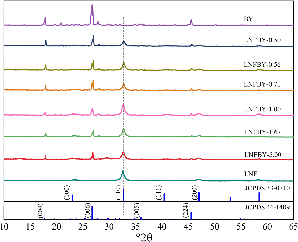
The XRD patterns of the LNFBY-x samples (Fig. 2) revealed that the muscovite sample (BY) has diffraction peaks characteristic of 2M 1-type muscovite (JCPDS 46-1409) (d 002 = 0.9835 nm, d 004 = 0.4989 nm, d 006 = 0.3307 nm, and d 115 = 0.3187 nm) and of quartz (JCPDS 79-1906) (d 100 = 0.4252 nm, d 011 = 0.3336 nm, and d 021 = 0.1991 nm) were present, indicating that the sample was 2M 1-type muscovite with a small amount of quartz impurity. The diffraction peaks of the LNF sample (d 100 = 0.3855 nm, d 110 = 0.2727 nm, d 111 = 0.2213 nm, d 200 = 0.1927 nm, and d 210 = 0.1704 nm) conformed to those of the standard spectrum of cubic perovskite phase (JCPDS 33-0710). The XRD peaks of all LNFBY-x composite samples were consistent with those of LNF and muscovite. The intensity of the muscovite diffraction peaks increased with increasing muscovite amount, which demonstrated that the introduction of LNF retained the muscovite structure.
XRD patterns of LNFBY-x samples over the range 10–65°2θ

Morphology, Structure, and Composition Characteristics
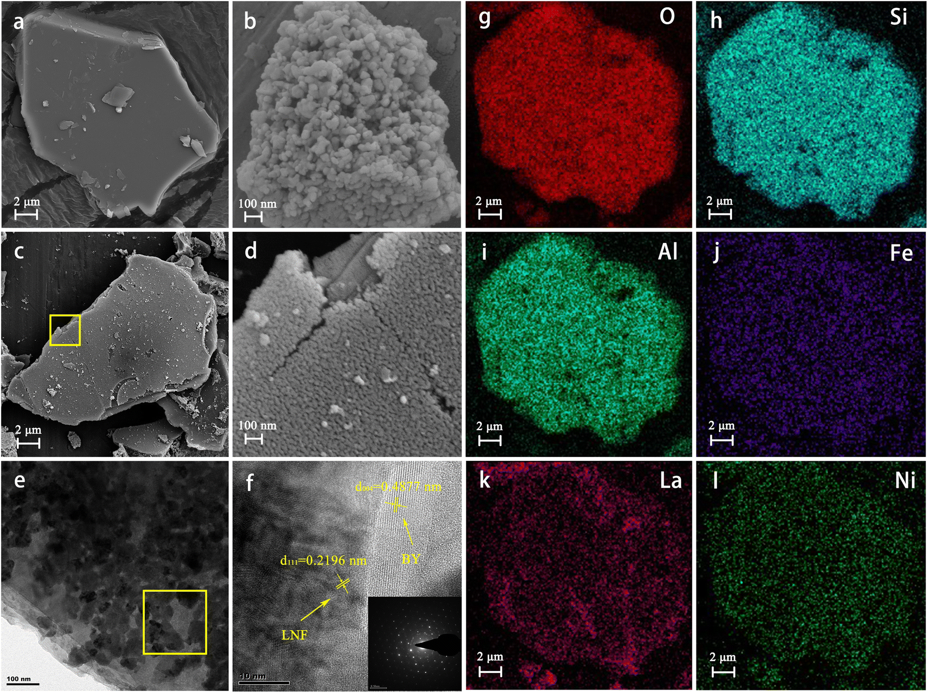
The SEM and TEM images of the BY, LNF, and LNFBY-1.00 samples (Fig. 3) showed that muscovite used in the experiment displayed a layered structure with a diameter of nearly 20 μm and a flake diameter to thickness ratio of ~2:1 (Fig. 3a). In addition, its surface was smooth and flat, laying a solid foundation for loading LNF. The pure LNF sample consisted of spherical particles with a particle size of ~50 nm with poor dispersion and obvious agglomeration (Fig. 3b). The SEM images of LNFBY-1.00 (Figs 3c and 3d) revealed that LNF nanoparticles were loaded successfully onto the surface of muscovite, forming a dense film layer. The particle size in the LNFBY-1.00 sample was close to 20 nm, which is significantly smaller than that of pure LNF, and agglomeration was significantly inhibited.
a SEM image of BY; b SEM image of LNF; c SEM image of LNFBY-1.00; d SEM image of LNFBY-1.00; e–f HRTEM images of LNFBY-1.00; g–l EDS mapping images of LNFBY-1.00

These observations were related to the abundant active sites on the surface of muscovite, assisting in controlling the growth of LNF grains (Tahir et al., Reference Tahir, Tahir, Zakaria and Muhammad2019). The HRTEM images (Figs 3e and 3f) revealed that the LNFBY-1.00 sample featured easily identifiable lattice stripes, which demonstrated the crystallinity of the sample. The measured lattice spacings of d = 0.4877 nm and d = 0.2196 nm corresponded to the (004) crystal plane of muscovite and the (220) crystal plane of LNF, respectively. Through energy-dispersive X-ray spectroscopy (EDS), the element proportions of the sample were determined (Table 2); in addition to the elements present in muscovite (O, Si, Al, and K) and Au generated during sample preparation, the LNFBY-1.00 sample also contained the elements (La, Ni, and Fe) present in LNF. EDS mapping of LNFBY-1.00 (Figs 3g–l) further verified that LNF was loaded successfully on the surface of muscovite without destroying the structure or changing the chemical composition of muscovite during the synthesis process.
EDS data for BY, LNF, and LNFBY-1.00 samples (at.%)

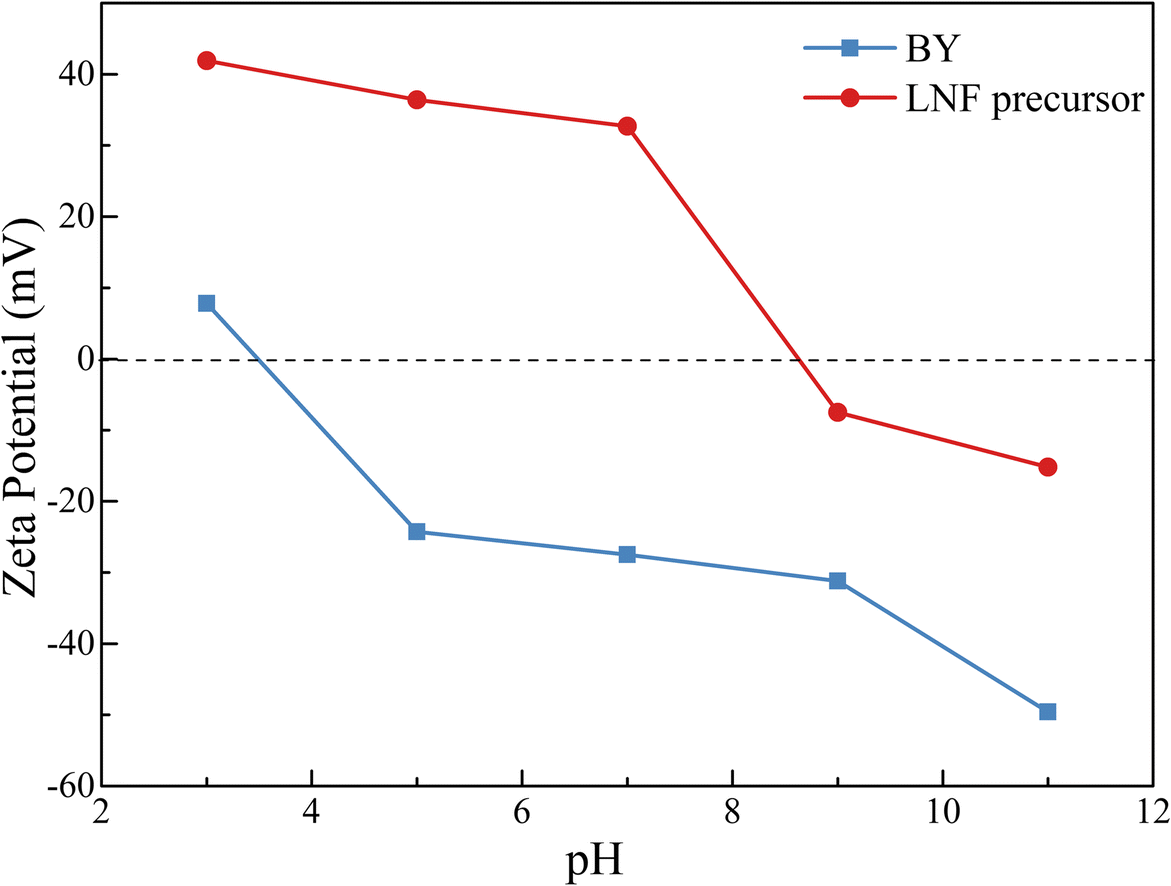
The surface zeta potential curves of muscovite and the LNF precursor over a range of pH values (Fig. 4) showed that the zeta potential of muscovite was negative at pH 7, the preparation stage, while that of the LNF precursor sol was positive. Consequently, when muscovite was suspended and dispersed in the LNF precursor sol, the two components, impacted by different surface charges, were attracted and bound to each other electrostatically. The positively charged LNF precursor sol nucleated and grew on the negatively charged muscovite surface, forming a coating layer that does not detach easily (Zhou et al., Reference Zhou, Lv, Guo, Xu, Wang, Zheng and Wu2012). Due to the chemical bonding, the two components were combined firmly, thereby improving utilization and recyclability.
pH and zeta potential diagram of the BY and LNF precursor

Nitrogen Adsorption-desorption Isotherms and NLDFT Pore-size Distribution
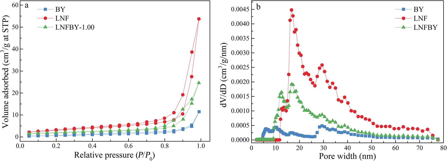
The nitrogen adsorption-desorption isotherms and Nonlocal-Density-Functional-Theory (NLDFT) pore-size distribution of the BY, LNF, and LNFBY-1.00 samples (Fig. 5a) showed that the adsorption-desorption isotherms of the muscovite sample had almost no hysteresis loop, while LNF and LNFBY-1.00 exhibited the typical type-II characteristics (Xu et al., Reference Xu, Maimaiti, Wang, Awati, Wang, Zhang and Chen2019), which demonstrated their mesoporous structure. The specific surface areas of the BY, LNF, and LNFBY-1.00 samples were calculated as 3.237 m2·g–1, 11.055 m2·g–1, and 8.412 m2·g–1, respectively. The NLDFT method was used to study further the pore-size distribution of the three samples (Fig. 5b), which showed that muscovite exhibited micropores and mesopores. The micropores may come from the surface of muscovite, and the mesopores may come from the laminar structure of muscovite. LNF had a wide pore-size distribution. The smaller pore size may originate from single LNF nanoparticles, while the larger pore size may be attributed to agglomeration of LNF nanoparticles. The average pore size of LNFBY-1.00 was concentrated at 14.7 nm, and the pore-size distribution was relatively reduced, probably because LNF nanoparticles were loaded on the surface of muscovite, which reduced agglomeration and occupied the original micropores on the surface of muscovite. Compared with LNF, LNFBY-1.00 demonstrated a moderate specific surface area and pore-size distribution, which could provide more adsorption sites and photocatalytic active sites, thereby improving photocatalytic activity.
a N2 adsorption-desorption isotherms and b pore-width distribution curves of the BY, LNF, and LNFBY-1.00 samples

Molecular Vibration and Bond-energy Changes
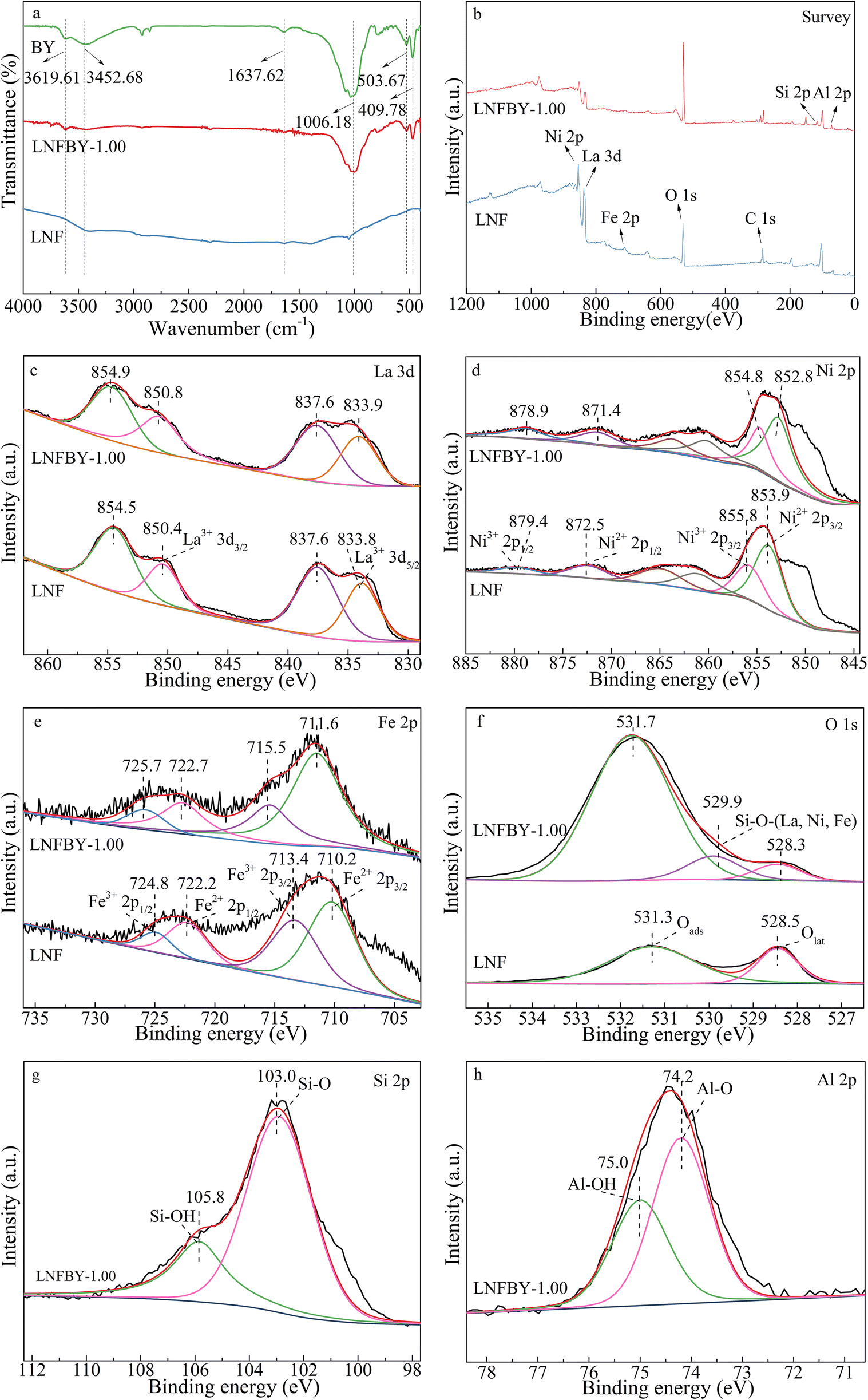
The FTIR absorption bands in the BY, LNF, and LNFBY-1.00 samples (Fig. 6a) at 3449.94 and 1637.62 cm–1 were attributed to the stretching and bending vibrations of adsorbed H2O, respectively (Khatri & Rana, Reference Khatri and Rana2020). The absorption bands at 3619.61 and 1006.18 cm–1 belonged to the stretching vibration of hydroxyl groups (–OH) (Edward, Reference Edward1982; Peng et al., Reference Peng, Ni, Zhou, Kou, Lu and Xu2018) and the Si–O bond (Asencios et al., Reference Asencios, Quijo, Marcos, Nogueira, Rocca and Assaf2019; Wang et al., Reference Wang, Mu, Hui and Wang2019), respectively. The bending vibration of Si–O produce the bands at 503.67 and 409.78 cm–1 (Farmer, Reference Farmer1968; Shi et al., Reference Shi, Li, Xia, Lu, Zuo, Luo and Yao2017). Compared with pure LNF, LNFBY-1.00 displayed the characteristic bands of silicate and Si–O bending vibrations, while other bands related to LNF remained unchanged. The results indicated that the presence of muscovite did not alter the structure of LNF.
a FTIR spectra of the BY, LNF, and LNFBY-1.00 samples; b–f XPS spectra of LNF and LNFBY-1.00 samples; b Survey, c La 3d, d Ni 2p, e Fe 2p, f O 1s; g–h XPS spectra of LNFBY-1.00; g Si 2p, h Al 2p

The full XPS spectra of the LNF and LNFBY-1.00 samples (Fig. 6b) indicated the presence of four elements (i.e. La, Ni, Fe, and O) in the two samples. In addition, Si and Al were also present in LNFBY-1.00. The binding energy obtained was corrected using the C 1s peak of 284.6 eV as a reference. According to the high-resolution spectrum of La 3d (Fig. 6c), La3+ 3d5/2 and La3+ 3d3/2 had binding energies of 833.8–833.9 eV and 850.4–850.8 eV (Schlapbach, Reference Schlapbach1981; Ye et al., Reference Ye, Yang, Zhang and Jiang2020), respectively. Due to the strong overlap of the Ni 2p3/2 and La 3d3/2 peaks, the core-level spectrum of Ni 2p3/2 was difficult to determine. Therefore, the XPS spectra show the range of the Ni 2p peak and a part of the La 3d3/2 peak (Fig. 6d). Peaks at 852.8–853.9 eV and 854.8–855.8 eV were attributed to Ni2+ 2p3/2 and Ni3+ 2p3/2, respectively, while those at 871.4–872.5 eV and 878.9–879.4 eV belonged to Ni2+ 2p1/2 and Ni3+ 2p1/2 (Hüfner & Wertheim, Reference Hüfner and Wertheim1975; Zhong et al., Reference Zhong, Jiang, Dang, He, Chen, Kuo, Kriz, Meng, Meguerdichian and Suib2018), respectively. As indicated in the high-resolution spectrum of Fe 2p (Fig. 6e), the binding energies at 710.2–711.6 eV and 722.2–722.7 eV corresponded to Fe2+ 2p3/2 and Fe2+ 2p1/2, and 713.4–715.5 eV and 724.8–725.7 eV to Fe3+ 2p3/2 and Fe3+ 2p1/2 (Liu et al., Reference Liu, Yi, Qin, Wu, Li, Chu, Jin, Li, Tong, Dong and Li2019), respectively. In the high-resolution spectrum of O 1s (Fig. 6f), the peak binding energy 531.3–531.7 eV belonged to oxygen in the corresponding hydroxyl group and surface adsorbed oxygen, while 528.3–528.5 eV was assigned to lattice oxygen (Brundle, Reference Brundle1977; Dong et al., Reference Dong, Sun, Zhang, Li and Zheng2018; Zhu et al., Reference Zhu, Chen, Liu and Liang2020). In the LNFBY-1.00 composite sample, the binding energy of 529.9 eV was attributed to oxygen in Si–O–La, Si–O–Ni, and Si–O–Fe (Chen et al., Reference Chen, Wu, Wang and Song2020; Li et al., Reference Li, Peng, Chen and Wang2018), which demonstrated that LNF nanoparticles and muscovite substrate were connected by ionic bonds; the bonding formed a strong interaction at the interface, promoted electron transfer between Si and La, Fe, and Ni via oxygen atoms, improved the ability of charge transfer (Chen et al., Reference Chen, Wu, Bu, Wang and Song2019a, Reference Chen, Wu, Liu, Wang and Songb), and simultaneously bound the LNF particles to prevent detachment from the muscovite carrier. In addition, the number of hydroxyl groups and surface-adsorbed oxygen in the LNFBY-1.00 composite sample exceeded that of the LNF sample, which indicated that more highly active oxygen species could be produced by the composite, thereby increasing the amount of active substances in the photocatalytic reaction and, more significantly, improving the photocatalytic activity.
Photocatalytic Activity
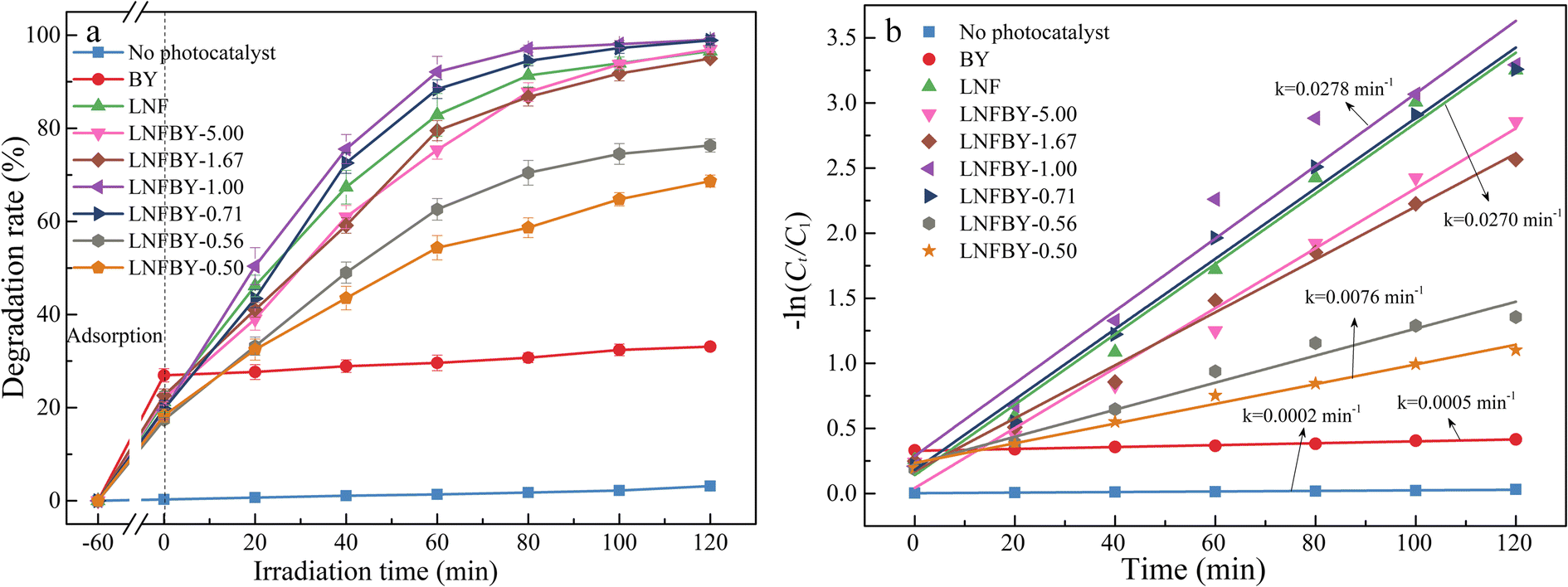
As can be seen from the degradation rate of MO of the LNFBY-x samples under visible-light irradiation with time (Fig. 7a), in the absence of catalyst, the extent of degradation was only 3% after 2 h of irradiation, which demonstrated that MO was relatively stable. With pure muscovite, the MO removal was only 33.1%, mainly due to physical adsorption. When the composite catalyst was added, the degradation of MO increased clearly. Using the extent of degradation of MO as an indicator to assess the photocatalytic activity, the photocatalytic activity decreased in the order LNFBY-1.00 > LNFBY-0.71 > LNF > LNFBY-5.00 > LNFBY-1.67 > LNFBY-0.56 > LNFBY-0.50 > BY. Therefore, one concludes that the enhancement in photocatalytic activity is related to the degree of dispersion of LNF nanoparticles and the characteristics of the muscovite carrier. At a mass ratio of LNF nanoparticles to muscovite of <1, some of the LNF nanoparticles did not compound completely with muscovite but formed agglomerated particles, some of which did not contribute effectively to the photocatalytic reactions. Such agglomeration resulted in the exposure of fewer photocatalytically active sites, thereby resulting in an insignificant improvement in the photocatalytic effect. When the mass ratio of LNF nanoparticles to muscovite reached a value of 1.00, however, LNF nanoparticles were dispersed evenly on the surface of muscovite, and the exposure of active sites was large. Furthermore, muscovite exhibited the unique adsorption characteristics of clay minerals, which could adsorb pollutants on the surface of the photocatalyst, achieving complete contact of the active sites with the pollutants and improving photocatalytic performance. Furthermore, the highly active oxygen-containing groups generated by the composite increased the amount of active substances in the photocatalytic reaction, which could be beneficial for improving photocatalytic activity. When the mass ratio of LNF nanoparticles to muscovite was >1.00, excessive muscovite blocked light to a certain extent, thereby reducing the transmittance of the system, the number of photons, and the photocatalytic activity. The fitting results of pseudo-first order kinetics of the experimental data of the photocatalytic degradation by the LNFBY-x samples (Fig. 7b) showed good linearity. LNFBY-1.00 demonstrated the greatest degradation-rate constant (0.0278 min–1), which was 1.03 times greater than that of LNF (0.0270 min–1). In recent years, the loading of photocatalytic materials on the surface of other carriers such as montmorillonite and kaolinite has, increasingly, been investigated (Fufa et al., Reference Fufa, Feyisa, Gultom, Kuo, Chen, Kabtamu and Zelekew2022; Lin et al., Reference Lin, Song, Bo, Wang and Qian2017; Yan et al., Reference Yan, Qiong, Li, Jun and Ytao2019). Although the specific surface area and dispersion of the photocatalytic materials have increased, their photocatalytic activity was still less than that of LNFBY-1.00. A possible reason was that these photocatalytic materials cannot form chemical bonds to the surfaces of these carriers, and cannot provide further catalytic reaction sites to enhance photocatalytic activity. In practice, its shedding and separation is also a key problem. Thus, muscovite with a natural flaky morphology could be an excellent carrier material for photocatalytic perovskite-type oxide materials, capable of controlling effectively the size of LNF nanoparticles, hindering agglomeration, improving dispersibility, exposing more catalytic reaction sites, and improving the utilization rate of photocatalytic materials, especially that of the highly active oxygen species produced by the composite, which could increase the number of active substances in the photocatalytic process. Thus, it could be conducive to photocatalytic reaction.
a Degradation rate of MO by LNFBY-x series samples; b photocatalytic degradation reactions of LNFBY-x series samples were fitted using quasi-first order kinetics

Stability and Recyclability
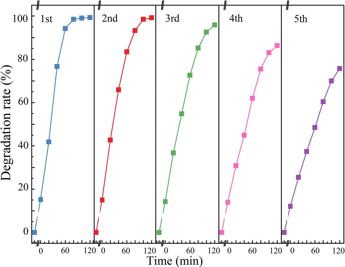
The degradation rate of MO of the LNFBY-1.00 sample under visible-light irradiation for 2 h after five consecutive cycles (Fig. 8) showed that the degradation rate of MO tended to decrease. After five cycles, the MO degradation rate by LNFBY-1.00 was 75.75%, which still showed high catalytic activity. Compared with pure LNF, the LNFBY-1.00 sample could be recovered more easily via filtration and natural sedimentation, which would significantly reduce the cost of catalyst recovery. As an explanation for the slight decrease in photocatalytic performance, residual MO, adsorbed on the catalyst during the photocatalytic degradation process, might be present, and some active sites remained occupied.
Recyclability of MO degradation in a LNFBY-1.00 sample after 2 h of visible light irradiation

Photocatalytic Mechanism
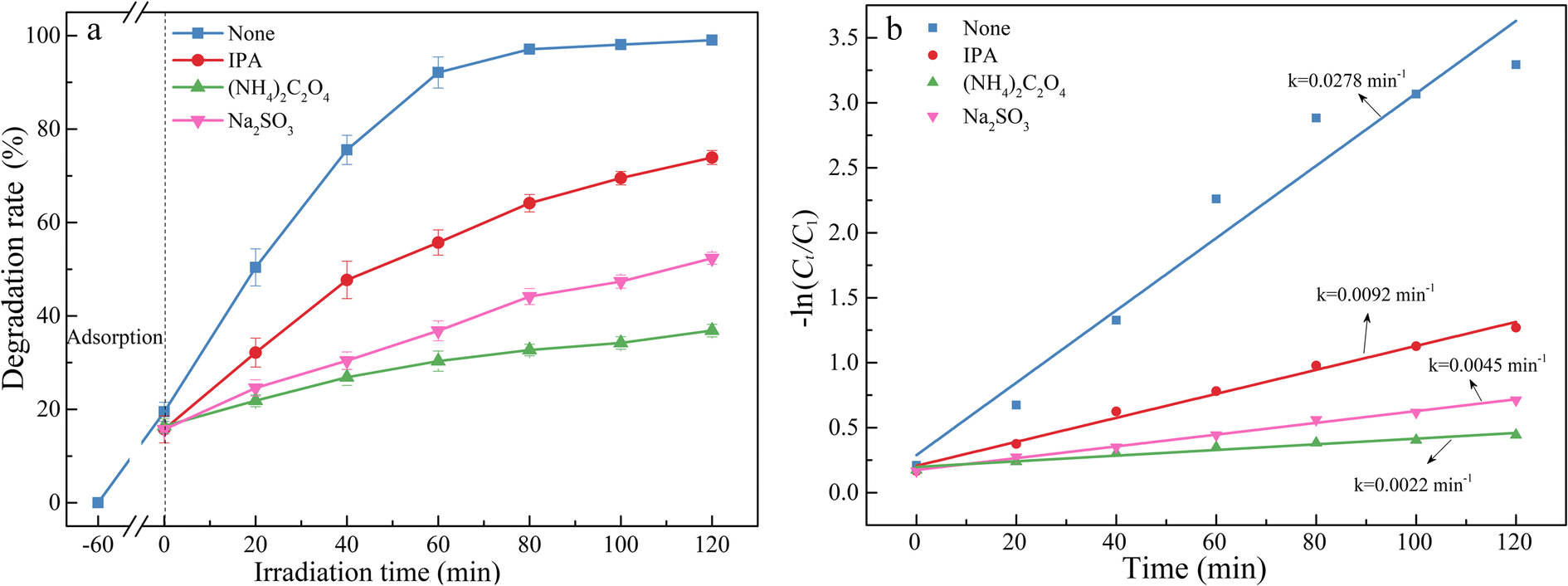
The MO degradation rates of LNFBY-1.00 with time after addition of various trapping agents (Fig. 9a) showed that the degradation rate of MO decreased after the addition of each scavenger. Compared to the other agents, the addition of IPA exerted less effect on the degradation rate of MO, probably because the production of ٠OH was lower. With the addition of Na2SO3 to the system, the degradation rate decreased, which demonstrated that e– contributed to a certain degree to MO degradation. In the presence of (NH4)2C2O4, the degradation rate dropped significantly, thereby demonstrating h+ as the major active substance in the photocatalytic degradation of MO. The contribution of the active free radicals decreased in the order h+ > e– > ٠OH.
a Effect of three different free radical capture agents on the degradation rates of MO; b photocatalytic degradation reactions were fitted with quasi-first order kinetics

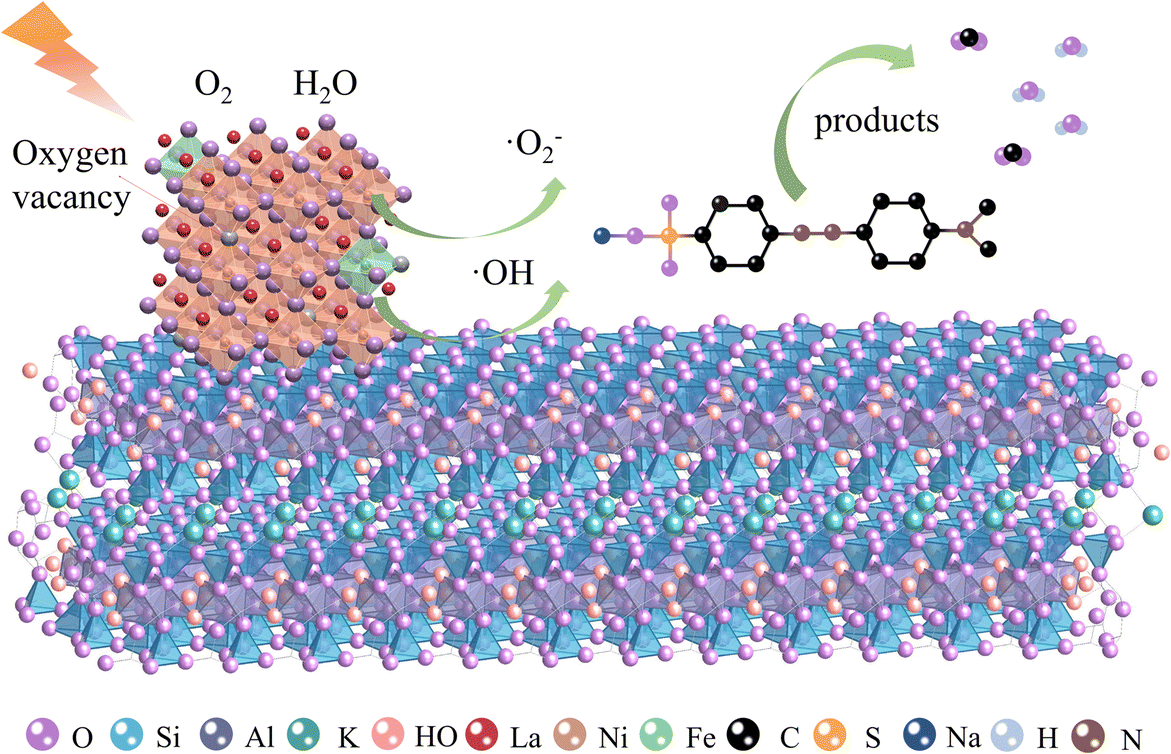
Based on the test results for the active species, the following degradation mechanism may be present (Fig. 10): LNF nanoparticles are excited by visible light with energy exceeding the threshold value to generate e–-h+ pairs, and the negatively charged e– reacts with O2 adsorbed on the catalyst surface to generate superoxide radicals (٠O2 -), thereby leading to MO degradation. Some h+ oxidized OH– and H2O in solution to produce hydroxyl radicals (٠OH), exhibiting high activity and participating in the degradation reaction. Some h+ interacted directly with MO. In such a process, the interfacial interaction between LNF nanoparticles and the muscovite substrate improved the charge transferability of the composite sample, thereby improving the photocatalytic reaction. In addition, the great adsorption performance by the muscovite support could result in a higher apparent MO concentration on the surface of the catalytic material, thereby further elevating the photocatalytic degradation rate.
Degradation mechanism diagram of the LNFBY-1.00 sample

Conclusions
(1) A series of LNFBY-x samples was synthesized by a sol-gel impregnation method from LNF nanoparticles and muscovite. The two components were connected firmly by ionic bonds without altering the phase and structure. Compounding with muscovite effectively controls the size of LNF nanoparticles and prevents agglomeration, thereby exposing more catalytic reaction sites and improving the utilization rate of the photocatalytic materials. The highly reactive oxygen species produced by the composite increased the amount of active substances in the photocatalytic process and enhanced the photocatalytic activity.
(2) Among the LNFBY-x samples, the LNFBY-1.00 (mass ratio of LNF to muscovite = 1.00) demonstrated the best structure and photocatalytic performance. The degradation rate of MO reached 99.03% after visible-light irradiation for 120 min. After five catalyst recycles, the MO degradation rate of the LNFBY-1.00 sample was 75.75%, which is still considered high catalytic activity. In the degradation process, the contribution of active free radicals decreased in the order h+ > e– > ٠OH.
(3) The reason for the enhanced photocatalytic activity was the binding of LNF nanoparticles to the muscovite substrate, thereby hindering the agglomeration of LNF nanoparticles. Therefore, the composite samples exhibited smaller particle sizes, larger specific surface areas, moderate pore size distributions, and strong adsorption capacity. In addition, the interfacial interaction between LNF nanoparticles and the muscovite substrate enhances the charge transferability of the composite sample, thereby improving the photocatalytic reaction. Furthermore, the concentration of MO around LNF nanoparticles was enhanced through adsorption by muscovite, thereby increasing the contact probability of LNF and MO molecules and ultimately improving the photocatalytic degradation rate.
Acknowledgments
This work was supported by the National Natural Science Foundation of China (grant numbers 41972042 and 42072048).
Funding
Funding sources are as stated in the Acknowledgments.
Declarations
Consent for publication
The authors declared their consent for publication.
Conflict of interest
The authors declare that they have no conflict of interest.