Introduction
Growth factor receptor-bound protein 14 (Grb14) is a member of the adaptor protein superfamily that includes Grb7 and Grb10. This family of proteins has similar structure and conservative molecular architecture that consists of an N-terminal proline rich region, a central pleckstrin homology (PH), a carboxyl-terminal SH2 domain, followed by a domain between the PH and SH2 domains termed the BPS region. The structure of the Grb7 family binding proteins allows the participation of these proteins in multiple cellular signalling transduction pathways (Lucas-Fernandez et al., Reference Lucas-Fernandez, Garcia-Palmero and Villalobo2008).
The Grb7 protein family is involved in metabolic processes in cells, triggered by growth factors that bind to tyrosine kinase receptors such as insulin-like growth factor I (IGF-I) and epidermal growth factor (EGF; Holt & Siddle, Reference Holt and Siddle2005). Upon tyrosine kinase receptor activation, Grb14 binds to phosphorylated sites on the β part of these receptors through SH2 and BPS subunits. This binding prevents the access of receptor substrates to the phosphorylated sites of tyrosine kinase receptors, resulting in a block in activation of mitogen-activated protein kinase (MAPK) and phosphatidylinositol-3 kinase (PI3K) intracellular pathways. MAPK and PI3K are kinases that have an important role in cell proliferation and differentiation (Han et al., Reference Han, Shen and Guan2001).
The Grb7 family of proteins have been associated with IGF-I-dependent tumors (Lim et al., Reference Lim, Riedel and Liu2004), insulin metabolism (Goenaga et al., Reference Goenaga, Hampe, Carre, Cailliau, Browaeys-Poly, Perdereau, Holt, Daly, Girard, Broutin, Issad and Burnol2009), diabetes (Holt et al., Reference Holt, Lyons, Ryan, Beale, Ward, Cooney and Daly2009) and growth abnormalities (McCann et al., Reference McCann, Zheng, Islam, Goodyer and Polychronakos2001; Charalambous et al., Reference Charalambous, Smith, Bennett, Crew, Mackenzie and Ward2003). In human breast cancer cells, the expression of Grb14 decreased when estradiol was added to cell culture in vitro (Kairouz et al., Reference Kairouz, Parmar, Lyons, Swarbrick, Musgrove and Daly2005). For reproductive physiology, reports on Xenopus oocytes have shown that Grb14 inhibited germinal vesicle breakdown (GVBD) and acted through tyrosine kinase receptors (Cailliau et al., Reference Cailliau, Le Marcis, Béréziat, Perdereau, Cariou, Vilain, Burnol and Browaeys-Poly2003; Goenaga et al., Reference Goenaga, Hampe, Carre, Cailliau, Browaeys-Poly, Perdereau, Holt, Daly, Girard, Broutin, Issad and Burnol2009; Browaeys-Poly et al., Reference Browaeys-Poly, Blanquart, Perdereau, Antoine, Goenaga, Luzy, Chen, Garbay, Issad, Cailliau and Burnol2010). To our knowledge, Grb14 protein has not been studied in mammalian oocytes to date.
In most mammals, the follicle-enclosed oocyte is arrested at the diplotene stage of first meiotic prophase. In cattle, oocytes develop competence to undergo GVBD when the follicle reaches 2–3 mm in diameter (Arlotto et al., Reference Arlotto, Schwartz, First and Leibfried-Rutledge1996; Lequarre et al., Reference Lequarre, Vigneron, Ribaucour, Holm, Donnay, Dalbies-Tran, Callesen and Mermillod2005). Oocytes undergo meiotic resumption after preovulatory leuteinizing hormone (LH) surge in vivo or, spontaneously, after oocyte removal from the follicular environment in vitro (Sirard et al., Reference Sirard, Florman, Leibfried-Rutledge, Barnes, Sims and First1989; Wu et al., Reference Wu, Ignotz, Currie and Yang1997). However, the resumption of meiosis is delayed when the oocytes are maintained with follicular hemisections in vitro (Richard & Sirard, Reference Richard and Sirard1996).
Cytoplasmic and molecular oocyte maturation is an event that occurs simultaneously with increase in follicular diameter, secretion of estradiol and establishment of dominance in mammals (Assey et al., Reference Assey, Hyttel, Greve and Purwantara1994; Campbell & McNeilly, Reference Campbell and McNeilly1996; Yang et al., Reference Yang, Kubota, Suzuki, Taneja, Bols and Presicce1998). These changes in the follicular environment are caused by follicular activity of the two gonadotrophic hormones, LH and follicle stimulating hormone (FSH), and by growth factors that act through tyrosine kinase receptors, such as IGF-I and EGF (Rivera & Fortune, Reference Rivera and Fortune2003; Reizel et al., Reference Reizel, Elbaz and Dekel2010). In cattle, the higher concentration of 17β-estradiol in larger follicles than in small follicles may be used for differentiation between dominant and atretic follicles (Meidan et al., Reference Meidan, Wolfenson, Thatcher, Gilad, Aflalo, Greber, Shoshani and Girsh1993).
Several mechanisms involved in the acquisition of developmental competence by the oocyte are not yet well understood. In vitro studies in different species have shown that growth factors that act through tyrosine kinase receptors increase maturation rates and blastocyst development (Li et al., Reference Li, Dai and Allen2004; Purohit et al., Reference Purohit, Brady and Sharma2005; Wang et al., Reference Wang, Feng, Ma, Cang, Li, Zh, Zhou, Wen, Shorgan and Liu2009; Zhang et al., Reference Zhang, Hansen and Ealy2010). The details of the intracellular routes activated by these growth factors are not fully understood, but there is evidence that these factors activate signal transduction pathways, such as MAPK and PI3K, to promote cell proliferation and differentiation (Sakaguchi et al., Reference Sakaguchi, Dominko, Yamauchi, Leibfried-Rutledge, Nagai and First2002; Sun et al., Reference Sun, Miao and Schatten2009). MAPK activation is an important intracellular signalling event during bovine oocyte maturation, and is necessary for GVBD and metaphase progression (Nurse, Reference Nurse1990; Fissore et al., Reference Fissore, He and Vande Woude1996;).
From this evidence, we hypothesized that Grb14 mRNA needs to be downregulated in cumulus–oocyte complexes (COCs) by estradiol during antral follicle development for the oocyte to acquire competence. The present study characterizes the expression of Grb14 mRNA in COCs with different ovarian follicular diameters. Furthermore, we demonstrated that the expression of Grb14 mRNA is not regulated by estradiol.
Materials and methods
All chemicals used were purchased from Sigma Chemicals Company, St. Louis, MO, USA, unless otherwise indicated in the text.
Cumulus–oocyte complexes for qRT-PCR
Abattoir ovaries were transported to the laboratory in saline solution (0.9% NaCl) plus 100 IU/ml penicillin and 50 μg/ml streptomycin, cooled (4°C) and stored in a thermal box. In the laboratory, follicles (1–3, 4–6, 6–8 or >8 mm in diameter) were aspirated with a vacuum pump (vacuum rate of 20 ml/min). COCs were recovered and selected (grades 1 or 2) under a stereomicroscope according to Leibfried & First (Reference Leibfried and First1979). For three replicates, 480 COCs of grades 1 or 2 (mean of 40 per group) were washed three times with phosphate-buffered saline (PBS) and put in 200 μl Trizol (Invitrogen, Carlsbad, CA, USA) for total RNA extraction and further study of the Grb14 gene expression by quantitative reverse transcription polymerase chain reaction (qRT-PCR).
Total RNA extraction and qRT-PCR
Total RNA was extracted from samples of 30–50 COCs using Trizol and according to the manufacturer's instructions. Total RNA was quantified by absorbance at 260 nm using a NanoDrop 1000 spectrophotometer (Thermo Scientific) and RNA integrity was verified electrophoretically on 1% agarose gels stained by ethidium bromide. Purity was measured by absorption rate based on OD260/OD280 ratios. Samples with ratios <1.7 were not used in this study.
Total RNA was treated with DNase (Promega, Madison, WI, USA) at 37°C for 5 min and 65°C for 10 min to digest any contaminating DNA. The reverse transcriptase (RT) reaction was performed with 1 μM oligo-dT primer, 4 U Omniscript RTase (Omniscript RT Kit, Qiagen, Mississauga, Canada), 0.5 mM dNTPs and 10 U RNase inhibitor (Amersham Biosciences, São Paulo, Brasil). Relative gene expression was assessed by qRT-PCR using the StepOnePlus™ RT-PCR system (Applied Biosystems, Foster City, CA, USA) and variability in the amount of mRNA was corrected by relative expression to that of the housekeeping gene glyceraldehyde-3-phosphate dehydrogenase (GAPDH). The calculation of relative expression was performed as recommended by Pfaffl (Reference Pfaffl2001). Primers for Grb14 (F-TTCCCAAAGCAAATTCAAGG and R-AGGATCAACAGCTGGCAAAC) and housekeeping gene GAPDH (F-GATTGTCAGCAATGCCTCCT and R-GGTCATAAGTCCCTCCACGA) were designed using Primer Express® Software v3.3 (Applied Biosystems) and synthesized by Invitrogen for quantitative analysis of the abundance of Grb14 mRNA.
Assessment of oocyte nuclear maturation to validate the experimental model
Ovaries obtained from the abattoir were transported to the laboratory in saline solution (0.9% NaCl) containing 100 IU/ml penicillin and 50 μg/ml streptomycin at 30°C stored in a thermal box. In the laboratory, the ovaries were washed three times with saline (pre-heated to 30°C). COCs were aspirated from follicles of 1–2 or 3–8 mm in diameter. Then, COCs were recovered and selected as described above. After recovery, the COCs were transferred randomly into 4-well culture plates (Nunc®, Roskilde, Denmark) containing 400 μl of the maturation medium consisting of TCM-199 with Earle's salts, l-glutamine, HEPES and sodium bicarbonate (Gibco Labs, Grand Island, NY, USA) supplemented with 100 IU/ml penicillin, 50 μg/ml streptomycin, 5 μg/ml LH (Lutropin-V®, Bioniche, Ontario, CA), 0.5 g/ml FSH (Follitropin®-V, Bioniche, Ontario, CA, USA), 0.2 mM sodium pyruvate and 0.4% bovine serum albumin (BSA). COCs were submitted to culture in an incubator at 39°C, in an atmosphere of 5% CO2 in air and saturated humidity as described by Stefanello et al. (Reference Stefanello, Barreta, Porciuncula, Arruda, Oliveira, Oliveira and Goncalves2006). After 20 h of culture in maturation medium, the cumulus cells were removed by vortexing and oocytes were fixed in 4% paraformaldehyde for 15 min and then were transferred into 0.5% Triton X-100 solution. Oocytes were stained with 10 μg/ml bisbenzimide (Hoechst 33342) and analyzed under a fluorescence microscope. Oocytes were classified in accordance with their nuclear maturation stage as immature: germinal vesicle (GV), GVBD, metaphase I (MI); or mature: anaphase I (AI), telophase I (TI), or metaphase II (M-II).
The estradiol effect on Grb14 mRNA expression
Grade 1 and 2 COCs were recovered and selected as described above. After recovery, the COCs were distributed randomly into 4-well culture plates that contained 200 μl of maturation droplets and cultured under the same conditions as described above but for 6 h. The maturation medium used was TCM-199 with Earle's salts, l-glutamine, HEPES and sodium bicarbonate (Gibco Labs, Grand Island, NY, USA) supplemented with 100 UI/ml penicillin, 50 μg/ml streptomycin, 0.5 g/ml FSH (Follitropin®-V, Bioniche, Ontario, CA), 0.2 mM sodium pyruvate and 0.4% BSA. The COCs were matured in the presence of two follicular hemisections per 50 μl of maturation medium (Richard & Sirard, Reference Richard and Sirard1996), in the presence or absence of 500 ng/ml 17β-estradiol (E2) and/or 100 μM fulvestrant (Fulv; an estrogen receptor antagonist). These concentrations were chosen based on previous studies (Ali & Sirard, Reference Ali and Sirard2002; Luo & Wiltbank, Reference Luo and Wiltbank2006; Wang et al., Reference Wang, Isobe, Kumamoto, Yamashiro, Yamashita and Terada2006). After culture for 6 h, COCs were recovered, placed in 200 μl Trizol and stored at –80°C for further total mRNA extraction.
The follicular hemisections were processed from follicles that measured 2–5 mm in diameter that were selected, isolated from the ovaries and dissected free of stromal tissue (Richard & Sirard, Reference Richard and Sirard1996). The follicles were sectioned into equal halves with a scalpel blade. The follicle halves were washed in TCM-199 and incubated for 2 h before addition of the COCs. This co-culture procedure has been used previously in our laboratory (Giometti et al., Reference Giometti, Bertagnolli, Ornes, da Costa, Carambula, Reis, de Oliveira, Emanuelli and Goncalves2005; Stefanello et al., Reference Stefanello, Barreta, Porciuncula, Arruda, Oliveira, Oliveira and Goncalves2006; Barreta et al., Reference Barreta, Oliveira, Ferreira, Antoniazzi, Gasperin, Sandri and Goncalves2008).
Statistical analysis
Gene expression results were submitted to analysis of variance (PROC GLM; General Linear Models Procedure). To ascertain the treatment effect, means were compared using least squares means (LSMEANS). All continuous variables were tested for normality using the Shapiro–Wilk test and normalized when necessary according to each distribution. The percentages of maturation were analyzed by chi-squared test, using PROC CATMOD. The multi-comparison among the different groups was performed by means of contrasts. Analyses were performed using the JMP statistical program (SAS Institute Inc., Cary, NC, USA) and level of significance was set at 5%. The results of gene expression were represented as mean ± standard error of the mean for each replication and nuclear maturation are represented in percentages.
Results
Validation of the experimental model
To validate our experimental model, we verified the meiotic progression of 120 oocytes from follicles of 1–2 mm or 3–8 mm in diameter (mean of 30 oocytes per treatment). In agreement of previous studies (Arlotto et al., Reference Arlotto, Schwartz, First and Leibfried-Rutledge1996), the number of competent oocytes that underwent meiotic progression was higher in oocytes from large (3–8 mm, 74.3%) than small follicles (1–2 mm; 32.1%; P < 0.05). We also assessed Grb14 mRNA expression in the different compartments of the COCs, observing that denuded oocyte and cumulus cells expressed Grb14 mRNA.
Profile of Grb14 mRNA expression in COCs during antral follicle development
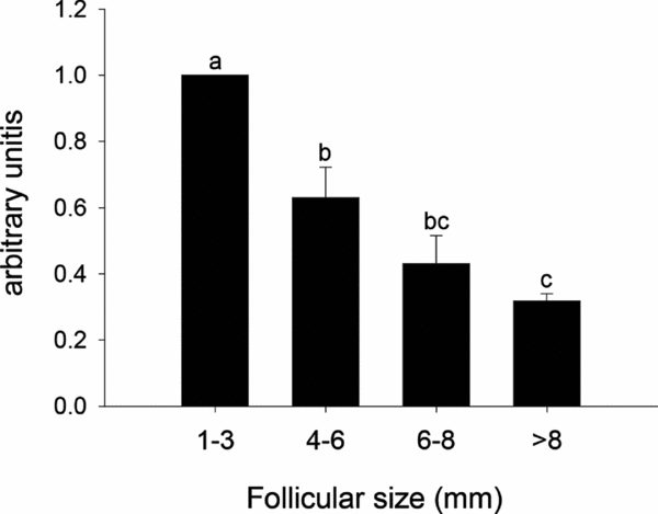
We assessed Grb14 mRNA expression in COCs from different follicle sizes relative to oocyte developmental competence acquisition (1–3, 4–6, 6–8 or >8 mm in diameter). Grb14 mRNA expression was higher in COCs from follicles of 1–3 mm in diameter and decreased significantly and gradually as the follicle size increased. The expression was significantly lower in COCs from the largest (>6 mm in diameter) than the smallest follicles (P < 0.05; Fig. 1).
Relative mRNA expression (mean ± standard error of the mean (SEM)) of Grb14 in cumulus-oocyte complexes (COCs) from different follicular sizes (1–3, 4–6, 6–8 or >8 mm). mRNA expression values were measured relative to the housekeeping gene GAPDH. In total, 480 COCs were evaluated (about 120 COCs/treatment). The experiment was conducted with three replicates. Different letters (a, b and c) indicate statistically significant differences between follicle sizes (P < 0.05).

The estradiol effect on Grb14 mRNA expression
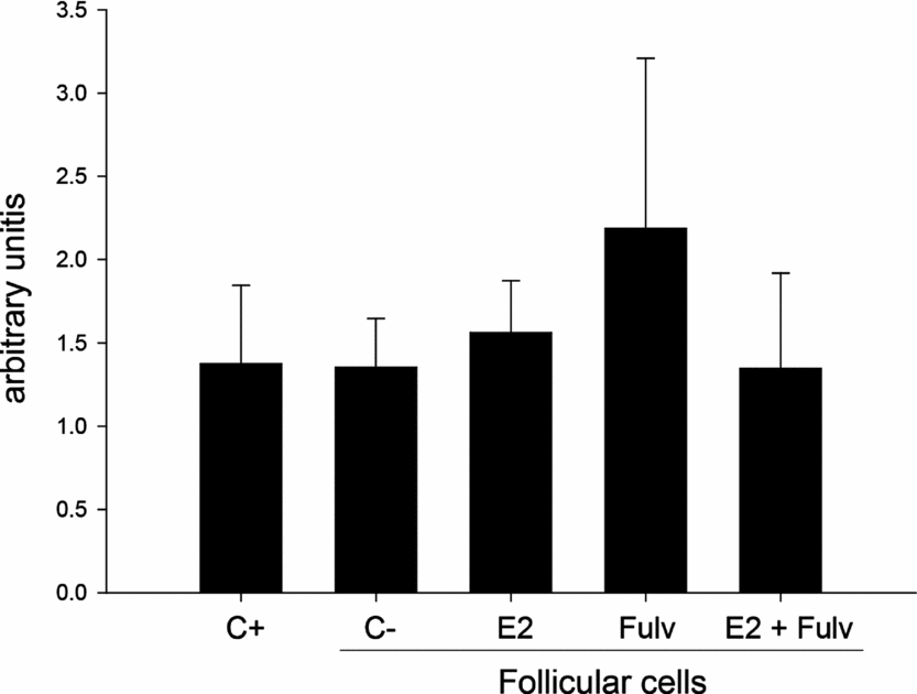
We investigated the role of estradiol in Grb14 mRNA expression in cattle COCs. Follicular cells and COCs were co-cultured in the presence or absence of E2 (500 ng/ml) and/or fulvestrant (100 μM; an estradiol receptor antagonist) for 6 h. The level of Grb14 mRNA expression in the treatment groups (E2, Fulv and/or E2 + Fulv) did not differ from that of the control groups cultured in the absence (positive control, C+) or presence (negative control, C) of follicular cells (P > 0.05; Fig. 2).
Effect of estradiol and fulvestrant on Grb14 mRNA expression in cumulus–oocyte complexes (COCs). COCs were cultured in the absence (positive control group, C+) or presence (negative control group, C–) of follicular cells for 6 h. The COCs in treatment groups were cultured in the presence of follicular cells with 17β-estradiol (E2; 500 ng/ml), fulvestrant (Fulv; 100 μM) or both (E2 + Fulv). In total, 600 COCs (from 3–8 mm follicles) were allocated into five groups. The experiment was conducted with three replicates. There was no statistical difference between groups (P > 0.05).
Discussion
In this study, we observed that the Grb14 mRNA is expressed in bovine COCs. To our knowledge, this is the first report to date of Grb14 mRNA expression in mammalian COCs. Moreover, we showed here that Grb14 is downregulated in COCs as follicle size increases and that the expression is not regulated by estradiol. Our results provide evidence of a new candidate gene involved in bovine oocyte maturation, probably one that has a role in controlling receptors with tyrosine kinase activity.
COCs were co-cultured with follicular hemisections to delay resumption of meiosis and to continue mRNA transcription (Richard & Sirard, Reference Richard and Sirard1996). This culture system has been used previously to study oocyte nuclear maturation (Giometti et al., Reference Giometti, Bertagnolli, Ornes, da Costa, Carambula, Reis, de Oliveira, Emanuelli and Goncalves2005; Stefanello et al., Reference Stefanello, Barreta, Porciuncula, Arruda, Oliveira, Oliveira and Goncalves2006; Barreta et al., Reference Barreta, Oliveira, Ferreira, Antoniazzi, Gasperin, Sandri and Goncalves2008). Under these condition, the effects of estradiol and fulvestrant (an estradiol antagonist) were evaluated based on Grb14 mRNA expression in COCs.
Cattle oocytes from follicles larger than 2 mm in diameter are able to undergo meiotic resumption (Fair et al., Reference Fair, Hyttel and Greve1995). With the validation of our experimental model, we demonstrated that oocyte competence for meiotic progression increased as follicle size developed, a finding that has been described previously (Lonergan et al., Reference Lonergan, Monaghan, Rizos, Boland and Gordon1994; Arlotto et al., Reference Arlotto, Schwartz, First and Leibfried-Rutledge1996). The findings for Grb14 mRNA expression observed in our study (Fig. 1) suggested that, in cattle, oocyte competence for meiosis resumption and metaphase progression is negatively related to Grb14 mRNA expression.
In Xenopus oocytes, increase in Grb14 mRNA expression inhibited the resumption of meiosis and prevented the activation of MAPK and PI3K intracellular signalling pathways. These signals are essential to promote oocyte GVBD by interaction with the phosphorylated tyrosine kinase receptor (Cailliau et al., Reference Cailliau, Le Marcis, Béréziat, Perdereau, Cariou, Vilain, Burnol and Browaeys-Poly2003). Taken together, our results and those reported previously both suggest that downregulation in Grb14 mRNA expression is an important intracellular mechanism to promote the activation of intracellular signalling pathways for acquisition of oocyte developmental competence to undergo GVBD and achieve M-II. Furthermore, we can infer that Grb14 is involved in molecular maturation because COCs derived from large follicles (>8 mm) had consistently less Grb14 mRNA than small follicles (<6 mm).
The increased level of estradiol in follicular fluid is a key characteristic for dominance during antral follicle development in cattle (Fortune et al., Reference Fortune, Rivera and Yang2004). Moreover, estradiol plays a role in cumulus cell proliferation and expansion (Sugiura et al., Reference Sugiura, Su, Li, Wigglesworth, Matzuk and Eppig2010). However, we did not observe any estradiol effect on Grb14 mRNA expression in COCs during follicle development. In contrast, studies using human breast cancer cells have shown that Grb14 protein expression is downregulated by estradiol and upregulated by the anti-estrogen ICI 182780 (Kairouz et al., Reference Kairouz, Parmar, Lyons, Swarbrick, Musgrove and Daly2005). These results led us to infer two possibilities. Firstly, estradiol might regulate Grb14 in COCs through a factor not present in our in vitro system. Secondly, Grb14 might be regulated by another steroid. Recently, an study demonstrated that dihydrotestosterone downregulated Grb10 (an adaptor protein structurally related to Grb14 and belonging to the Grb7 superfamily) mRNA expression in mouse skeletal muscle (Svensson et al., Reference Svensson, Movérare-Skrtic, Windahl, Swanson and Sjögren2010).
Growth factor signalling by receptor tyrosine kinase induced an increase in oocyte competence and subsequent embryo development (Purohit et al., Reference Purohit, Brady and Sharma2005; Zhang et al., Reference Zhang, Hansen and Ealy2010). As Grb14 inhibits factors with tyrosine kinase activity via MAPK and PI3K (Goenaga et al., Reference Goenaga, Hampe, Carre, Cailliau, Browaeys-Poly, Perdereau, Holt, Daly, Girard, Broutin, Issad and Burnol2009), the present results provide evidence that Grb14 mRNA expression has to decrease in COCs for the oocyte to acquire developmental competence.
In conclusion, Grb14 mRNA expression in COCs was downregulated during follicle development, suggesting a role for this receptor-bound protein in oocyte competence acquisition in cattle. Moreover, we demonstrated that Grb14 mRNA downregulation is not dependent upon the action of estradiol in cattle COCs.
Acknowledgements
We thank the Silva abattoir for providing bovine ovaries. We also thank CNPq and CAPES for their financial support.