Dietary fatty acids (FA) influence synthesis of molecules and cytokines responsible for regulation of major components of the immune system. Among long chain fatty acids, ω-3 and ω-6 polyunsaturated fatty acids (PUFA) play a major role in modulation of inflammatory responses (Miles & Calder, Reference Miles and Calder1998). Omega-6 PUFA such as linoleic acid (18:2 ω-6) results in production of arachidonic acid (ARA; 20:4 ω-6) whereas ω-3 PUFA such as linolenic acid (αLA: 18:3 ω-3) results in eicosapentaenoic acid (EPA: 20:5 ω-3) formation. Eicosanoids can be synthesised from both precursors; however, eicosanoids synthesised from EPA have a lower biological activity and thus are less pro-inflammatory (e.g. prostaglandin E3 or leukotriene B5) than those synthesised from ARA, such as prostaglandin E2 and leukotriene B4 (Yaqoob & Calder, Reference Yaqoob and Calder1995; Wanten & Calder, Reference Wanten and Calder2007). Feeding high dietary sources of ω-3 PUFA has been shown to affect aspects of immune function including lowered lymphocyte response to mitogens and natural killer cell ability (Wanten & Calder, Reference Wanten and Calder2007). Furthermore, feeding dietary sources of ω-3 PUFA, as compared with ω-6 PUFA, reduced peripheral blood mononuclear cell (PBMC) production of pro-inflammatory markers interleukin (IL)-1β, IL-6, and tumour necrosis factor (TNF)-α in some animal models (Meydani, Reference Meydani1996). Evidence also suggests that ω-3 PUFA affect inflammatory responses in an eicosanoid-independent manner, namely through altered gene expression of inflammatory mediators (Calder, Reference Calder2002, Reference Calder2006). Specific effects on different aspects of immune system and inflammatory markers, however, have been dose-dependent in majority of cases but inconsistent, overall (Field et al. Reference Field, Johnson and Schley2002).
Feeding whole flaxseed, an ω-3 enriched feed ingredient, to dairy cows has been associated with some alterations in eicosanoid production, reproductive performance, and aspects of immune functions (Petit et al. Reference Petit, Dewhurst, Scollan, Proulx, Khalid, Haresign, Twagiramungu and Mann2002; Lessard et al. Reference Lessard, Gagnon and Petit2003). Anti-inflammatory effect of αLA-enriched flaxseed on prostaglandins production in dairy cows has been investigated (Petit et al. Reference Petit, Dewhurst, Scollan, Proulx, Khalid, Haresign, Twagiramungu and Mann2002). Altered cell membrane fluidity and structure, signal transduction pathways (e.g. protein kinases), and altered inflammatory gene expression are among eicosanoid-independent mechanisms of action (Calder, Reference Calder2006).
Camelina (Camelina sativa) is a member of the mustard family and another dietary source of ω-3 as it has a similar FA profile to that of flaxseed (αLA-rich; Hurtaud & Peyraud, Reference Hurtaud and Peyraud2007). Feeding camelina to dairy cows caused significant alterations in milk FA profile especially 18-carbon FA (Hurtaud & Peyraud, Reference Hurtaud and Peyraud2007); however, to our knowledge, no study has been conducted to examine the potential effects of αLA-enriched camelina meal on markers of inflammation in dairy cows. We hypothesised that expression of pro-inflammatory markers would be reduced by increased dietary intake of αLA through feeding ω-3 enriched camelina meal. Therefore, the objectives of the present study were to determine the effect of camelina meal on (1) αLA content of serum phospholipids, and (2) mRNA expression of inflammatory mediators at local (milk cells; MC) and systemic (peripheral blood mononuclear cells) levels.
Materials and methods
Animals and treatments
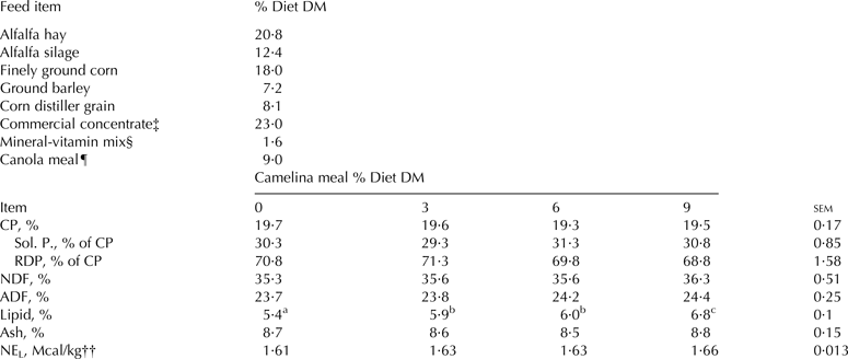
All animal procedures were approved by the University of Idaho Animal and Care Use Committee. Experimental design and rations are reported in detail elsewhere (Hatch, Reference Hatch2013). Briefly, 18 primiparous Holstein lactating cows (60 ± 15 d in milk; DIM) were randomly assigned to a treatment sequence in a 4 × 4 Latin square design. Periods lasted 16 d after which cows were switched to the next round of treatment without any washout interval. Rations were formulated to provide incremental dietary concentration of αLA, via inclusion of camelina meal (Omega Meal, Great Northern Growers Chester, MT, USA) for canola meal at 0 (control), 3, 6, or 9% of ration dry matter (DM). Camelina meal contained (on a DM basis) 35·6% CP, 18·8% ether extract, 24·1% ADF, 40·5% NDF, and 5·8% ash. Major fatty acids included 41·3% αLA, 28·5% linoleic acid, 19·9% oleic acid, 2·3% stearic acid, and 7·1% palmitic acid. Cows were fed individually using Calan gates (American Calan Inc., Northwood, NH, USA). Dietary items and nutrient composition of rations are listed in Table 1. On the last 2 d of each period (d 15 and d 16), blood samples were obtained via jugular vein. On the same days composite milk samples, proportional to morning and evening yields, were also obtained. These time points were used based on our previous experimental observations and that of others, which allowed sufficient time for adaption to new rations in Latin square designs (Griinari et al. Reference Griinari, Dwyer, McGuire, Bauman, Palmquist and Nurmela1998; Eigersma et al. Reference Eigersma, Ellen, van der Horst, Boer, Dekker and Tamminga2004; Roy et al. Reference Roy, Ferlay, Shingfield and Chilliard2006; Mosley et al. Reference Mosley, Mosley, Hatch, Szasz, Corato, Zacharias, Howes and McGuire2007).
Nutrient composition of treatment diets on a dry matter basis to varying dietary inclusion of camelina meal in place of canola meal†

† Values represent the mean of 4 samples composited by treatment within each period
‡ Containing 13·7% soybean meal, 9·4% molasses, 33·5% beet pulp, 42·1% wheat middlings, and 1·3% Di-calcium phosphate
§ Contained 11·6% Ca, 0·18% P, 5·12% Mg, and 131·4 IU vitamin A, 20·2 IU vitamin D, and 832·3 IU vitamin E /kg DM
¶ Canola meal was replaced with incremental amount of camelina meal (0, 3, 6, and 9%)
†† Calculated based on NRC 2001
Sample processing and analysis
Milk samples were processed to obtain MC, as previously reported (Feng et al. Reference Feng, Salter, Parr and Garnsworthy2007) with modifications. Briefly, milk samples (~1 l) were processed in a sequential centrifugation as follows: to 1 l milk, 1 ml of 0·5 mm EDTA was added and sample was centrifuged at 2700 g for 15 min at 4 °C. Supernatant was removed and cell pellet was washed twice with ice-cold phosphate buffer solution (PBS; pH 7·2) containing 0·5 mm EDTA. The cell pellet was then re-suspended in 4 ml PBS and centrifuged at 2700 g for 15 min at 4 °C in a 15-ml conical tube. Supernatant was removed and the cell pellet was re-suspended in 200 µl 1X PBS, transferred into a 1·5 ml centrifuge tube and centrifuged at 2700 g for 15 min at 4 °C. After removing supernatant, the cell pellet was treated with 600 µl of RLT PLUS and β-mercaptoethanol (Qiagen Inc., Valencia, CA, USA) and homogenate was obtained using Qiashredder (cat. # 79654; Qiagen Inc., Valencia, CA, USA), centrifuged at 12 000 g for 2 min at room temperature and stored at −80 °C.
Peripheral blood mononuclear cells were isolated via the Accu-Paque gradient method (cat. # AN3100; density 1·086 ± 0·0002 g/ml; Accurate Chemical & Scientific Corp., Westbury, NY, USA) using routine methods (Park et al. Reference Park, Fox, Hamilton and Davis1993). The PBMC homogenate was obtained as described above for MC's homogenate. Both MC and PBMC homogenates were processed to extract total RNA following manufacturer's protocol (cat. # 74134 Qiagen Inc., Valencia, CA, USA). Concentrations of total RNA were determined using a NanoDrop 1000 (Thermo Scientific, Wilmington, DE, USA).
Serum lipids were extracted using chloroform : methanol (2 : 1) as previously described (Clark et al. Reference Clark, Ferris, Fey, Brown, Hundricser and Jensen1982). The extracted serum lipids (1 mg) were fractionated into cholesterol esters, triacylglycerol, mono- and diacylglycerols, non-esterified fatty acids, and phospholipids (PL) as previously described by Hamilton & Comai (Reference Hamilton and Comai1988). Fatty acid composition of the PL fraction was determined using gas chromatography as previously described (Mosley et al. Reference Mosley, Shafii, Moate and McGuire2006) with minor modification. Briefly, plasma PL were methylated using base-catalyzed transesterification (Christie, Reference Christie1982) with a reaction time of 1 h. The fatty acid methyl esters (FAME) were analysed on a gas chromatograph (Hewlet-Packard 6890 Series with auto injector) fitted with a flame ionization detector and a 100 m × 0·25 mm, with 0·2 µm film capillary column coated with CP-Sil 88 (Varian). Initially, the oven temperature was 70 °C (for 4 min); it was then increased to 175 °C at a rate of 4 °C/min and held for 3 min. The oven temperature was then increased to 185 °C at a rate of 1 °C/min and held for 20 min, increased to 220 °C at a rate of 3 °C/min, and then increased to 230 °C at a rate of 10 °C/min and held for 10 min.
Quantitative real time PCR
Quantitative real-time polymerase chain reaction (rt-PCR) was carried out in a 7500 Fast Real-Time PCR system (Applied Biosystems, Foster City, CA, USA) using custom designed TaqMan MGB probes as described previously (Watts et al. Reference Watts, Rezamand, Sevier, Price and McGuire2013) except for glyceraldehyde 3-phosphate dehydrogenase (GAPDH). Briefly, the RNA isolated was processed using the RNeasy Mini Plus Kit (Qiagen) according to the manufacturer's instructions. The concentration of RNA was determined using a NanoDrop ND-1000 spectrophotometer (NanoDrop Technologies, Rockland DE, USA). Total RNA was converted into complementary DNA using Applied Biosystems High Capacity Reverse Transcriptase Kit (Applied Biosystems, Foster City CA 94404, USA). Single-stranded cDNA was synthesised by reverse transcription using a PCR Sprint Thermal Cycler (Thermo Electron Corp., Milford MA 02139, USA). The single-stranded cDNA was then used as the template for quantitative PCR to evaluate relative expression of target genes. The forward and reverse primer sequences for the analysis of GAPDH were CTGCCACCCAGAAGACTGT and GCCAGTAGAAGCAGGGATGATATTC, respectively. Analysis for genes of interest was performed in triplicate with bovine ribosomal protein-9 (RPS9) and GAPDH used as housekeeping (HK) genes. The thermal cycling conditions for regular PCR were as follows: stage one enzyme activation 50 °C for 2 min, stage two denature 95 °C for 10 min, stage three anneal 95 °C for 15 s, with 40 replications through stage 3. Steps 1 and 2 were done only at the beginning. The abundance of target genes normalised to HK genes (geometrical mean of RPS9 and GAPDH for MC; GAPDH for PBMC) and relative to a calibrator, is illustrated by fold change method (2−ΔΔCt). Average abundance of target genes for cows fed control ration (0% camelina meal) was considered as the calibrator.
Statistical analyses
All statistical analyses were conducted using SAS, version 9.2 for Windows (SAS Institute Inc., Cary, NC, USA). The effect of dietary treatments (αLA) on relative expression of mRNA in the PBMC and MC was tested by the MIXED procedure of SAS using original delta Ct values (ΔCt = Cttarget gene − Cthouse-keeping gene). The effect of dietary treatments (αLA) on the serum content of FA was also tested by the MIXED procedure of SAS. Cow was considered as a random factor. Protected least significance difference was used to compare least squares means (LSM) and data are reported as LSM ± standard error of means (sem). Linear and quadratic contrasts were also tested. For mRNA data, LSM ± sem are illustrated in fold change (2−∆∆Ct method) relative to control (0% camelina meal). Significant differences were declared at P ⩽ 0·05.
Results
The current study was designed to evaluate immuno-modulatory effects of camelina meal fed at incremental rates (0 to 9% DM basis) in place of canola meal to early-lactating (60 ± 15 DIM) primiparous Holstein cows in a Latin square design. All animals completed the study and no cases of clinical mastitis or other infectious diseases were observed throughout the study. The camelina meal fed contained 24·2% αLA (DM basis), which is drastically lower than the 41·3% αLA content (DM basis) reported by Hurtaud & Peyraud (Reference Hurtaud and Peyraud2007).
An estimate of intake (DMI × dietary fat per cent × dietary FA as per cent of FAME) in current study shows that as inclusion rate of dietary camelina meal increased the intake of αLA increased (46·4, 72·4, 90·0, and 113·2 g/d for 0, 3, 6, and 9% camelina meal diets, respectively) whereas that of linoleic acid did not change drastically (371·3, 374·4, 389·4, and 385·8 g/d for 0, 3, 6, and 9% camelina meal diets, respectively).
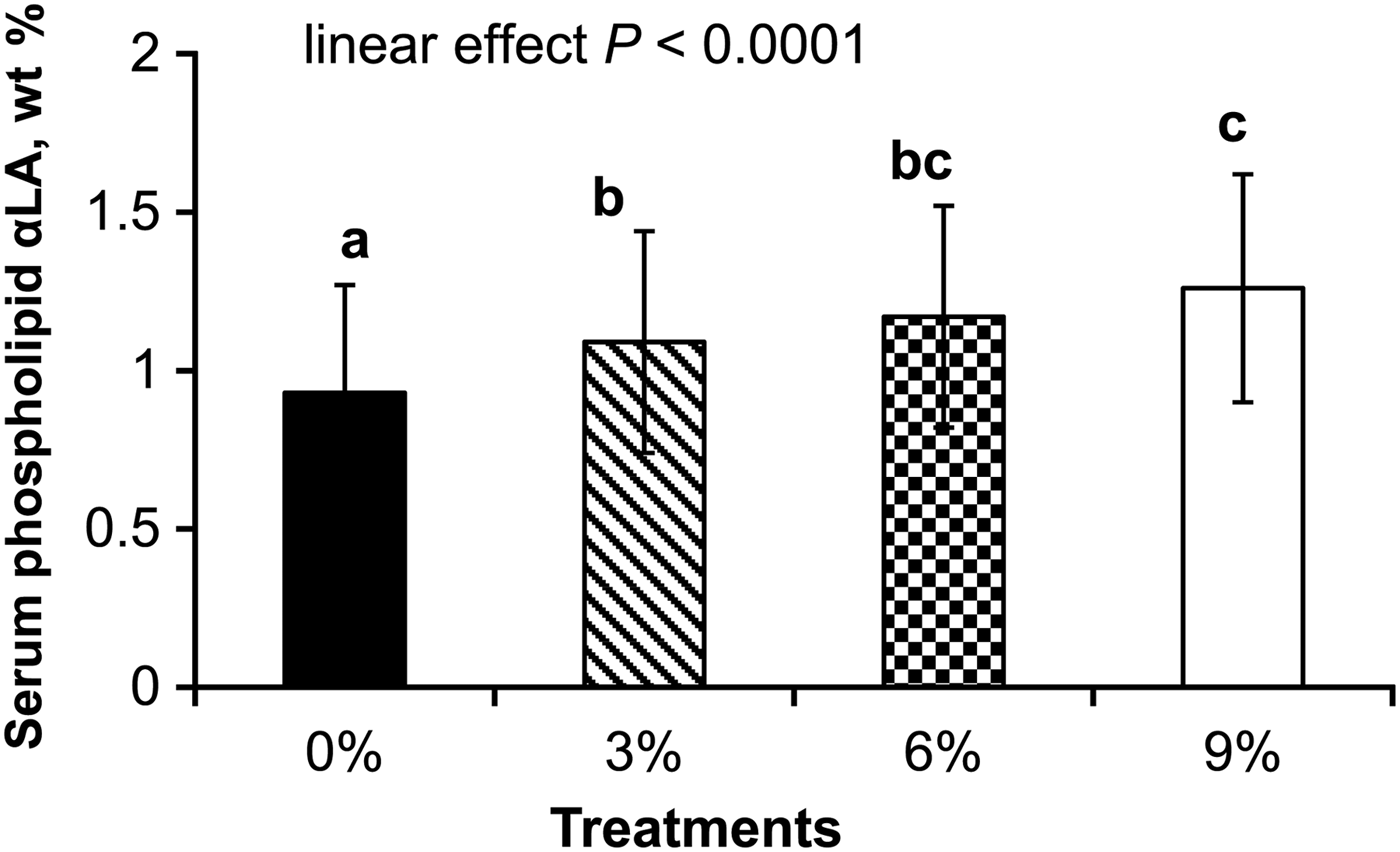
Feeding camelina meal caused a significant difference in αLA content of serum PL (Table 2); as the feeding rate increased the αLA content of serum PL was increased in a linear fashion. Furthermore, whereas no significant effect was detected for palmitic and oleic acids, both stearic and linoleic acids were decreased as the feeding rate of camelina increased (Table 2).
Serum phospholipids content of α-Linolenic acid and other important fatty acids for cows receiving diets fed varying dietary inclusion of camelina meal in place of canola meal
† Linear effect (P < 0·001)
‡ Linear effect (P = 0·002)
§ Linear effect (P < 0·001)
Gene expression of pro-inflammatory cytokines
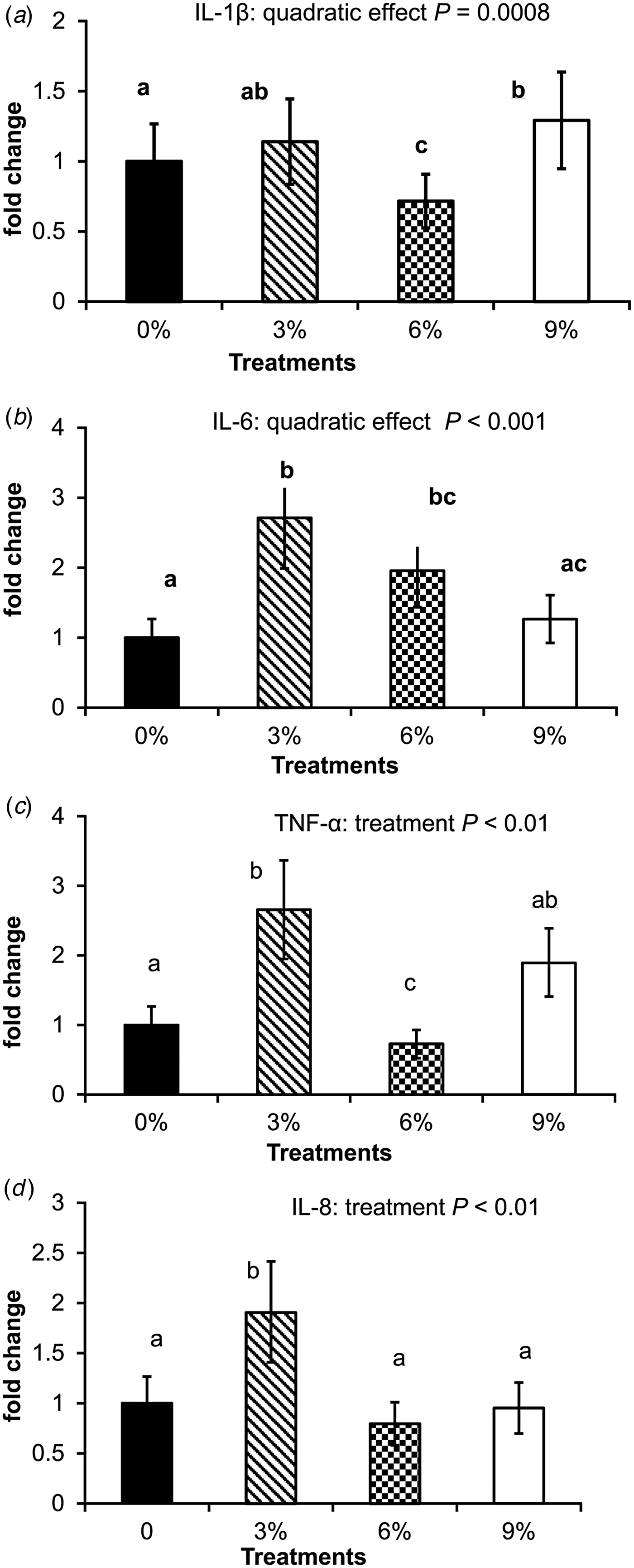
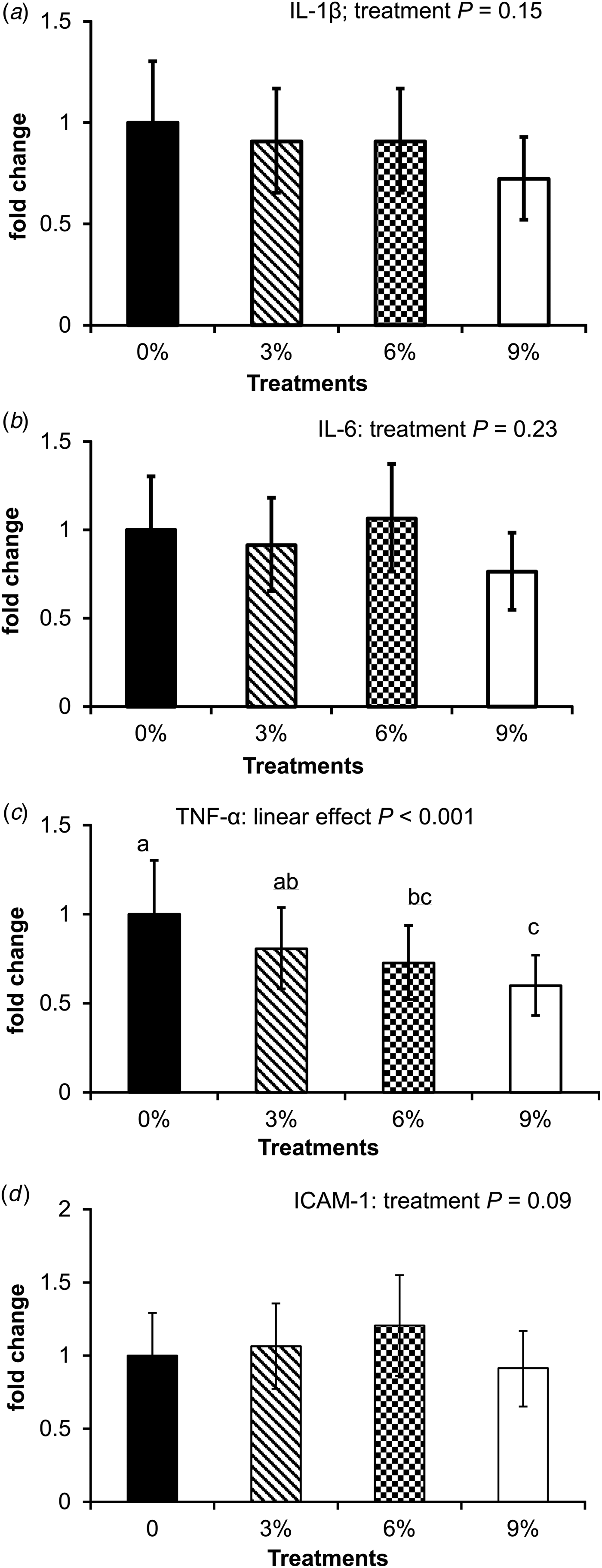
Feeding camelina meal at 6% DM (1·45% αLA from camelina meal) caused a down-regulation (P = 0·0008) of mRNA expression of IL-1β in PBMC when compared with that of control (Fig. 2a). Expression of IL-1β was however, up-regulated in PBMC from cows fed 9% camelina meal. Feeding camelina meal caused no detectable difference in mRNA expression of IL-1β in MC (Fig. 3a). No difference in mRNA expression of ICAM-1 of MC was detected (Fig. 3d). Relative expression of IL-1β was reduced by 6% camelina meal but increased when camelina meal was fed at 9% in PBMC.
α-Linolenic acid (αLA) content of serum phospholipids (wt%) for lactating primiparous Holstein cows fed αLA-enriched rations at 0, 3, 6 or 9% camelina meal (DM basis). Feeding camelina meal caused a linear increase in αLA content of serum phospholipids content (P < 0·0001). Columns with common superscripts do not differ significantly at P < 0·05.
Gene expression of inflammatory markers in peripheral blood mono-nuclear cells isolated from lactating primiparous Holstein cows fed α-linolenic acid (αLA)-enriched rations at 0, 3, 6, or 9% camelina meal (dry matter basis). (a) mRNA expression of interleukin (IL)-1β: a quadratic effect (P = 0·0008) was observed. (b) mRNA expression of IL-6: a quadratic effect (P < 0·001) was observed. (c) mRNA expression of tumour necrosis factor (TNF)-α: a significant treatment effect (P < 0·001) was observed. (d) mRNA expression of IL-8: a significant treatment effect (P < 0·001) was observed. Columns with common superscripts do not differ significantly at P < 0·05.
Gene expression of inflammatory markers in milk cells isolated from lactating primiparous Holstein cows fed α-linolenic acid (αLA)-enriched rations at 0, 3, 6, or 9% camelina meal (dry matter basis). (a) mRNA expression of interleukin (IL)-1β: no significant change by dietary treatments was detected (P = 0·15). (b): mRNA expression of IL-6: no significant change by dietary treatments was detected (P = 0·23).
(c) mRNA expression of tumour necrosis factor (TNF)-α: a significant linear down-regulation by dietary treatments (P = 0·0008) was observed. (d) mRNA expression of intra-cellular adhesion molecule (ICAM)-1: no significant change by dietary treatments was detected (P = 0·23). Columns with common superscripts do not differ significantly at P < 0·05.
Feeding camelina meal was associated with a quadratic response of IL-6 mRNA relative expression in isolated PBMC (Fig. 2b), with greatest increase from cows fed 3% camelina meal that gradually returned to the level of the control as feeding rate of camelina meal increased to 9%. No difference, however, was detected for IL-6 mRNA relative expression in isolated MC (Fig. 3b).
Relative expression of TNF-α mRNA in PBMC isolated from cows fed 6% camelina meal was lowest among treatments whereas that for cows fed 3% camelina meal showed greater expression compared with control (Fig. 2c). Expression of TNF-α mRNA in MC was down-regulated linearly as feeding rate of camelina meal increased (Fig. 3c).
Feeding camelina meal was associated with an increase in IL-8 mRNA relative expression in isolated PBMC (Fig. 2d) from cows fed 3% camelina meal relative to that from control. As feeding rate of camelina meal increased up to 9%, no difference in IL-8 mRNA expression was detected relative to the 0% camelina meal (control) treatment.
Discussion
Camelina fed in the present study had lower αLA content relative to the camelina meal reported by Hurtaud & Peyraud (Reference Hurtaud and Peyraud2007). This may be in part related to an inefficient oil extraction method used for the meal in the current study.
The difference observed in FA profiles between the current study and that of Hurtaud & Peyraud (Reference Hurtaud and Peyraud2007) could be attributed to the oil extraction processes or variety of camelina that may have differed between the two experiments, which results in different total lipid contents as well as FA profiles observed. As shown in Fig. 1, concentration of αLA in serum PL (wt%) increased linearly as αLA-enriched rations at 0, 3, 6 or 9% camelina meal was fed. Feeding other dietary sources of ω-3 enriched feeds such as flaxseed has been shown to cause significant increases in plasma and milk αLA contents (Petit et al. Reference Petit, Dewhurst, Scollan, Proulx, Khalid, Haresign, Twagiramungu and Mann2002; Lessard et al. Reference Lessard, Gagnon and Petit2003). Even though plasma PL content of αLA was not measured when cows were fed camelina meal or camelina seed, milk αLA was increased because of feeding camelina in a corn silage-based ration (Hurtaud & Peyraud, Reference Hurtaud and Peyraud2007). Although the FA profile of isolated PBMC was not measured in the current study, due to small amount of samples, an assumption may be made that alterations in serum PL FA profile would possibly affect FA profile of immune cell membranes. It has been previously speculated that changes in dietary FA may cause alterations in circulating FA profile and similarly in those of the immune cells in circulation (Skeaff et al. Reference Skeaff, Hodson and McKenzie2006; Lacetera et al. Reference Lacetera, Scalia, Mashek, Bernabucci and Grummer2007). In support of this view, we recently reported changes in serum phospholipids FA profile of early lactating Holstein cows because of various types of dietary fat fed for a short amount of time (Watts et al. Reference Watts, Rezamand, Sevier, Price and McGuire2013).
Diets rich in ω-3 may down-regulate pro-inflammatory markers possibly through inhibition of transcription factors such as nuclear transcription factor (NF) κB (Calder, Reference Calder2006; Field et al. Reference Field, Johnson and Schley2002) in a dose-dependent manner. For instance, a moderate amount of dietary ω-3 PUFA (<10% of fat fed) was not associated with reduced pro-inflammatory markers such as IL-1, IL-6, and TNF-α whereas a greater rate of dietary ω-3 PUFA (>10% of total fat) would have an immunosuppressive effect (see review by Field et al. Reference Field, Johnson and Schley2002). The mechanisms responsible involve alterations of inflammatory cell membrane PL fatty acid profile with greater incorporation of ω-3 PUFA at the expense of ARA (Calder, Reference Calder2006) as was observed in the present study (Fig. 1). Furthermore, inflammatory responses may be suppressed by PUFA such as αLA and other long chain ω-3 PUFA that activate peroxisome proliferator-activated receptor-γ (Lee et al. Reference Lee, Plakidas, Lee, Heikkinen, Chanmugam, Bray and Hwang2003).
Feeding camelina affected mRNA expression of various genes related to inflammation in several ways. The up-regulation of mRNA expression of IL-1β in PBMC isolated from cows fed 9% camelina meal (2·2% αLA from camelina meal) may have been related to over-production of intermediate trans FA in the rumen, given that milk fat percentage (3·51, 3·39, 3·32, and 3·01% for 0, 3, 6, 9% camelina diets, respectively) and fat yield (0·98, 0·96, 0·91, and 0·82 kg/d for 0, 3, 6, 9% camelina diets, respectively) was significantly reduced for cows fed 9% camelina meal. Milk content of C18:1 trans FA (both trans 10 and trans 11) as well as C18:2 trans 9, trans 12 were, however, increased as the inclusion rate of camelina meal increased (Hatch, Reference Hatch2013). This in turn could be associated with greater expression of pro-inflammatory IL-1β in circulation. Findings on mammary gland inflammation suggest that IL-1β is produced in greater abundance by monocytes than by macrophages (Alluwaimi, Reference Alluwaimi2004). Further, an elevated level of IL-1β is correlated with an increased influx of neutrophils, indicating a possibility of IL-1β serving as a chemoattractant in natural or experimentally-induced mammary inflammation (Alluwaimi, Reference Alluwaimi2004). Intra-mammary administration of exogenous IL-1 is also associated with drastic elevation of milk somatic cells, mostly neutrophils (Nickerson et al. Reference Nickerson, Owens, Rejman and Oliver1993), further supporting the notion that IL-1β may indirectly function as a chemoattractant.
The quadratic response of IL-6 mRNA relative expression in isolated PBMC shown in cows fed camelina below 9% could have implications on pro-inflammatory response in dairy cattle. Interleukin-6 is one of the important pro-inflammatory cytokines involved in regulating an appropriate immune response possibly via balancing neutrophils and monocytes influxes (Alluwaimi, Reference Alluwaimi2004). Further, elevated IL-6 has been detected in both endotoxin (LPS)-stimulated mammary epithelial cells as well as in milk obtained from cows with coliform mastitis (Alluwaimi, Reference Alluwaimi2004). Studies in humans have reported a reduction of IL-6 abundance in PBMC isolated from subjects who were fed diets enriched in ω-3 PUFA (Meydani, Reference Meydani1996). Increased systemic IL-6 mRNA expression (PBMC) in the present study is in disagreement with previous cell culture and human studies, but may be explained, in part, by the observation that IL-1β was reduced in PBMC isolated from cows fed 6% camelina meal. It has been postulated that IL-1 and IL-6 may play different roles in inflammation and that elevated IL-6 would be associated with reduced neutrophil influx (Kaplanski et al. Reference Kaplanski, Marin, Montero-Julian, Mantovani and Farnarier2003; Alluwaimi, Reference Alluwaimi2004), an event supported by reduced IL-1β expression in the present study.
Tumour necrosis factor-α is an important mediator of the inflammatory response that regulates recruitment and activation of neutrophils, and induces hepatic acute phase proteins such as haptoglobin (Alluwaimi, Reference Alluwaimi2004). Studies have demonstrated that feeding ω-3 PUFA decreased soluble TNF-α production in rodent macrophages as well as in equine and rats blood monocytes (Field et al. Reference Field, Johnson and Schley2002), similar to the present study in cows fed the 3% camelina meal diet. Further, studies have demonstrated that human subjects fed ω-3 PUFA enriched diets (either 1·2 or 2·4 g EPA + DHA/d) had decreased TNF production as well as IL-1 and IL-6 by mononuclear cells (see review by Field et al. Reference Field, Johnson and Schley2002). Stronger anti-inflammatory response observed in MC as compared with PBMC in the present study may be related to site specificity indicating the mammary glands may have responded differently to elevated incoming ω-3 PUFA. This observation may also be related in part to the frequency with which various samples (milk vs. bold) were obtained.
Interleukin-8 is produced upon stimulation by bacterial components in different cell types such as monocytes and macrophages, and is known as a strong neutrophil chemoattractant (Alluwaimi, Reference Alluwaimi2004). Interleukin-8 has been detected in mid- and late lactation with no change over time in mammary glands (Alluwaimi et al. Reference Alluwaimi, Farver and Cullor2003). Several stimuli such as endotoxin or Escherichia coli have been shown to cause increased mRNA expression and protein abundance of IL-8 in a mammary epithelial cell line (Alluwaimi, Reference Alluwaimi2004). Studies in humans have demonstrated reductions of circulating IL-8 by inclusion of dietary ω-3 PUFA. For instance, oesophageal cancer patients who received ω-3 PUFA at 1·5 mg/ml had a reduced plasma IL-8 as compared with that of patients who received enteral solutions containing lower (0·46 mg/ml) ω-3 PUFA (Aiko et al. Reference Aiko, Yoshizumi, Tsuwano, Shimanouchi, Sugiura and Maehara2005). In a follow-up study, when amounts of ω-3 PUFA were increased to 4·0 vs. 1·5 mg/ml, a similar observation on reduced plasma IL-8 was reported (Aiko et al. Reference Aiko, Yoshizumi, Ishizuka, Horio, Sakano, Kumano, Kanai and Maehara2008). No conclusive evidence currently exists, however, to provide an explanation for the elevated IL-8 mRNA expression in PBMC isolated from cows fed moderate amount of an αLA-enriched feed in the present study. It should be noted that no clinical or sub-clinical cases of mammary inflammation were detected for cows included in this study.
A main function of intra cellular adhesion molecule (ICAM)-1, a transmembrane protein expressed by lymphocytes and macrophages as well as endothelial cells, is to bind with integrins and facilitates transmigration of leukocytes (Yang et al. Reference Yang, Froio, Sciuto, Dvorak, Alon and Luscinskas2005). Elevated ICAM-1 increases the adhesiveness of circulating immune cells to endothelia and thus promotes an elevated state of extravasation (Corl et al. Reference Corl, Gandy and Sordillo2008). Recently, it was shown that exposure to LPS (E. coli O111:B4) caused up-regulation of ICAM-1 in bovine primary mammary endothelial cells as early as 1 h post-challenge that peaked at 4 h, after which expression of ICAM-1 started to decline (Corl et al. Reference Corl, Gandy and Sordillo2008). As stated earlier, no cases of mastitis were observed in this study. Fatty acids have been shown to affect ICAM-1 as increased trans FA intake was positively related to an elevated plasma concentration of soluble ICAM-1 in healthy human subjects (Lopez-Garcia et al. Reference Lopez-Garcia, Schulze, Meigs, Manson, Rifai, Stampfer, Willett and Hu2005). Elevated IL-1β can affect expression of adhesion molecules such as ICAM-1 (von der Thüsen et al. Reference von der Thüsen, Kuiper, van Berkel and Biessen2003). Because there was no detectable alteration in MC with regards to expression of IL-1β, this suggests that the immunosuppressive effect of αLA-enriched camelina meal may not have been strong enough at the local (MC) level to drastically modulate ICAM-1 expression without presence of an exogenous stimulus.
In the current study, it was observed that feeding αLA-enriched camelina meal, similar to other αLA-enriched feeds, caused an increase in αLA content of serum PL in a linear fashion. The present study also documents, for the first time, both pro- and anti-inflammatory effects of αLA-enriched camelina meal on some of the local (MC) and systemic (PBMC) inflammatory markers at transcription level. The lack of anti-inflammatory effects by αLA-enriched on few of the markers measured in this study may be, in part, related to the notion that in general long chain PUFA are more sensitive to lipid peroxidation than are saturated or mono-unsaturated FA. This could be associated with increased requirements for anti-oxidants in immune cells, to which these PUFA are incorporated whereas dietary intake of anti-oxidants may not be sufficiently increased (Field et al. Reference Field, Johnson and Schley2002). In addition, expression of inflammatory markers in MC may be affected by the proportions of different cell types in the milk cells collected (Shields et al. Reference Shields, Rezamand, Sevier, Seo, Price and McGuire2011). Shifts from macrophages to secretory cells or other immune cell types would influence expression of inflammatory markers. However, the method used is a non-invasive, inexpensive means to study the expression of genes of interest originating from mammary gland. Further research is warranted to elucidate mechanisms involved in potential alterations to functional capacities of the bovine innate immunity when cattle are fed αLA-enriched feed ingredients such as camelina meal.
Conclusion
Various aspects of the immune system may be affected by dietary ω-6 and ω-3 FA. For instance, both ARA (ω-6) and EPA (ω-3) can be used as substrates for eicosanoid production; however, eicosanoids synthesised from ARA are thought to have greater biological and pro-inflammatory activities than those synthesised from EPA. Furthermore, ω-3 PUFA can independently alter gene expression of pro- as well as anti-inflammatory mediators. Therefore, feeding ω-3 enriched feeds may have anti-inflammatory effects on the bovine immune system at local and/or systemic levels. Results from the present study support the notion that feeding camelina meal improves αLA content of milk. It also lends support to the idea that inflammatory mediators in blood and mammary secretions are affected at the gene level by feeding αLA-enriched diets. Our data also show that when fed up to 6% DM, dietary camelina meal down-regulated expression of some of the key local (milk; e.g., TNF-α) and systemic (blood; e.g. IL-1β, IL-8, and TNF-α) pro-inflammatory markers. Response from IL-6 was not, however, in line with the rest of inflammatory mediators tested and warrants further investigation. Our findings and those of others about similar αLA-enriched sources indicate that feeding αLA-enriched feeds such as camelina meal may be a useful tool to improve baseline expression of some of the inflammatory mediators as well as milk αLA content for human consumption.
Funding for this project was provided by the United Dairymen of Idaho. Authors would like to thank J. Williams, K. M. Yahvah and J. Peak for technical assistance.