

Introduction
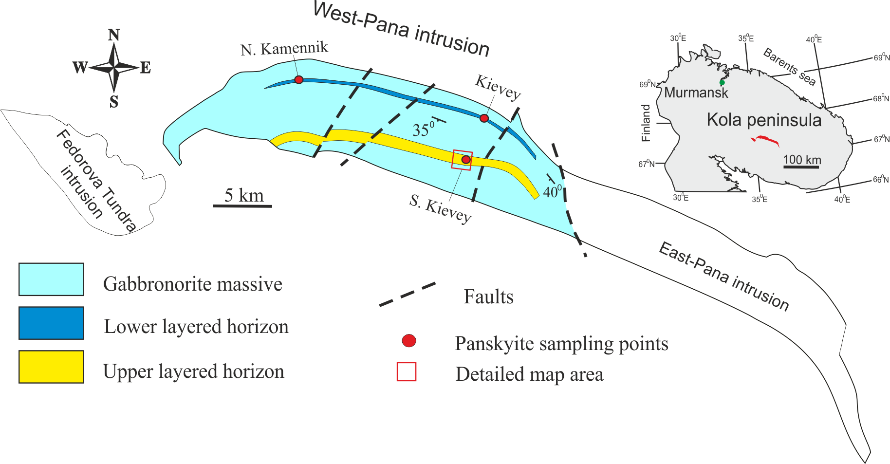
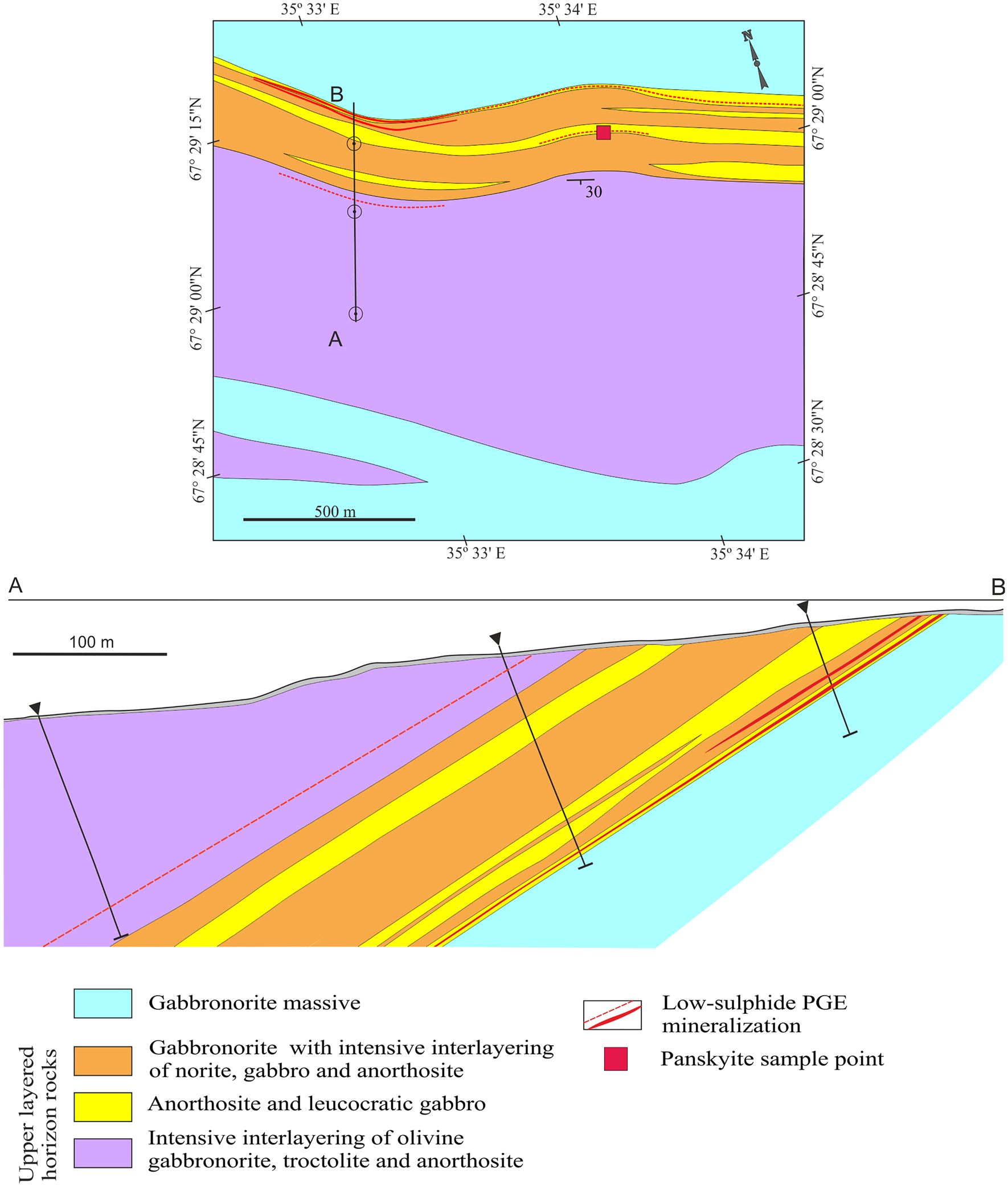
Panskyite, Pd9Ag2Pb2S4, is a new platinum-group mineral (PGM) discovered in a sample taken from an outcrop of mineralised anorthosites containing irregular disseminations of sulfides with platinum-group element (PGE) mineralisation in the area of the Southern Kievey ore occurrence Fedorova–Pana layered intrusion, Kola Peninsula, Russia (67°29′04.9″N, 35°35′02″E), Figs 1 and 2. The mineral was also found in low-sulfide ores from the deposits of Kievey and Northern Kamennik of the same intrusion (Fig. 1).
Schematic geological map of the Fedorova-Pana intrusion (adapted from Subbotin et al., Reference Subbotin, Vymazalová, Laufek, Savchenko, Stanley, Gabov and Plášil2019).

Geological map and cross-section (A–B) of the Southern Kievey area (adapted from Korchagin and Mitrofanov, Reference Korchagin and Mitrofanov2008).

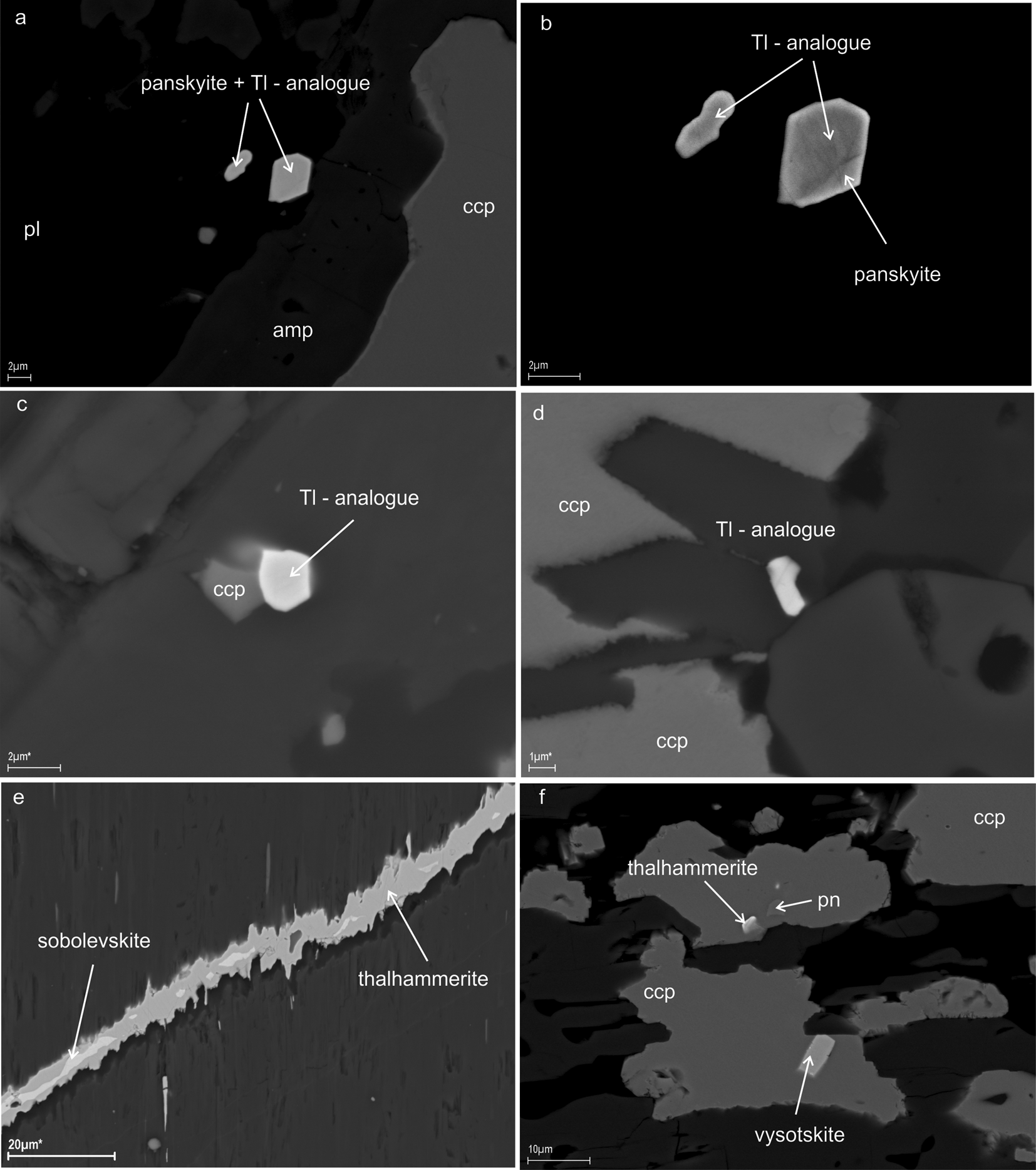
Panskyite is a Pb analogue of thalhammerite (Pd9Ag2Bi2S4). In addition a Tl analogue Pd9Ag2(Tl,Pb)2S4 was observed in samples from Southern Kievey Figs 3 and 4. The Fedorova–Pana layered intrusion is the second known occurrence for thalhammerite and the existence of the Tl analogue is reported for the first time.
Back-scattered electron images of panskyite, thalhammerite and its thallium analogue (unnamed phase Pd9Ag2(Tl,Pb)2S4) from West-Pana intrusion: (a) panskyite in an intergrowth with unnamed phase Pd9Ag2(Tl,Pb)2S4; (b) a detail; (c,d) unnamed phase Pd9Ag2(Tl,Pb)2S4; (e,f) thalhammerite. ccp – chalcopyrite, pn – pentlandite, pl – plagioclase and amp – amphibole. Samples are from: (a–d, f) Southern Kievey, (e) Northern Kamennik. For the location see Fig. 1; chemical composition is plotted on Fig. 4.

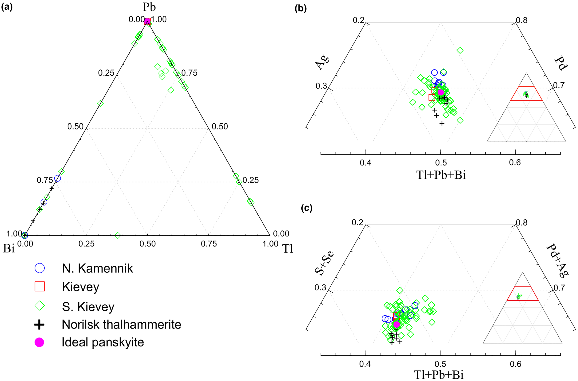
Plot of compositional data for panskyite, thalhammerite and their thallium analogue (unnamed phase Pd9Ag2(Tl,Pb)2S4) from Northern Kamennik, Kievey and Southern Kievey, compared with thalhammerite from Noril`sk (Vymazalová et al., Reference Vymazalová, Laufek, Sluzhenikin, Kozlov, Stanley, Plášil, Zaccarini, Garuti and Bakker2018). The location of the samples studied is shown in Fig. 1.

A mineral with a similar composition to panskyite was preliminarily described as an unnamed phase Pd5AgPbS2 by Subbotin et al. (Reference Subbotin, Gabov, Korchagin and Savchenko2017). No phase of similar composition is listed among the unnamed minerals of Smith and Nickel (Reference Smith and Nickel2007).
Both the mineral and its name were approved by the International Mineralogical Association Commission on New Minerals, Nomenclature and Classification (IMA-CNMNC), under the number 2020-039 (Vymazalová et al., Reference Vymazalová, Subbotin, Laufek, Savchenko, Stanley, Gabov and Plášil2020). The mineral name is for the locality, the Pansky massif (Cyrillic: Панский массив) of the Fedorova–Pana layered intrusion in the Kola Peninsula, Russia. The name Pansky intrusive massif is the most commonly used term in Russia. It has developed historically and comes from the geographical name of the mountain range – Panskie Tundra Ridge. It is now believed that the Pansky massif unites two independent intrusions: West-Pana and East-Pana (Korchagin and Mitrofanov, Reference Korchagin and Mitrofanov2008). Previously, these two intrusions were considered as tectonic blocks of one intrusion – the Pansky massif (see Fig. 2, Schissel et al., Reference Schissel, Tsvetkov, Mitrofanov and Korchagin2002).
Holotype material (polished section), along with its synthetic analogue (experiment number Exp 51), is deposited at the Department of Earth Sciences of the Natural History Museum, London, UK, catalogue number BM 2020,2.
Occurrence
The Fedorova–Pana layered intrusion has a plate-like shape of 80 km long and 0.5 to 6 km wide, with its layering dipping in a south-west direction (Fig. 1). It consists of three separate intrusions: Fedorova Tundra, West-Pana and East-Pana. The Fedorova–Pana complex belongs to a group of Paleoproterozoic layered intrusions distributed in the north-eastern part of the Fenno–Scandian Shield.
The Southern Kievey area, where the holotype specimen was found, is located in the south-eastern part of the West-Pana intrusion in the zone of the Upper Layered Horizon (Fig. 2). It consists of interlayered gabbronorites, norites, leucogabbro and anorthosites and has a thickness of up to 300 m. It contains 0.1–2% disseminated Cu–Ni sulfides and PGE-mineralisation. The main minerals are chalcopyrite, bornite, pentlandite, millerite, vysotskite, braggite, kotulskite, keithconnite, telluropalladinite, sperrylite, stillwaterite, törnroosite and Au–Ag–Pd alloys. Zones containing disseminated sulfides and PGMs form concordant lenses 0.5–2.0 m thick and up to a few hundred metres long near the top and base of the anorthosite bodies. The actual sulfide mineralisation is erratic and discontinuous along strike and dip. This particular level of mineralisation is called the Southern PGE reef (Korchagin and Mitrofanov, Reference Korchagin and Mitrofanov2008).
In other samples, panskyite was found in low-sulfide PGE ores of the Kievey and Northern Kamennik deposits. These deposits are located in the Northern PGE reef in the Lower Layered Horizon zone (Fig. 1). Disseminated sulfides form extended (0.9–6.1 km) layers of variable thickness (0.2–6.5 m) explored to a depth of 250–300 m. The ores contain 0.5–3% base-metal sulfides consisting mainly of chalcopyrite, pentlandite, pyrrhotite and PGM, mainly kotulskite, moncheite, vysotskite, braggite and Au–Ag–Pd alloys. For more information on the geology of these deposits and the mineral composition of the ores, see the publications of Korchagin et al. (Reference Korchagin, Subbotin, Mitrofanov and Mineev2009) and Korchagin et al. (Reference Korchagin, Goncharov, Subbotin, Groshev, Gabov, Ivanov and Savchenko2016).
In the holotype specimen, panskyite is associated with millerite, pentlandite, violarite, chalcopyrite, bornite, vysotskite, braggite, laflammeite, keithconnite, zvyagintsevite, Au–Ag and Au–Ag–Pd alloys, rock-forming minerals of the plagioclase (labradorite) group, minerals of the amphibole and chlorite groups, clinozoisite and quartz. Other samples (from Southern Kievey, Kievey and Northern Kamennik) contain in addition: magnetite, pyrrhotite, pyrite, siegenite, chalcocite, clausthalite, coldwellite, thalhammerite, laurite, hollingworthite, irarsite, sperrylite, arsenopalladinite, törnroosite, kotulskite, telluropalladinite, telargpalite, lukkulaisvaaraite, Pt–Fe alloy, unnamed phase Pd9Ag2(Tl,Pb)2S4 (the Tl analogue of panskyite) and thalhammerite (Figs 3 and 4).
Panskyite was found in the disseminated low-sulfide PGE-ore. This type of ore was formed in reef zones of the layered intrusions of the Fedorova–Pana complex as a result of processes of liquation and segregation of sulfides during the cooling of the basic melt (Naldrett, Reference Naldrett2004; Schissel et. al., 2002). Panskyite was probably formed as a result of relatively low-temperature (<500°C) post-magmatic alterations of low-sulfide PGE ore, in a similar manner to thalhammerite in galena–pyrite–chalcopyrite and millerite–bornite–chalcopyrite vein-disseminated ores from the Komsomolsky mine of the Talnakh and Oktyabrsk deposits, Noril'sk region (Vymazalová et al., Reference Vymazalová, Laufek, Sluzhenikin, Kozlov, Stanley, Plášil, Zaccarini, Garuti and Bakker2018).
Appearance and physical and optical properties
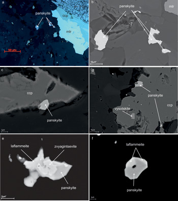
In polished sections, panskyite occurs as small individual anhedral grains of 0.5–10 μm in the interstices of rock-forming silicates (Fig. 5). It often intergrows or forms tiny inclusions in base-metal sulfides (millerite, chalcopyrite, bornite and chalcocite) and forms complex intergrowths with other PGM (zvyagintsevite, laflammeite and vysotskite). Reflected light and back-scattered electron images of panskyite and associated minerals are shown in Fig. 5. Panskyite in intergrowths with its Tl-rich Pb analogue thalhammerite is shown in Fig. 3a,b.
Microphotographs of panskyite from the West-Pana intrusion: (a–d) in association with base-metal sulfides; (e,f) in intergrowths with other PGM (laflammeite and zvyagintsevite). (a) Reflected light, (b–f) back-scattered electron images. ccp – chalcopyrite, mlr – millerite, pn – pentlandite and vl – violarite. Samples are from Southern Kievey apart from (d) which is from Kievey; for sample locations see Fig. 1.

Due to the small grain size and intergrowths with other ore minerals, the physical properties of panskyite could not be determined on natural or synthetic material. Panskyite is opaque and has a metallic lustre. The powder of the synthetic analogue is grey in colour and has a grey streak. The mineral is brittle. The density calculated on the basis of the empirical formula and cell dimensions of panskyite is 9.81 g.cm–3.
In plane-polarised light, panskyite is creamy white. It is weakly bireflectant, weakly pleochroic and it has a distinct anisotropism with brown to grey rotation tints. Internal reflections were not observed.
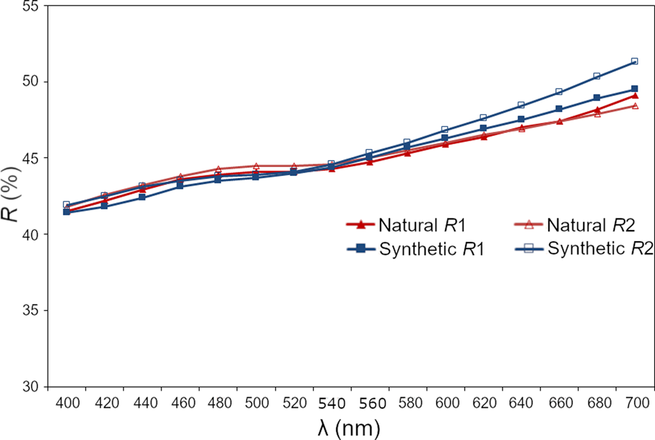
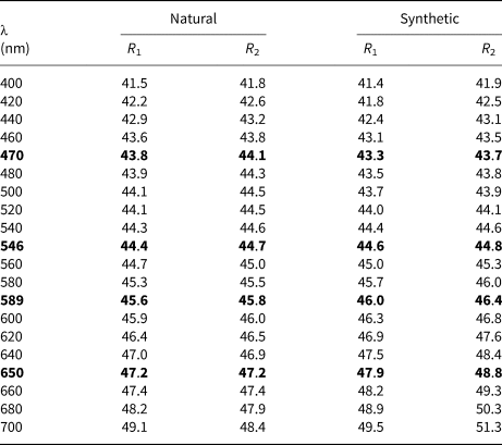
The reflectance measurements were made in air relative to a WTiC standard on both panskyite and its synthetic analogue using a J & M TIDAS diode array spectrometer attached to a Zeiss Axiotron microscope. The results are given in Table 1 and illustrated in Fig. 6.
Reflectance data for panskyite compared to its synthetic analogue, Pd9Ag2Pb2S4. The reflectance values (R, %) are plotted versus wavelength (λ, nm).

Reflectance data for panskyite and its synthetic analogue, Pd9Ag2Pb2S4.

The values required by the Commission on Ore Mineralogy are given in bold.
Synthetic analogue
The size (0.5–10 μm) of inclusions of panskyite, embedded in chalcopyrite or intergrown with other PGM, prevented its extraction in an amount required for relevant crystallographic and structural investigations. Therefore, these investigations were performed on its synthetic analogue.
Synthetic Pd9Ag2Pb2S4 phase was prepared using an evacuated silica glass tube method in the Laboratory of Experimental Mineralogy of the Czech Geological Survey in Prague. Palladium (99.95%), silver (99.999%), lead (99.9999%) and sulfur (99.999%) were used as starting materials for synthesis. The evacuated tube with its charge was sealed and annealed. After cooling by cold-water bath, the charge was ground into powder in acetone using an agate mortar, and thoroughly mixed to homogenise. The pulverised charge was sealed in an evacuated silica-glass tube again, and heated at 400°C for 7 months. The experimental product was quenched rapidly in cold water.
Chemical composition
Electron probe micro-analyses (EPMA) were performed with a CAMECA SX-100 electron probe microanalyser in wavelength-dispersive mode using an electron beam focussed to 1–2 μm. The standards used were pure elements for Pd, Ag and Ni; and galena for Pb and S. Concentrations were quantified on the PdLα, AgLα, PbMα and SKα, with an accelerating voltage of 15 keV, and a beam current of 10 nA on the Faraday cup. Other elements were below the detection limit.
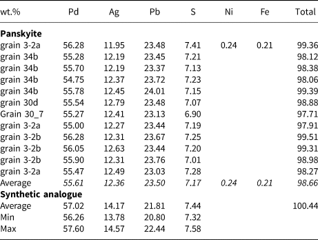
The EPMA results are given in Table 2. The EPMA data of natural material were collected on five different grains. The empirical formula, calculated for the mean composition (n = 12) on the basis of a total of 17 atoms per formula unit is (Pd9.05Fe0.07Ni0.07)Σ9.19Ag1.98Pb1.96S3.87 for panskyite and Pd9.07Ag2.22Pb1.78S3.93 for its synthetic analogue, with an ideal formula Pd9Ag2Pb2S4.
EMPA data for panskyite and its synthetic analogue Pd9Ag2Pb2S4.

Panskyite has a Bi analogue: thalhammerite; in addition we have observed its Tl-rich analogue from South Kievey. The compositional data of panskyite, thalhammerite and Tl analogues from North Kamennik, Kievey and South Kievey, compared with thalhammerite from Noril`sk are plotted in Fig. 4 (in total 88 analyses from SEM EDS and EPMA).
X-ray crystallography
Single-crystal X-ray diffraction
A small fragment of synthetic Pd9Ag2Pb2S4 was mounted on a glass fibre and examined using a Rigaku Super Nova single-crystal diffractometer with an Atlas S2 CCD detector utilising MoKα radiation, provided by the microfocus X-ray tube and monochromatised by primary mirror optics. The ω rotational scans were used for the collection of three-dimensional intensity data. From 639 reflections, 186 were classified as unique observed with I > 3ρ(I). Corrections for background, Lorentz effects and polarisation were applied during data reduction with CrysAlis software. Empirical absorption correction was performed using the same software yielding R int = 0.0308. The crystal structure was solved with a charge-flipping method using the program Superflip (Palatinus and Chapuis, Reference Palatinus and Chapuis2007) and subsequently refined by the full-matrix least-squares algorithm of JANA2006 (Petříček et al., Reference Petříček, Dušek and Palatinus2014). Because of the similarity of the atomic number of Pd and Ag (46 and 47, respectively), it is nearly impossible to directly distinguish between these atoms from single-crystal (MoKα radiation) diffraction data. The refinement indicated five metallic positions, of which one was assigned as Pb and the remaining four as Pd. The Pd sites show multiplicities 2:8:8:4. Considering the empirical chemical composition Pd9.07Ag2.22Pb1.78S3.93 (Z = 2) and coordination environment of the 4e site, which was very different from the others (see structure description), the 4e site was refined as the Ag position. An analogous situation was observed during the refinement of isostructural thalhammerite, Pd9Ag2Bi2S4 (Vymazalová et al., Reference Vymazalová, Laufek, Sluzhenikin, Kozlov, Stanley, Plášil, Zaccarini, Garuti and Bakker2018).
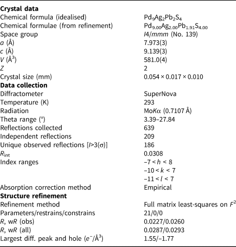
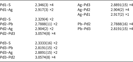
In subsequent refinement cycles, occupancy of all metallic sites was left free to vary (Pd vs. vacancy, Ag vs. vacancy and Pb vs. vacancy). While Pd and Ag sites were found to be fully occupied, refinement of the Pb position indicated 0.957(6) occupancy. Final anisotropic refinement in I4/mmm space group for 21 parameters converged smoothly to an R 1 = 0.0227 and wR = 0.0260 for 186 observed reflections. Details of data collection, crystallographic data and refinement are given in Table 3. Atom coordinates and displacement parameters are listed in Table 4. Table 5 shows selected bond lengths.
Crystallographic data for the selected crystal of the synthetic analogue of panskyite, Pd9Ag2Pb2S4.

Fractional coordinates and anisotropic displacement parameters (Å2) for the synthetic analogue of panskyite, Pd9Ag2Pb2S4.

*Refined with 0.96(1) occupancy.
Selected bond distances (Å) in the synthetic analogue of panskyite, Pd9Ag2Pb2S4.

Powder X-ray diffraction
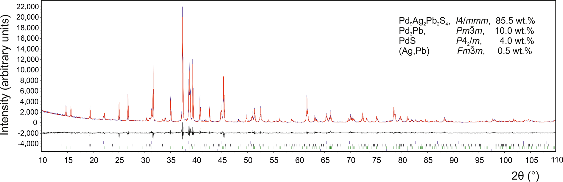
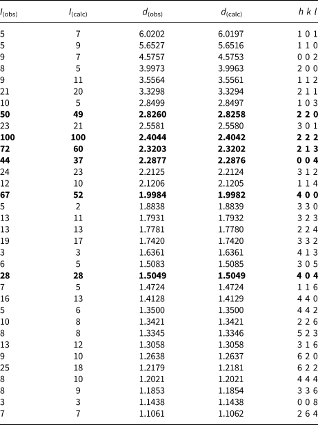
The powder X-ray diffraction pattern of synthetic panskyite was collected in Bragg–Brentano geometry on a Bruker D8 Advance diffractometer equipped with the LynxEye XE detector and CuKα radiation. Data were collected in the range from 10° to 145°2θ with a step size of 0.005°2θ and 2 second counting time per step. The structure model obtained from a single-crystal X-ray study of panskyite was used in the subsequent Rietveld refinement. The program Topas 5 (Bruker AXS, 2014) was used. The refinement involved refinement of scale factors, unit-cell parameters, isotropic size and strain. The background was determined by means of a Chebyshev polynomial function of the 5th order. No fractional coordinates were refined. The final cycles of refinement converged to the agreement factors R p = 7.92% and R wp = 10.33%, the refinement indicated 0.5, 4 and 10 wt.% impurities of (Ag,Pb) alloy, PdS and Pd3Pb, respectively. Powder X-ray diffraction data of the synthetic analogue of panskyite are summarised in Table 6. Fig. 7 shows the final Rietveld plot. The crystallographic information file has been deposited with the Principal Editor of Mineralogical Magazine and is available as Supplementary material (see below).
The final Rietveld fit of the synthetic analogue of panskyite, Pd9Ag2Pb2S4. The sample contains ca. 10, 4.0 and 0.5 wt.% Pd3Pb, PdS and (Ag,Pb) alloy as impurities, respectively.

Powder X-ray diffraction data of synthetic analogue of panskyite (CuKα radiation, Bruker D8 Advance, Bragg-Brentano geometry, 0.36° fixed divergence slit). Only reflections with I (obs) ≥ 1 are listed.

The strongest lines are given in bold.
Structure description
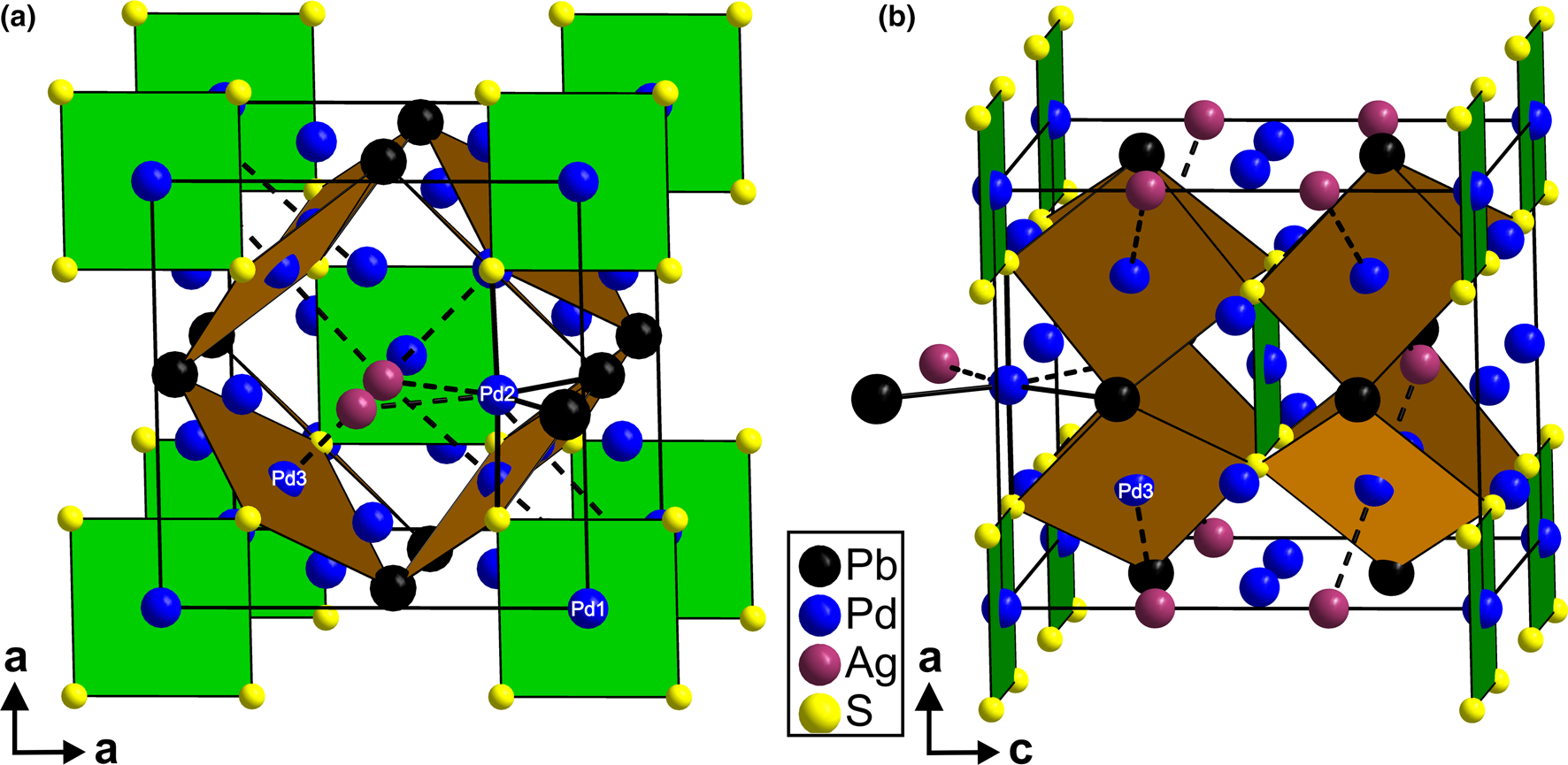
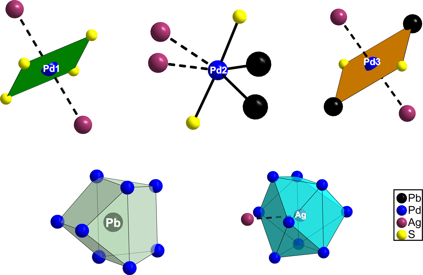
The crystal structure of panskyite contains three distinct palladium sites and one silver, lead and sulfur site. Its crystal structure is shown in Fig. 8, selected coordination polyhedra in Fig. 9.
Crystal structure of the synthetic analogue of panskyite showing [Pd1S4] (green) an [Pd3Pb2S2] (orange) squares. (a) View along c and (b) view along a axis. Ag–Pd interactions are indicated by dashed lines, coordination environment of one Pd2 is shown in (a).

Coordination polyhedra of Pd1, Pd2, Pd3, Pb and Ag sites in the panskyite crystal structure. Weak Ag–Pd and Ag–Ag interactions are indicated by dashed lines.

In panskyite, the Pd1 position forms square–planar coordination of S atoms at a distance of 2.346(3) Å. This coordination is perfectly planar and resembles that observed in vysotskite, PdS (Brese et al., Reference Brese, Squattrito and Ibers1985). In addition to four S contacts, Pd1 atoms have two long contacts with Ag atoms oriented perpendicular to the [Pd1S4] plane at a distance of 2.917(3) Å. The Pd2 site shows two short S and two Pb contacts at 2.329(4) and 2.7888(11) Å, respectively. Sulfur and Pb atoms make up a disphenoid, where Pd2 is placed in the middle of an S–S edge. This coordination is further completed by two Ag atoms at a distance of 2.904(2) Å. Consequently, the complete coordination sphere of Pd2 atoms can be described as an elongated tetragonal bipyramid [Pd2S2Pb2Ag2] with the Ag atoms placed at a cis position (i.e. where Ag atoms are mutually adjacent). The central atom Pd2 is shifted slightly from the centre of the bipyramid towards the Pb–Pb edge. The coordination of Pd3 is similar to that of Pd2. Pd3 is surrounded by two S and Pb atoms forming a [Pd3S2Pb2] square and two additional Ag atoms at a distance of 2.8891(15) Å. The Ag atoms are oriented in a trans position (i.e. perpendicular to the [S2Pb2] square). Thus, the complete coordination sphere of the Pd3 site can be viewed as a compressed tetragonal bipyramid [Pd3S2Pb2Ag2].
Silver atoms are coordinated by nine Pd atoms at distances forming a monocapped tetragonal antiprism. The Ag–Pd distances are within the range from 2.8891(15) to 2.917(2) Å, which is comparable to those observed in kravtsovite, PdAg2S (Vymazalová et al., Reference Vymazalová, Laufek, Sluzhenikin, Stanley, Kozlov, Chareev and Lukashova2017) and lukkulaisvaaraite Pd14Ag2Te9 (Vymazalová et al., Reference Vymazalová, Grokhovskaya, Laufek and Rassulov2014). The Ag atoms show weak Ag–Ag bonds at 3.304(4) Å across the shared tetragonal face (Fig. 9) of the antiprism. A lead atom has eight Pd contacts arranged in the form of bi-capped trigonal prism and showing a 4 + 4 bonding scheme. Observed Pd–Pb distances 2.7888(16) and 2.8191(15) Å are slightly shorter than corresponding Pd–Bi distances [2.808(1) and 2.8378(1) Å] observed in isostructural thalhammerite Pd9Ag2Bi2S4 (Vymazalová et al., Reference Vymazalová, Laufek, Sluzhenikin, Kozlov, Stanley, Plášil, Zaccarini, Garuti and Bakker2018). By analogy with thalhammerite, no short (<3.5 Å) Pb–S and Pb–Ag contacts occur in the panskyite crystal structure.
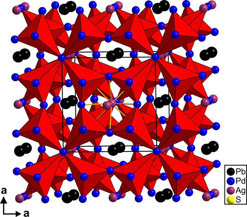
In terms of anion-centred polyhedra (Fig. 10), the S site has deformed trigonal–bipyramidal coordination with two Pd2, one Pd1 sites in equatorial and Pd3 in axial positions. While all Pd–S bonds are approximately equal (2.32–2.34 Å), the S atom is shifted from the polyhedra centre towards the Pd1 position thereby, reducing the Pd2–Pd2 distance in the equatorial plane to 3.37 Å and extending the Pd2–Pd1 distance to 4.29 Å. Consequently, the Pd2–Pd2–Pd1 atoms form an isosceles triangle instead of an equilateral one as in the case of regular coordination. The bipyramids share only corners (i.e. Pd atoms), forming a three-dimensional framework. While Pd3 and Pd2 vertices are shared with two adjacent bipyramids, four bipyramids are linked by sharing Pd3 vertices, producing four-fold ‘iron crosses’. Lead and Ag atoms are placed in the voids in the framework forming Pb–Pd, Pd–Ag and Ag–Ag bonds. No short S–S bonds were observed. The shortest S–S distance of 3.316(5) Å is forced by the coordination geometry of adjacent cations.
Crystal structure of panskyite showing a corner-sharing anion-based trigonal-bipyramids [SPd]. Ag–Ag and Ag–Pd interactions are indicated in the middle of the Figure. Note the four-fold ‘iron crosses’ of [SPd5] coordination bipyramids.

Relation to other minerals
Panskyite is the Pb analogue of thalhammerite Pd9Ag2Bi2S4 (Vymazalová et al., Reference Vymazalová, Laufek, Sluzhenikin, Kozlov, Stanley, Plášil, Zaccarini, Garuti and Bakker2018). The main structural difference between these two minerals is that panskyite shows slightly shorter Pd–Pb bonds (2.803 Å on average) than the corresponding Pd–Bi bonds (2.822 Å on average) observed in thalhammerite. This is also reflected by a slightly smaller unit-cell volume of panskyite (581.0 Å3) in comparison with thalhammerite (589.7 Å3). Based on the chemical composition, there is also a Tl analogue (Fig. 3, 4); however, its crystal structure has not yet been studied.
Proof of identity of panskyite and its synthetic analogue
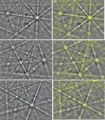
A TESCAN Mira 3GMU scanning electron microscope combined with an electron back-scatter diffraction (EBSD) system (Nordlys Nano detector, Oxford Instruments) was used for the measurements. The sample surface was prepared for EBSD by polishing with colloidal silica (OP-U) for 30 min. Acquisition conditions were: accelerating voltage 20 kV, beam current ~3 nA, no binning (full EBSP resolution is 1344 × 1024). Indexing conditions were: refined accuracy mode, 8 bands, 44 reflectors. The EBSD patterns were collected and processed using a proprietary computer program: AZtec HKL (Oxford Instruments). The solid angles calculated from the patterns were compared with a structural model proposed match containing 44 reflectors to index the patterns. EBSD patterns (also known as Kikuchi patterns) obtained from the natural material (20 measurements on different grains of panskyite) were found to match the patterns generated from our refined structural model for Pd9Ag2Pb2S4 (Fig. 11). The values of the mean angular deviation (MAD, i.e. the goodness of fit of the solution) between the calculated and measured Kikuchi bands range between 0.22° and 0.44°. These values reveal a very good match; as long as values of mean angular deviation are <1°, they are considered as indicators of an acceptable fit (HKL Technology v. 2004).
Electron back-scattered diffraction patterns (EBSPs) showing different orientations of grains of panskyite; in the right pane, the Kikuchi bands are solved (indexed).

Acknowledgments
The authors acknowledge Ritsuro Miyawaki, Chairman of the IMA-CNMNC and its members, for helpful comments on the submitted data. The reviewer's comments of Louis J. Cabri, an anonymous reviewer, Structures Editor Peter Leverett and the Principle Editor Stuart Mills are greatly appreciated. This research was supported by the Grant Agency of the Czech Republic (project No. 18-15390S to A.V.), by the Czech Geological Survey (DKRVO/ČGS 2018–2022), and by the Operational Programme Research, Development and Education financed by European Structural and Investment Funds and the Czech Ministry of Education, Youth and Sports (Project No. SOLID21 CZ.02.1.01/0.0/0.0/16_019/0000760). C.J. Stanley acknowledges Natural Environment Research Council grant NE/M010848/1 Tellurium and selenium cycling and supply. We are also grateful to Dr. Alexey U. Korchagin, General Director of Pana JSC, for his assistance in conducting research and preparing the publication.
Supplementary material
To view supplementary material for this article, please visit https://doi.org/10.1180/mgm.2020.100