Introduction
Chronic diseases in childhood can lead to learning disorders during school life. Allergic rhinitis is one of the important impairments that occur in childhood, because it causes nasal obstruction-induced disturbed breathing. In addition, nasal symptoms, including a sensation of itch and rubbing of the nose, sneezing, rhinorrhoea, and congestion reduce health-related quality of life by causing loss of sleep and limiting the activity of the patient.Reference Meltzer1 Allergic rhinitis is believed to cause learning disorders by causing reduced attention, disturbed night sleep, consequent daytime sleepiness, etc.Reference Simons2 Various treatments are administered to ameliorate allergic rhinitis and its symptoms; anti-histamine therapy is the most commonly used treatment. Clinical trials with anti-histamines have shown that 36–80 per cent of patients experience good to excellent symptom relief (approximately twice of that noted in the placebo group) in seasonal allergic rhinitis and 55–86 per cent experience moderate to excellent symptom relief in perennial allergic rhinitis.Reference Scadding3 However, the possibility of learning disability due to the sleepiness induced as a side effect of the anti-histamines remains a concern. Ketotifen, an anti-histamine, reduces the accuracy of preschool children to perform working memory tasks.Reference Tsujii, Yamamoto, Ohira, Takahashi and Watanabe4 However, the mechanism underlying the learning disability induced in patients with allergic rhinitis remains unclear.
The hippocampus, an important region in the brain, is responsible for learning and memory. It is reportedly associated with cognitive mapping, detection of spatial configuration, and contextual learning and declarative memory.Reference O'Keefe5 Furthermore, synapse formation in the hippocampus might be closely related to learning and memory. Significant age-related impairments in hippocampus-dependent learning have been assessed using water maze and fear-conditioning tests.Reference von Bohlen6 Yang et al. Reference Yang, Pan and Gan7 reported that learning and novel sensory experience lead to spine formation and elimination by a protracted process in mice, and that the extent of spine remodelling correlates with the behavioural improvement observed after learning.
Despite these studies, it is still not known whether allergic rhinitis can induce anatomical changes during synapse formation in the hippocampus. Therefore, in this study, we examined the possible alterations in the expression patterns of synapse-related molecules in the hippocampus of mice models of allergic rhinitis using the following markers: glutamic acid decarboxylase (GAD) 65 and 67, synaptophysin and spinophilin. Our results indicate that the synaptic structure present in the hippocampus may remain unaltered in the allergic rhinitis patients.
Materials and methods
Twelve seven-week-old virgin female BALB/c mice (Japan Crea Inc., Hamamatsu, Japan) were housed in separate standard cages. They were bred under the specific pathogen-free conditions at our Institute and were allowed to feed and drink ad libitum. On day 0, six mice were intra-peritoneally sensitised with 30 µg ovalbumin (OVA) diluted in 100 µl of alum solution. This injection was repeated on day 7. On the other hand, the control (non-sensitised) mice were intra-peritoneally injected with phosphate-buffered saline mixed with 100 µl of alum solution on day 0 and the injection was repeated on day 7. On days 24, 25, 26, 27, 28, 31, 32, 33, 34 and 35 after the first sensitisation, the OVA-sensitised mice were challenged with an intra-nasal injection of 10 µl of 10 µg/ml OVA solution prepared in phosphate-buffered saline and administered into both the nostrils. The control mice were challenged with an intra-nasal injection of 10 µl of phosphate-buffered saline into both the nostrils. After the last challenge, the mice were placed in the observation cages, and the number of sneezing attacks and nasal rubbing movements were counted for 10 minutes by the method proposed by Sugimoto et al. Reference Sugimoto, Kawamoto, Chen and Kamei8 On day 36, three mice from each group were anaesthetised with ether and transcardial perfusion was performed with 4 per cent paraformaldehyde. The brains of these mice were removed and soaked overnight at 4°C in the same solution. Serial 25-μm-thick coronal sections were cut on a vibratome (VT1000S; Leica Microsystems, Wetzlar, Germany); these sections were blocked overnight at 4°C in 1 per cent bovine serum albumin (BSA) prepared in phosphate-buffered saline containing 0.3 per cent Triton X-100 and 0.05 per cent sodium azide. The blocked sections were incubated for 3 days at 4°C with the following primary antibodies raised in different species: mouse monoclonal anti-synaptophysin (1:20 000; a gift from Dr Fujita), mouse monoclonal anti-GAD65 (1:100 000; MAB5406; Chemicon, Temecula, California, USA), rabbit polyclonal anti-spinophilin (1:10 0000; a gift from Dr Greengard) and rabbit polyclonal anti-GAD67 (1:5000; G5038; Sigma-Aldrich, St. Louis, Missouri, USA). These sections were washed with phosphate-buffered saline and then incubated at 4°C with a mixture of fluorescein isothiocyanate-conjugated donkey anti-rabbit immunoglobulin (Ig) G antibody (1:250; Jackson ImmunoResearch Laboratories, West Grove, Pennsylvania, USA) and cyanine 3-conjugated donkey anti-mouse IgG antibody (1:500; Jackson ImmunoResearch Laboratories) for 3 h. Finally, these sections were mounted in Vectashield (Vector Laboratories, Peterborough, UK) and examined under a confocal laser-scanning microscope (TCS-SP2; Leica Microsystems). All these experiments were performed in accordance with the Guidelines for Animal Experiments practiced at our Institute and the Law (No. 105) and Notification (No. 6) of the Government of Japan. We assert that all procedures contributing to this work comply with the ethical standards of the relevant national and institutional guides on the care and use of laboratory animals (A22–143–0 and A26–148–0).
Results
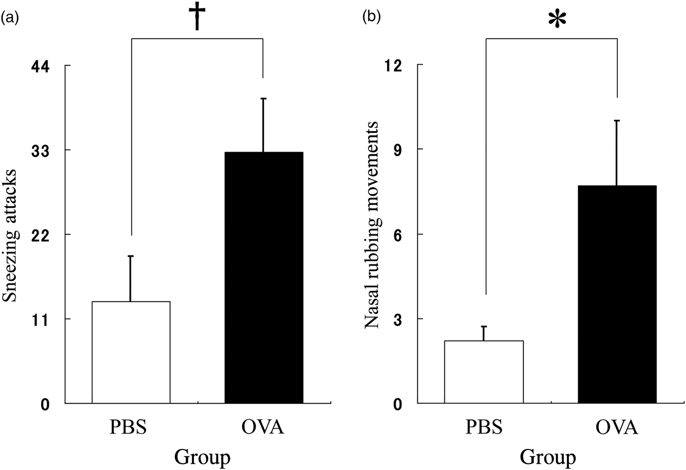
The observed nasal symptoms are shown in Figure 1. The number of sneezing attacks and nasal rubbing movements were higher in the allergic rhinitis group than in the control group (p < 0.1 and < 0.05, respectively). The results indicate that an animal model of allergic rhinitis was successfully developed.
Behavioural assessment of allergic response. (a) The number of sneezing attacks was greater in the ovalbumin (OVA) group (black column) than in the phosphate-buffered saline (PBS) group (white column). (b) The number of nasal rubbing movements was greater in the OVA group (black column) than in the phosphate-buffered saline group (white column). (n = 6 in each group) †p < 0.1; *p < 0.05.

Immunofluorescence double labelling for synaptophysin and spinophilin showed no differences in the expression patterns between the AR and control groups (Figure 2). Similarly, we did not observe any difference in the distribution patterns of GAD65 and GAD67 between the two groups (Figure 3).
Representative patterns of expression of spinophilin and synaptophysin in the hippocampus of control and rhinitis animals. (a) There are no apparent differences in immunolabelling for spinophilin and synaptophysin in the whole area of the hippocampus. (b) Higher magnification (×400) images of the cornu ammonis 1 area of the hippocampus immunostained for spinophilin and synaptophysin.
Representative patterns of expression of glutamic acid decarboxylase (GAD) 65 and GAD67 in the hippocampus of control and rhinitis animals. (a) There are no apparent differences in immunolabelling for GAD65 and GAD67 in the whole area of the hippocampus. (b) Higher magnification (×400) images of the cornu ammonis 1 area of the hippocampus immunostained for GAD65 and GAD67.
Discussion
Our study provides the first evidence that allergic rhinitis may not alter the synaptic structures in the hippocampus, at least during a two-week exposure time. Neuroimmunomodulation plays a crucial role in homoeostasis because the central nervous system is closely related to the immune system. Several evidences have validated the effect of allergy on brain function. Costa-Pinto et al. reported that OVA-immunised mice entered the open arm in lesser time than non-immunised mice in the elevated plus-maze test, and that the number of Fos-immunoreactive neurons in the paraventricular nucleus and the serum corticosterone levels were higher in OVA-immunised mice.Reference Costa-Pinto, Basso, DeSá-Rocha, Britto, Russo and Palermo-Neto9 Tonelli et al. showed that in an open field test, the total distance moved in the allergic mice was the same as that moved in the control mice, and that the duration of social contact in the allergic mice was shorter than that observed in the control mice.Reference Tonelli, Katz, Kovacsics, Gould, Joppy and Hoshino10 The evidence suggests that the allergic response induces anxiety, social withdrawal, and hyperactivity of the hypothalamic–pituitary–adrenal axis. To the best of our knowledge, no morphological or physiological research of the brain has been conducted thus far to show the effect of allergic response on cognitive functions such as learning or memory.
Synaptic reconstruction is thought to be a critical morphological change occurring in the brain during learning. The spine apparatus, which is considered a post-synaptic structure, is closely associated with learning and memory. Deller et al. used the radial arm maze test to show that absence of the spine apparatus decreased the ability of mice to acquire spatial learning; they also showed a decrease in the long-term potentiation of the field excitatory post-synaptic potential in synaptopodin-deficient mice.Reference Deller, Orth, Del Turco, Vlachos, Burbach and Drakew11 Another group of researchers showed that the administration of oestradiol benzoate improved spatial learning and increased the level of hippocampal synaptophysin.Reference Frick, Fernandez and Bulinski12 These findings led us to hypothesise that learning impairment in patients with allergic rhinitis is due to changes in the synapse formation in the hippocampus. However, the absence of changes in the levels of synaptophysin and spinophilin refuted this hypothesis.
Allergic response is one of the commonly known stressors that affect a living body, especially the hippocampal morphology. Exposure to elevated serum levels of corticosterones and prolonged stress induced by restraint and immobilisation induces the shrinkage of apical dendrites of cornu ammonis 1 and 3 (CA1 and CA3, respectively) neurons, and reduces the spine number.Reference Alfarez, Wiegert and Krugers13 An increase in the corticosterone levels in animals with allergic rhinitis may alter the synaptic structure in the hippocampus. However, in our study, we found that the synaptic structure in the hippocampus was stable in allergic rhinitis mice. However, further studies are required to elucidate these discrepancies.
The hippocampal γ-aminobutyric acid-ergic (GABAergic) interneurons play a crucial role in regulating the activity of the principal neurons. This regulation mainly consists of three processes.Reference Bernard, Cossart, Hirsch, Esclapez and Ben-Ari14 First, the activation of post-synaptic GABA receptors produces direct hyperpolarising or shunting effects and then counteracts the membrane depolarisation induced by excitatory inputs. Second, GABA receptor-mediated synaptic responses directly block the action potential firing. Finally, the hippocampal interneurons lead to the synchronisation of the firing of principal neurons during network oscillations. The pharmacological blockade of cholinergic systems, which disrupts the low-frequency theta rhythm of the oscillations in the hippocampus, exerts delayed effect on the acquisition rate of the behavioural conditioned response.Reference Berry and Seager15 In our study, the intra-nasal challenge with OVA did not affect the immunoreactivity of GAD65/67, thereby proving that the hippocampal GABAergic interneurons are not responsible for the learning impairment in allergic rhinitis patients.
• This is a research article describing stability of the synaptic structure in the hippocampus of BALB/c mice with allergic rhinitis
• Immunofluorescence double labelling for synaptophysin and spinophilin showed no differences in the expression patterns
• We did not observe any difference in the distribution patterns of GAD65 and GAD67
• The present study reports that allergic rhinitis may not alter the synaptic structure in the hippocampus, at least during a short-term exposure
An increase has been observed in the already high prevalence rate of allergic rhinitis across the world.Reference Sakashita, Hirota, Harada, Nakamichi, Tsunoda and Osawa16–Reference Bauchau and Durham18 Marshall et al. showed that during ragweed seasons, allergic patients experience a subtle decrease in their cognitive processing; some patients also express difficulties in working memory.Reference Marshall, O'Hara and Steinberg19 Moreover, Shaaban et al. reported that the adjusted relative risk for asthma was 1.63 for atopy alone, 2.71 for non-allergic rhinitis and 3.53 for allergic rhinitis, and that only allergic rhinitis with sensitisation to mites was associated with an increased risk of asthma; this risk was independent of sensitisation to other allergens.Reference Shaaban, Zureik, Soussan, Neukirch, Heinrich and Sunyer20 Therefore it is very important to take strong countermeasures againt allergic rhinitis. In this study, intra-nasal short-term exposure to the allergen did not produce an abnormal change in the synaptic structure of the hippocampus. There is a high possibility that allergic rhinitis induced learning impairment is independent of structural changes in the brain; instead, it could be based on the subsequent effects of allergic rhinitis such as reduced attention and sleepiness during the daytime. These factors indicate that adequate treatment of allergic rhinitis can relieve patients and prevent subsequent complications. The present study reports that allergic rhinitis may not alter the synaptic structure in the hippocampus, at least during a short-term exposure. Active treatment of allergic rhinitis in childhood may be recommended in order to prevent learning disorders.
Acknowledgement
We thank Madoka Kasao for her valuable technical assistance.
Financial support
This research received no specific grant from any funding agency, commercial or not-for-profit sectors.