The anthracycline chemotherapeutic agents have been widely used in the treatment of paediatric malignancies. The use of these agents has significantly improved the long-term survivors of the childhood cancers.Reference Mertens, Yasui and Neglia1 However, these agents have well-known cardiotoxic effects which is dose related. Cardiotoxic effects can occur both in acute and chronic patterns. Acute cardiotoxicity is characterised by arrhythmia, various conduction abnormalities and an abrupt deterioration in left ventricular function.Reference Arola, Saraste and Pulkki2 Chronic dose-related cardiotoxicity is the most common and dangerous and can manifest as cardiac failure, months or even years after the completion of treatment and is thought to be related to myocardial toxicity.Reference Giantris, Abdurrahman and Hinkle3 As a result, mortality related to cardiac events in childhood cancer survivors is about tenfold higher than age-matched control patients.Reference Mertens, Yasui and Neglia1, Reference Lipshultz, Lipsitz and Sallan4, Reference Lipshultz5 For many years, left ventricular function has been estimated by calculating the fractional shortening and ejection fraction, which is readily obtained from M-mode echocardiography. However, fractional shortening and ejection fraction lack sensitivity to detect subtle changes in cardiac function. Fractional shortening measures only the anterior and posterior myocardial motion at the mid-ventricular level. Similarly, ejection fraction measures fractional changes in chamber volume.Reference Lipshultz, Orav, Sanders, McIntosh and Colan6, Reference Borow, Neumann, Marcus, Sareli and Lang7 For this reason, during the last decade, new echocardiographic techniques for evaluating subtle changes in myocardial function have been introduced.Reference Jurcut, Wildiers, Ganame, D’Hooge, Paridaens and Voigt8 Tissue Doppler imaging (TDI) is a sensitive technique for the evaluation of diastolic function based on measurement of wall motion velocities, a relatively pre-load-independent method but angle dependent, and is known to exhibit many technical limitations.Reference Lipshultz, Orav, Sanders, McIntosh and Colan6 More recently, speckle tracking echocardiography (STE) has been proposed to be useful and a valid imaging modality in the diagnosis of global and regional systolic and diastolic myocardial dysfunction. It provides an objective and quantitative evaluation of global and regional myocardial function independently of any geometrical assumption.Reference Reisner, Lysyaskky, Agman, Mutlak, Lessick and Friedman9, Reference Korinek, Wang and Sengupta10 The aim of this study was to detect late cardiotoxic effects of anthracycline therapy in asymptomatic long-term survivors of childhood cancers, based on myocardial strain imaging by STE. We have hypothesised that paediatric cancer survivors treated with varying doses of anthracycline have subtle left ventricular dysfunction with non-conventional echocardiographic methods (TDI and STE) who have normal cardiac functions with conventional echocardiographic methods. Also this subtle cardiac dysfunction can have positive relationship with total anthracycline dose.
Patients and methods
This cross-sectional prospective study was conducted between January, 2016 and December, 2016 in the paediatric oncology outpatient clinic of Dr. Sami Ulus Children’s Hospital and included 45 asymptomatic long-term survivors of childhood cancers. All patients had completed cancer treatment at least 1 year prior to enrolment and had no evidence of current malignancy, known cardiac symptoms, or cardiomyopathy. To focus our analysis on the effects of anthracyclines, we excluded patients if they had a history of radiotherapy with potential impact to the heart. We stratified our study group based on their cumulative lifetime anthracycline exposure such as < 300 mg/m2 and ≥ 300 mg/m2. All patients, or their legal guardians, provided written informed consent. The echocardiographic findings of these patients were compared with those of 50 control patients, who were children with functional heart murmur, non-specific chest pain attending the cardiac outpatient clinic, for which no underlying organic causes were identified. The study was approved by the local ethics and research committee of the hospital.
Echocardiography
All echocardiographic studies were performed by one physician (T.Y.) using a commercially available echocardiographic system (S5-1 probe, IE33, Philips, Andover, Massachusetts, United States of America) by monitoring DII derivation, in the back-to-top or left lateral decubitus position. All images were digitally stored from three cardiac cycles as standard apical four-chamber and parasternal short axis views.
Conventional echocardiography and TDI measurements
Left ventricular end-diastolic dimension, inter-ventricular septum and left ventricle posterior wall thickness dimensions in systole and diastole, ejection fraction, and fractional shortening were calculated from two-dimensional guided M-mode echocardiographic tracings obtained at the parasternal long axis position using Teicholz formula. Left ventricular mass was calculated with the anatomically validated formula of Devereux.Reference Lang, Bierig and Devereux11 Left ventricular mass index adjusted for body size and is taken as left ventricular mass (g) divided by body surface area (m2). The following Doppler echocardiography parameters of left ventricle diastolic performance were evaluated: peak mitral flow velocities during early and late diastole, their ratio and mitral deceleration time. TDI measurements were performed on the left ventricle septal mitral annulus and lateral mitral annulus. The maximal systolic myocardial velocity and early and late diastolic myocardial velocity were measured. The isovolumic contraction time was calculated from the beginning of QRS in the echocardiogram until the beginning of systolic myocardial velocity wave, whereas the isovolumic relaxation time was calculated from the end of systolic myocardial velocity wave until the beginning of early diastolic myocardial velocity wave. Ejection time was measured from the beginning to the end of S wave. Myocardial performance index, a Doppler-derived time interval index, combines both systolic and diastolic time intervals to generate a combined index of global ventricular function. Myocardial performance index (Tei index) was calculated as the sum of the isovolumetric contraction time and the isovolumetric relaxation time divided by the ejection time.Reference Nagueh, Middleton and Kopelen12
Two-dimensional speckle tracking analyses
Myocardial deformation parameters (strain and strain rate) were measured using commercially available software (QLAB ver. 6.0, TMQ, Philips Medical Systems) on standard two-dimensional grayscale left ventricle images from standard apical four-chamber view for longitudinal strain and standard parasternal short axis at the papillary muscle plane for circumferential strain. Five consecutive heart beats synchronised to a continuous electrocardiography were recorded with frame rate set > 50 frames per second. The endocardial border was manually traced, and a region of interest was drawn to include the entire myocardium in all. The software algorithms automatically segmented the left ventricular planes into equidistant segments and then performed speckle tracking on a frame-to-frame basis. Tracking quality was visually checked, corrected when needed, and accepted by the operator. Strain values of left ventricle were assessed automatically in 13 segments, and then averaged manually by the operator as mean value of each strain and presented as global longitudinal strain and global circumferential strain.Reference Cerqueira, Weissman and Dilsizian13 Because the software does not calculate the peak values of strain rate automatically, the peak strain rate and average global rate were calculated manually. All two-dimensional speckle tracking analysis were performed by the same investigator to avoid inter-observer variability. To obtain intra-observer variability off-line two-dimensional speckle tracking analysis were performed by same investigator blinded to patients’ data.
Statistical analysis
The statistical analysis was performed by SPSS (Statistical Package for the Social Sciences, Chicago, Illinois, United States of America, and version 21.0). Numeric variables are expressed as mean ± SD and categorical ones are expressed as percentages (%). Normal distribution of samples was detected by Kolmogorov–Smirnov analysis. Comparisons of demographic data and echocardiographic parameters between patients and controls were performed using two independent samples t-test for normally distributed variables and by Mann–Whitney U-test for non-normally distributed ones. The correlations between continuous variables were assessed with the Spearman rank correlation coefficient. Statistical significance for all analyses was assumed at p < 0.05.
Results
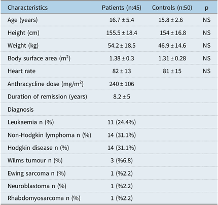
The demographic characteristics of the study population are summarised in Table 1. There were no significant differences between the patient and control groups with respect to age, gender, body surface area, and heart rate.
Demographic data of studied populations

NS = not significant
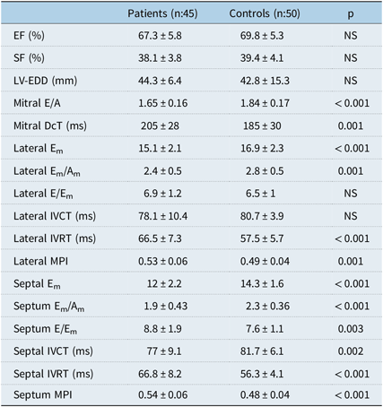
In the conventional echocardiographic measurement of left ventricle; the M-mode left ventricle dimensions, wall thicknesses, left ventricle ejection fraction and fractional shortening, and left ventricle mass index were similar between two groups. Ratio of peak mitral flow velocities during early and late diastole were in normal range but according to control group significantly lower in patient group. Similarly mitral deceleration time was in normal range but according to control significantly longer in patient group.
TDI assessment of left ventricle showed normal range systolic, early and late diastolic myocardial velocities, Doppler-derived time intervals, and Tei index in patient group, but some TDI values were abnormal according to controls. These relatively abnormal TDI values were as follows: lower peak septal and lateral maximal systolic myocardial velocity and early diastolic myocardial velocity waves, longer septal isovolumetric relaxation time and lateral isovolumetric relaxation time values, longer isovolumetric contraction time value, higher septal and lateral myocardial performance index values, higher ratio of the peak early diastolic Doppler mitral velocity to peak early diastolic lateral and septal myocardial velocities, lower septal, and lateral ratios of early and late diastolic myocardial velocities in patients compared to controls. Standard echocardiographic parameters are summarised in Table 2.
Conventional and tissue Doppler echocardiographic measurements of left ventricle

NS = not significant; EF = ejection fraction; Em = early diastolic velocity; Em/Am = ratio of early and late diastolic myocardial velocity; E/Em = ratio of early and late diastolic myocardial velocity; Dct = deceleration time; IVCT = isovolumetric contraction time; IVRT = isovolumetric relaxation time; LV-EDD = left ventricle end diastolic diameter; MPI = myocardial performance index; SF = fractional shortening.
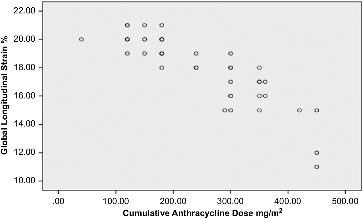
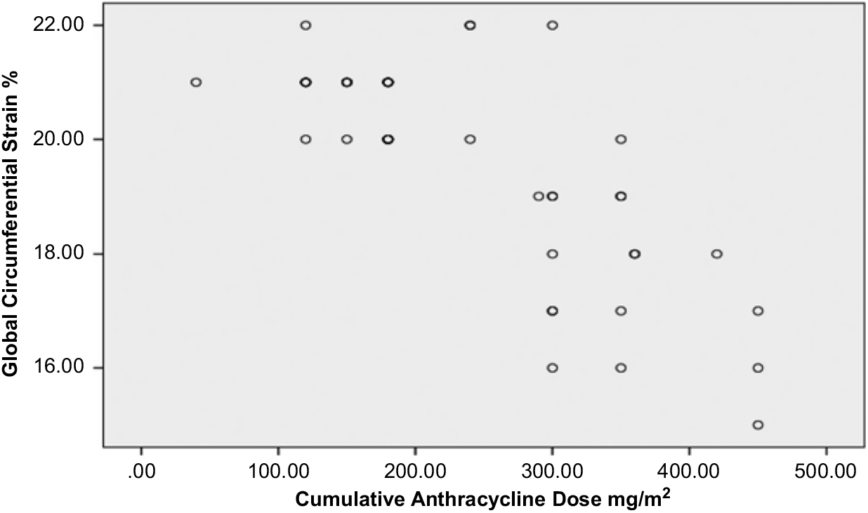
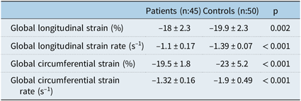
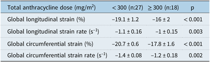
The patient group had statically significant lower global longitudinal strain and global circumferential strain values compared to controls. Also global longitudinal strain rate and global circumferential strain rate values were significantly lower in patient group. There was significant negative correlation between cumulative and global longitudinal strain and global circumferential strain (r: –0.86 p < 0.001 and r- –0.7 p < 0.001 respectively) (Figs 1 and 2). Global longitudinal and circumferential strain and strain rate values were showed in Table 3. Compression of global longitudinal and circumferential strain and strain rate values according to total anthracycline dose showed significantly lower values in patients who had ≥ 300 mg/m2 total anthracycline dose compared to in patients who had < 300 mg/m2 total anthracycline dose (Table 4).
Correlation between GLS and total anthracycline dose.

Correlation between GCS and total anthracycline dose.

Global longitudinal and circumferential strain and strain rate values of the study population

Compression of global longitudinal and circumferential strain and strain rate values according to total anthracycline dose

Discussion
In the present study we showed that in most instances the early detection of sub-clinical anthracycline cardiotoxicity in paediatric cancer survivors cannot be achieved with conventional echocardiographic tools. The long-term cardiac adverse effects of the anthracycline chemotherapy can be identified with non-conventional echocardiographic measures as a reduction left ventricle global longitudinal and circumferential strain and strain rate.
Regular evaluation for early detection of sub-clinical cardiotoxic effects of anthracyclines has a pivotal role in ongoing follow-up for cancer survivors receiving this treatment. Because the cardiac adverse effects of anthracyclines remain one of the most important causes of non-cancer-related morbidity and mortality.Reference Grenier and Lipshultz14–Reference Trachtenberg, Landy and Franco17 Anthracycline-induced cardiotoxicity has been associated with progressive left ventricle dilatation and systolic dysfunction over time which most commonly observed as decrease in the left ventricle ejection fraction. In current clinical practice, the two-dimensional and M-mod echocardiographies continue to remain most commonly used modality for detection of left ventricle dysfunction. But these methods have well-known limitations to evaluate subtle contractility changes of left ventricle myocardium. Because cardiac motion and deformation occur three dimensionsal. The heart rotates and repositions (circumferential), shortens and dilates in longitudinal direction and thickens and relaxes in radial orientation during cardiac cycle. However, conventional echocardiography and TDI provide information in a single direction from a fixed transducer position and dependent on the angle between the beam and the myocardial motion. Also changes in pre-load and after-load can influence conventional echocardiography parameters. The novel echocardiographic methods to evaluate regional and global left ventricle function such as two-dimensional speckle tracking analysis have the advantage in early detection of subtle myocardial systolic dysfunction. Anyway two-dimensional speckle tracking analysis has the advantage that it is accurate, reproducible, and angle independent, thus eliminating the major limitation of TDI and it enables a complete assessment of regional and global function in three directions.
Previous echocardiographic studies in cancer survivors reported that diastolic function impairment occurs earlier than systolic dysfunction and can be a predictor of further deterioration.Reference Iarussi, Galderisi and Ratti18–Reference Christiansen, Hamre and Richard Massey21 Consistent with these findings we also showed some early sub-normal changes in myocardial relaxation (lower ratio of trans-mitral flow velocities during early and late diastole, longer mitral deceleration time and lower ratios of septal and lateral early and late diastolic myocardial velocities in patients group when compared to control group, compatible with disturbance of the left ventricle diastolic function) in the present study. Mitral septal early diastolic TDI velocity integrates trans-mitral driving pressure and myocardial relaxation which were higher in patient group compared to controls. This indicates relatively higher left ventricle filling pressure in patient group. Additionally mean lateral and septal myocardial performance index values were relatively higher in the patient group, and this indicates relatively impaired left ventricle systolic and diastolic functions in the patient group. Actually all these Doppler and tissue-Doppler-derived parameters were in normal range but when compared to control group all these parameters were relatively sub-normal. However, there is no data about diastolic functional impairment can predict subsequent cardiotoxicity. Furthermore, several studies have demonstrated that systolic strain changes occur prior to or in the absence of diastolic dysfunction.Reference Sawaya, Sebag and Plana22–Reference Tsai, Gjesdal and Wethal24
The two-dimensional speckle tracking analysis-based strain has been used to detect early myocardial changes in patients receiving anthracycline chemotherapy. Cheung et al showed impaired left ventricle myocardial deformation in all of three dimensions (longitudinal, circumferential, and radial) and mechanical dyssynchrony despite normal left ventricle fractional shortening in children with acute lymphoblastic leukaemia who had been off treatment for at least 1 year. Additionally, they found significant negative correlation between total anthracycline dose and global longitudinal and circumferential strain rate.Reference Cheung, Hong, Chan, Wong and Ha23 Another study among adult survivors of childhood, adolescent, and young adult cancer observed reduced global longitudinal strain ( ≤ 16%) in 18% of total patients despite normal left ventricle ejection fraction. Anyway, this study found no significant association between left ventricle systolic and diastolic function and cumulative anthracycline dose.Reference Yu, Raikhelkor and Zabor25 Mavinkurve-Groothuis et al showed that both conventional echocardiographic parameters (left ventricle ejection fraction and fractional shortening) and global myocardial strain, strain rate, and time to peak systolic strain in asymptomatic survivors of childhood cancer (approximately 15 years after the last anthracycline therapy) were significantly lower compared with healthy controls. Additionally they found no significant relation between global longitudinal strain and global radial strain (mid-cavity) measurements and gender, age at diagnosis, follow-up duration, and age at follow-up and cumulative anthracycline dosage.Reference Mavinkurve-Groothuis, Groot-Loonen and Marcus26 Recently published study of Çetin et al also showed lower global longitudinal and radial strain values compared to control in asymptomatic childhood cancer survivors with normal left ventricle ejection fraction and fractional shortening.Reference Çetin, Babaoğlu, Başar, Deveci and Çorapçıoğlu27 Majority of these findings of two-dimensional speckle tracking analysis-based strain studies are consistent with the present study’s result such as reduced global longitudinal and circumferential strain and strain rate values in survivors of childhood cancer compared to controls and negative impact of total anthracycline dose on the left ventricle function. Based on the recommended imaging surveillance frequencies in the long-term follow-up guidelines published by the Children’s Oncology Group, patients were classified as high risk who had ≥ 300 mg/m2 cumulative anthracycline dose at any age.Reference Landier, Bhatia and Eshelman28 In the present study, we found significantly lower global longitudinal strain, strain rate and global circumferential strain, strain rate in patients who had ≥ 300 mg/m2 cumulative anthracycline dose at any age compared to patients who had < 300 mg/m2 total anthracycline dose.
Conclusion
The results of this study reinforce previous studies and showed that long-term cardiac side effects of anthracycline chemotherapy in survivors of childhood cancer may not be detected by conventional echocardiographic methods (such as left ventricle ejection fraction and fractional shortening, TDI assessment of left ventricle). Sub-clinical left ventricle systolic and diastolic dysfunction may be more likely detected by more sensitive echocardiographic method such as two-dimensional speckle tracking strain. Periodic follow-up with more sensitive echocardiographic methods provides early detection of left ventricle dysfunction and may help to prevent the progression of the left ventricle dysfunction. Cumulative dose of anthracycline chemotherapy appears to be an independent risk factor for the development of left ventricle dysfunction in survivors of childhood cancer, especially patients who had ≥ 300 mg/m2 cumulative anthracycline dose at any age, should be followed closer, and additional studies are needed to further explore the effect of possible confounders.
Acknowledgements
None.
Financial Support
This research received no specific grant from any funding agency, commercial or not-for-profit sectors.
Conflicts of Interest
None.
Ethical Standards
The authors assert that all procedures contributing to this work comply with the Helsinki declaration of 1975, as revised in 2008, and has been approved by the Institutional Ethics Committee.