INTRODUCTION
Leishmaniasis caused by the intracellular protozoan Leishmania is one of the world's most neglected diseases (WHO, 2016).
Although the immunology, biology and genetics of the parasites causing these diseases have been studied extensively, there are no effective human vaccines for their prevention, and treatment of kinetoplastid infections is hampered by outdated drugs (Uliana et al. Reference Uliana, Trinconi and Coelho2017). The use of these drugs has been limited due to their elevated cost, side-effects, variable degree of efficacy, route of administration, long treatment duration and the emergence of drug-resistant strains. Therefore, research aiming at the development of new therapeutic tools to fight leishmaniasis remains a crucial goal today (Menezes et al. Reference Menezes, Guedes, Petersen, Fraga and Veras2015).
The design of new potential drugs for Leishmania treatment claims to understand the essential metabolic biochemical pathways and crucial parasite-specific enzymes. In this context, enzymes that can help to avoid the damage caused by oxidative stress have emerged as interesting targets (Coimbra et al. Reference Coimbra, Antinarelli, Silva, Souza, Meinel, Rocha, Soares and da Silva2016; Singh et al. Reference Singh, Garg and Ali2016). The most interesting ones are those that present biochemical and structural differences with their human counterparts (Hunter et al. Reference Hunter, Alphey, Bond and Schuttelkopf2003; Piacenza et al. Reference Piacenza, Zago, Peluffo, Alvarez, Basombrio and Radi2009; Menna-Barreto and de Castro, Reference Menna-Barreto and de Castro2014). It has been shown that superoxide dismutase (Fe-SOD) enzyme plays an important role in the defence of trypanosomatids against oxidative agents. It is exclusive to the parasite, and parasitic protozoan survival is closely related to the ability of this enzyme to evade toxic radical damage originated by their host (Turrens, Reference Turrens2004; Bodyl and Mackiewicz, Reference Bodyl and Mackiewicz2008; Sanz et al. Reference Sanz, Gomez-Contreras, Navarro, Sanchez-Moreno, Boutaleb-Charki, Campuzano, Pardo, Osuna, Cano, Yunta and Campayo2008; Sánchez-Moreno et al. Reference Sanchez-Moreno, Sanz, Gomez-Contreras, Navarro, Marin, Ramirez-Macias, Rosales, Olmo, Garcia-Aranda, Campayo, Cano, Arrebola and Yunta2011).
From a chemical point of view, thiophene entity is a promising scaffold in medicinal chemistry due to its broad spectrum as an anti-inflammatory, analgesic or antibacterial (Issa et al. Reference Issa, Mohamed, El-Batran, Abd El-Salam and El-Shenawy2009; Arun et al. Reference Arun, Balakrishna and Sridhar2010; Puterová and Krutosilová, Reference Puterová, Krutosiková, Nylund and Johansson2010). Moreover, the potential of thiophene derivatives as leishmanicidal agents only or in combination with other moieties has also been reported (Félix et al. Reference Félix, de Souza, de Lima, Frade, de Serafim, Rodrigues, Néris, Ribeiro, Scotti, Scotti, de Aquino, Mendonça and de Oliveira2016), and the leishmanicidal properties for a range of benzodioxole derivatives have also been described (Parise-Filho et al. Reference Parise-Filho, Pasqualoto, Magri, Ferreira, da Silva, Damião, Tavares, Azevedo, Auada, Polli and Brandt2012; Fernandes et al. Reference Fernandes, de Almeida, Ferreira, Marques, Rocha, Coelho, Carvalho and Viegas2015). Naphthalene derivatives have already been described for their antileishmanial activity (Mori-Yasumoto et al. Reference Mori-Yasumoto, Izumoto, Fuchino, Ooi, Agatsuma, Kusumi, Satake and Sekita2012; Manzano et al. Reference Manzano, Cochet, Boucherle, Gomez-Perez, Boumendjel, Gamarro and Peuchmaur2016). The interest in Mannich base-type derivatives as drugs or drug candidates is well known, and their antitrypanosomal action has been reported (Lee et al. Reference Lee, Bauer, Melchers, Ruppert, Rattray, Yardley, Davioud-Charvet and Krauth-Siegel2005; Wenzel et al. Reference Wenzel, Wong, Maes, Müller, Krauth-Siegel, Barrett and Davioud-Charvet2009; Mahal et al. Reference Mahal, Ahmad, Schmitt, Lockhauserbäumer, Starz, Pradhan, Padhye, Sarkar, Koko, Schobert, Ersfeld and Biersack2017). Moreover, Mannich reaction is an important tool for C–C bond formation in organic chemistry, widely used for the preparation of β-aminoketones used as antiparasitic agents. So, taking into account the potential of these scaffolds, we decided to explore the antitrypanosomal capacity of a new family of Mannich base derivatives.
Recently, our research group has described the in vitro and in vivo anti-Trypanosoma cruzi activity of 20 arylaminoketone Mannich base-type compounds obtained by condensation of the corresponding arylamines and different aromatic rings with interest in medicinal chemistry, including thiophene, benzothiophene, benzodioxole and naphthalene (see Supplementary Material) (Moreno-Viguri et al. Reference Moreno-Viguri, Jiménez-Montes, Martín-Escolano, Santivañez-Veliz, Martin-Montes, Azqueta, Jimenez-Lopez, Zamora Ledesma, Cirauqui, López de Ceráin, Marín, Sánchez-Moreno and Pérez-Silanes2016). This family of compounds has shown promising activity in the infective forms of the parasites, and no genotoxicity or mutagenicity was observed in the primary screening. The mechanism of action of these compounds has been studied at metabolic levels by 1H nuclear magnetic resonance (1H NMR), and the study has been completed by testing their activity against Fe-SOD. These molecules could be potential candidates for Leishmania therapy (Turrens Reference Turrens2004; Sanchez-Moreno et al. Reference Sanchez-Moreno, Gomez-Contreras, Navarro, Marin, Ramirez-Macias, Rosales, Campayo, Cano, Sanz and Yunta2015) because they show selectivity over Fe-SOD. Therefore, we decided to test these molecules against promastigote and amastigote forms of Leishmania infantum, Leishmania donovani and Leishmania braziliensis strains.
MATERIALS AND METHODS
Chemistry
The synthesis of the arylaminoketone Mannich base compounds (1–20) was previously described (Moreno-Viguri et al. Reference Moreno-Viguri, Jiménez-Montes, Martín-Escolano, Santivañez-Veliz, Martin-Montes, Azqueta, Jimenez-Lopez, Zamora Ledesma, Cirauqui, López de Ceráin, Marín, Sánchez-Moreno and Pérez-Silanes2016). The desired compounds were prepared by condensation of the corresponding methylketone with the appropriate arylamine via Mannich reaction in acidic medium and using 1,3-dioxolane as the solvent and the formaldehyde source. Purification of the compounds was performed in all cases using flash column chromatography eluting in gradient with CH2Cl2/methanol. Spectroscopic data were the same as those described in (Moreno-Viguri et al. Reference Moreno-Viguri, Jiménez-Montes, Martín-Escolano, Santivañez-Veliz, Martin-Montes, Azqueta, Jimenez-Lopez, Zamora Ledesma, Cirauqui, López de Ceráin, Marín, Sánchez-Moreno and Pérez-Silanes2016), and the adequate purity of the compounds was confirmed by the analytical data.
Parasite strain and culture
Promastigote forms of L. infantum (MCAN/ES/2001/UCM-10), L. braziliensis (MHOM/BR/1975/M2904) and L. donovani (LCR-L 133 LRC), Jerusalem (Israel), were cultured in vitro in medium trypanosomes liquid (MTL) supplemented with 10% inactive fetal calf serum and kept in an air atmosphere at 28 °C in Roux flasks (Corning, New York, USA) with a surface area of 75 cm2, following the methodology described by González et al. (Reference González, Marín, Rodríguez-González, Hitos, Rosales, Reina, Díaz, González-Coloma and Sánchez-Moreno2005).
In vitro activity assays
The tested compounds were first dissolved in dimethyl sulfoxide (Panreac, Barcelona, Spain) at a final concentration of 0·1% and then assayed for toxicity and inhibitory effects on parasite and mammalian cell growth as previously described by González et al. (Reference González, Marín, Rodríguez-González, Hitos, Rosales, Reina, Díaz, González-Coloma and Sánchez-Moreno2005).
Cell culture and cytotoxicity tests
The macrophage line J774.2 [European Collection of Authenticated Cell Cultures (ECACC) number 91051511] was used for the cytotoxicity test. The macrophages were cultured and the cytotoxicity testing was performed by the flow cytometry analysis according to a method previously described by Kirkinezos and Moraes (Reference Kirkinezos and Moraes2001)
Promastigote and amastigote assay
The compounds were dissolved in the culture medium to give final concentrations of 100, 50, 25, 10 and 1 µ m. The effects of each compound against the promastigote forms at the different concentrations were tested according to the methodology described by González et al. (Reference González, Marín, Rodríguez-González, Hitos, Rosales, Reina, Díaz, González-Coloma and Sánchez-Moreno2005). The inhibition effect was expressed as the IC50 value, i.e. the concentration required to result in 50% inhibition, calculated by the linear regression analysis.
In the case of amastigote forms, J774.2 macrophage cells were cultured and seeded at a density of 1 × 10⁴ cells per well in 24-well microplates (Nunc) with rounded coverslips on the bottom and cultured for 2 days, according to the method described by Sánchez-Moreno et al. (Reference Sánchez-Moreno, Gómez-Contreras, Navarro, Marín, Ramírez-Macías, Olmo, Sanz, Campayo, Cano and Yunta2012).
Infectivity assay
Adherent macrophage cells grown as described above were infected in vitro with promastigote forms of L. infantum, L. braziliensis and L. donovani at a ratio of 10 : 1. The tested compounds (IC25 concentrations) were added immediately after infection, and incubated for 12 h at 37 °C in 5% CO2 (Gonzalez et al. Reference González, Marín, Rodríguez-González, Hitos, Rosales, Reina, Díaz, González-Coloma and Sánchez-Moreno2005). Compounds and non-phagocytozed parasites were removed by washing, and then the infected cultures were cultured for 10 days in fresh medium. Cultures were washed every 48 h and fresh culture medium was added. Compound activity was determined on the basis of both the percentage of infected cells and the number of amastigotes per infected cell in treated and untreated cultures in methanol-field and Giemsa-stained preparations. The percentage of infected cells and the mean number of amastigotes per infected cell were determined by analysing more than 200 host cells distributed in randomly chosen microscopic fields.
Metabolite excretion
Cultures of L. infantum, L. braziliensis and L. donovani promastigotes (initial concentration 5 × 10⁵ cells mL−1) received the IC25 dose of each compound (except for control cultures). The methodology used was described by Fernandez-Becerra et al. (Reference Fernandez-Becerra, Sánchez-Moreno, Osuna and Opperdoes1997).
Superoxide dismutase (SOD) inhibition studies
Promastigotes of Leishmania spp. were grown in tissue-culture flasks and an axenic medium, as described above, until reaching a population of approximately 1 × 10⁷ parasites mL−1. Cells were harvested at the logarithmic growth phase by centrifugation (1500 g for 10 min at room temperature). The pellet of cells was washed twice in the MTL medium without serum, and the cells were counted, distributed into aliquots of 5 × 10⁹ parasites mL−1 in MTL medium without serum, and allowed to grow for 24 h.
After 24 h, the promastigote culture was centrifuged (1500 g for 10 min) and the supernatant was filtered (Minisart®, Φ 20 µm). The filtered supernatant was subjected to ice-cold ammonium sulphate precipitation at 35% salt concentration. Following centrifugation, the resultant supernatant was then treated with 85% ice-cold ammonium sulphate and the second precipitate was collected. The resulting precipitate was finally dissolved in 2·5 mL of distilled water and desalted by chromatography in a Sephadex G-25 column (GE Healthcare Life Sciences®, PD 10 column), previously equilibrated with 25 mL of distilled water, bringing it to a final volume of 3·5 mL (Fraction P85e).
The protein content was quantified using the Sigma Bradford test, which uses bovine serum albumin as a standard (Bradford, Reference Bradford1976). Iron and copper–zinc Fe-SOD activities were determined using a previously described method (Beyer and Fridovich, Reference Beyer and Fridovich1987) that measures the reduction in nitroblue tetrazolium (NBT) by superoxide ions. According to the protocol, 845 µL of stock solution [3 mL of L-methionine (300 mg, 10 mL−l), 2 mL of NBT (1·41 mg, 10 mL−1) and 1·5 mL of Triton X-100 1% (v/v)] were added to each well, along with 30 µL of the parasite homogenate fraction, 10 µL of riboflavin (0·44 mg, 10 mL−l) and an equivalent volume of the different concentrations of the compounds being tested. Seven different concentrations were used for each agent, from 0·1 to 100 µ m. In the control experiment, the volume was made up to 1000 µL with 50 mm potassium phosphate buffer (pH 7·8, 3 mL), and 30 µL of the parasite homogenate fraction were added to the mixtures containing the compounds. Next, the absorbance (A 0) was measured at 560 nm in a UV spectrophotometer. Afterward, each well was illuminated with UV light for 10 min under constant stirring and the absorbance (A 1) was measured again. The human CuZn-SOD and substrates used in these assays were obtained from Sigma-Aldrich®. The resulting data were analysed using the Newman–Keuls test.
RESULTS
In vitro antileishmanial evaluation
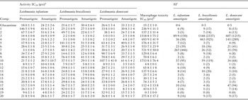
In a first step, we assayed the in vitro antileishmanial activity of compounds 1–20 on both extra- and intracellular forms of the parasites. Table 1 shows the IC50 values obtained after 72 h of exposure when compounds 1–20 were tested on extra- and intracellular forms of L. infantum, L. braziliensis and L. donovani. Toxicity values against J774.2 macrophage after 72 h of culture were also calculated and Selectivity Index (SI) values for the amastigote form have also been included in Table 1. Results obtained for the reference drug glucantime were included in all cases for comparison. An overall analysis of the biological data evidenced that nine of the screened compounds (3, 4, 7, 11, 12, 14, 17, 18 and 19) showed high activity against at least one of three Leishmania species in both promastigote and amastigote forms. For example, the SI of compound 3 exceeded that of the reference drug in L. infantum by 150-fold, by 2337-fold in L. braziliensis and by 1215-fold in L. donovani. Different authors have claimed that compounds having SI values >20 can be considered ideal candidates for further development as leishmanicidal drugs (Nwaka and Hudson, Reference Nwaka and Hudson2006). This requirement is satisfied by compounds 3, 4, 6, 7, 10 and 17 (17 only in L. donovani).
In vitro activity and toxicity for the arylaminoketone derivatives on extra- and intracellular forms of Leishmania spp.

Results are averages of four separate determinations, ±s.d.
a IC50: is the concentration required to give 50% inhibition, calculated by linear regression analysis.
b Against J774.2 macrophages after 72 h of culture.
c Selectivity Index (SI) = IC50 macrophages toxicity/IC50 activity on extracellular or intracellular forms of the parasite. In parenthesis: compound SI/reference drug SI.
Infectivity assay
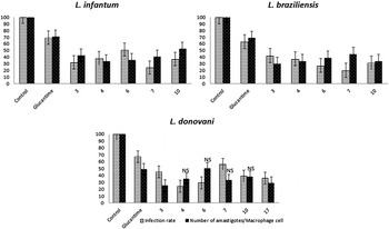
In order to gain a better insight into the activities of the lead compounds 3, 4, 6, 7, 10 and 17, their effect on the infectivity and intracellular replication of amastigotes was subsequently determined. Macrophage cells were grown and infected with promastigotes in the stationary phase. The parasites invaded the cells and underwent morphological conversion to amastigotes within 1 day after infection. On day 10, the rate of host cell infection reached its maximum (control experiment). We used the IC25 of each product as the test dosage. Figure 1 shows the effect of the studied derivatives on the infection and growth rates of the three Leishmania species. A measure of the average number of amastigotes per infected macrophage led to similar conclusions: in the case of L. infantum (Fig. 1A), all compounds were more effective than glucantime. Amastigote numbers obtained on L. braziliensis (Fig. 1B) also showed that all compounds were clearly more effective than glucantime under the tested conditions. It was also observed that the infection rate decreased with respect to the control and, furthermore, the six compounds (3, 4, 6, 7, 10 and 17), were also remarkably more effective in decreasing parasite infectivity than glucantime at an IC25 dose.
Effect of arylaminoketone derivatives 3, 4, 6, 7 and 10 on the infection and growth rates and mean numbers of amastigotes per infected J774.2 macrophage cell (at IC25 concentration) of Leishmania infantum (A), Leishmania braziliensis (B) and Leishmania donovani (C). Values are the means of three separate experiments. All compounds are statistically significant against glucantime at a P value < 0·05, except compounds labelled as NS.

Metabolite excretion
Trypanosomatids are unable to completely degrade glucose to CO2, so that they excrete part of the hexose skeleton into the medium as partially oxidized fragments. The nature and percentage of the oxidized fragments depend on the pathway used for glucose metabolism (Turrens, Reference Turrens2004). The catabolism products in Leishmania species are principally succinate, acetate, D-lactate and L-alanine (Kirkinezos and Moraes, Reference Kirkinezos and Moraes2001).
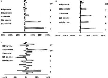
In order to acquire information regarding the effects of 3, 4, 6, 7, 10 and 17 on the glucose metabolism of the parasite, we obtained the 1H NMR spectrum of three species of Leishmania (L. infantum, L. braziliensis and L. donovani) promastigotes treated with the test compounds (compound 17 only in L. donovani); the final excretion products were qualitatively and quantitatively identified. Figure 2 shows the results obtained and the comparison with those found for untreated control promastigotes.
Variation percentages in the area of the peaks corresponding to excreted catabolites by Leishmania infantum (A), Leishmania brasilienzis (B) and Leishmania donovani (C) promastigotes in the presence of compounds 3, 4, 6, 7, 10 and 17 at their IC25 compared with a control sample after 96 h of incubation.

All the compounds induce an increase in succinate production in the three species of Leishmania ranging from 16·1 to 251·3% (compound 17 only in L. donovani) as can be observed in Fig. 2. This effect is observed in L. donovani to a lesser extent (Fig. 2C) except for compound 10 that presents a higher accumulation of succinate in L. donovani than in L. infantum and L. braziliensis.
SOD enzymatic inhibition in the Leishmania parasites and in human erythrocytes
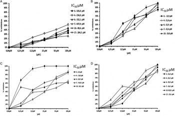
Considering the obtained results, we decided to test the effects of these compounds on Fe-SOD isolated from L. infantum, L. braziliensis and L. donovani over a range of concentrations, from 0·1 to 100 µ m. We used promastigote forms of both species, which excrete Fe-SOD when cultured in a medium lacking inactive FBS (Kirkinezos and Moraes, Reference Kirkinezos and Moraes2001). The inhibition data obtained are shown in Fig. 3(A–C), and the corresponding IC50 values are included for easier evaluation of the displayed graphs; for comparison, Fig. 3A shows the effects of the same compounds on CuZn-SOD obtained from human erythrocytes.
(A) In vitro inhibition of CuZn-SOD in human erythrocytes by compounds 3, 4, 6, 7, 10 and 17. (B–D) In vitro inhibition (%) of Fe-SOD of Leishmania infantum (B), Leishmania braziliensis (C) and Leishmania donovani (D) promastigotes by compounds 3, 4, 6, 7, 10 and 17. Values are the average of three separate determinations. Differences between the activities of the control homogenate and those incubated with the tested compounds were obtained according to the Newman–Keuls test. IC50 was calculated by linear regression analysis.

Regarding the SOD enzymatic inhibition in the Leishmania parasites and in human erythrocytes (Fig. 3), the most remarkable result was the inhibitory effect on Fe-SOD found for the highly antileishmanial compounds 3, 4 and 7 in the three species tested, whereas their inhibition of human CuZn-SOD was clearly lower. If we consider the IC50 calculated for L. infantum, inhibition of Fe-SOD by compounds 3, 4 and 7 was 25-, 11- and 29-fold higher, respectively, than inhibition of CuZn-SOD. Compound 3 showed a Fe-SOD inhibition 25, 66 and 10 times higher than CuZn-SOD inhibition in L. infantum, L. braziliensis and L. donovani, and compound 7 showed the respective values of 29, 14 and 36. Therefore, compounds 3 and 7 could be considered the most selective inhibitors of Fe-SOD.
DISCUSSION
As explained above (Moreno-Viguri et al. Reference Manzano, Cochet, Boucherle, Gomez-Perez, Boumendjel, Gamarro and Peuchmaur2016), previous studies have indicated that arylaminoketone Mannich base-type compounds may be considered prospective chemotherapeutic drugs in the treatment of Chagas disease caused by T. cruzi parasites (Turrens, Reference Turrens2004). We now comment on the results obtained regarding the antiparasitic activity of compounds 1–20 (Table 1) against three significant species of Leishmania: L. infantum, L. braziliensis and L. donovani. It was shown that the inhibition activities against intracellular forms of the parasites of studied compounds (17 with efficacy only against L. donovani) were higher than those found for the reference drug glucantime, whereas the effect on extracellular forms was more random. Regarding the toxicity in mammalian cells, the tested compounds were found to be much less toxic for macrophages than the reference drug. Therefore, compounds 3, 4, 6, 7, 10 and 17 were considered the lead ones due to their excellent antileishmanial activity and were selected for subsequent studies.
Interestingly, the best SI results for the more representative intracellular forms were obtained in L. infantum and L. donovani, two species forming part of the L. donovani complex, pointing towards a greater specificity towards parasites causing the particularly harmful visceral leishmaniasis in both its European and American versions.
With regard to the structure–activity relationship, in general, derivatives with the benzo[b]thiophene scaffold are less cytotoxic than the rest of the derivatives.
In the infectivity assay (Fig. 1), all the compounds were more effective in relation to IC25 than glucantime. The infection rates decreased with respect to the control and the reference drug glucantime. The measure of the average number of amastigotes per infected macrophage led to similar conclusions. All these data seem to be in line with results previously described for T. cruzi (Moreno-Viguri et al. Reference Moreno-Viguri, Jiménez-Montes, Martín-Escolano, Santivañez-Veliz, Martin-Montes, Azqueta, Jimenez-Lopez, Zamora Ledesma, Cirauqui, López de Ceráin, Marín, Sánchez-Moreno and Pérez-Silanes2016).
Regarding the studies to elucidate the possible mechanism of action, the studied compounds produce greater glucose metabolism alteration because they increase succinate excretion (Fig. 2). Detection of large amounts of succinate as a major end product is a usual feature, because it relies on glycosomal redox balance, enabling re-oxidation of the NADH produced in the glycolytic pathways. It is interesting to mention that the increase in succinate with these compounds indicates catabolic changes that could be related to mitochondria malfunction, due to the redox stress produced by inhibition of the mitochondrion-resident Fe-SOD enzyme (Marín et al. Reference Marín, Ramírez-Macías, López-Céspedes, Olmo, Villegas, Díaz, Rosales, Gutiérrez-Sánchez and Sánchez-Moreno2011).
In addition, these compounds led to greater levels of Fe-SOD inhibition. All these data appear to confirm some type of relation between the antileishmanial activity and the Fe-SOD inhibition, coinciding with the results described in previous work (Ginger, Reference Ginger2005). Fe-SOD inhibition could also, at another level, be related to the catabolic changes discussed above because a mitochondrial malfunction, originated from the redox stress produced by inhibition of the mitochondrion-resident Fe-SOD enzyme (Marín et al. Reference Marín, Ramírez-Macías, López-Céspedes, Olmo, Villegas, Díaz, Rosales, Gutiérrez-Sánchez and Sánchez-Moreno2011) should result in severe alteration of pyruvate metabolism, and consequently, a decrease in the production of succinate. Because the Fe-SOD present in mitochondria is an essential part of the antioxidant protective response of the parasite, its inhibition would be related to a decrease in the rate of survival for the parasite.
SUPPLEMENTARY MATERIAL
The supplementary material for this article can be found at https://doi.org/10.1017/S0031182017001123.
FINANCIAL SUPPORT
This work has been carried out with the financial support of Fundación Caja Navarra (Project no. 70314), Fundación María Francisca de Roviralta, FIMA (Fundación para la Investigación Médica Aplicada; Project ISTUN-API-2011/01) and from the former Spanish Ministry of Science and Innovation (MICINN) and now from the Ministry of Economy and Competitiveness (MINECO) (project Consolider Ingenio CSD2010-00065). M.J. is indebted to the University of Navarra for a grant. R.M-E. is grateful for a FPU Grant (no. FPU14/01537) from the Ministry of Education of Spain.