Pulmonary atresia with intact ventricular septum has been reported to occur in from 4.5 to 5.4 cases in each 100,000 live births.Reference Daubeney, Delany and Anderson1, Reference Walmsley, Sandor, Potts, Imai and Momma2 Despite improved mortality in recent years,Reference Ashburn, Blackstone and Wells3 the combination remains a frequently lethal condition, not least because of associated anomalies. These include hypoplasia of the tricuspid valve or right ventricle, and severe coronary arterial anomalies, especially the presence of a right ventricular-dependent type of coronary arterial circulation. Coronary arterial abnormalities include anomalous origins and patterns of distribution, stenoses, dilations, and occlusions. We undertook this retrospective study to elucidate the prevalence and types of such anomalies, also reviewing cineangiocardiograms to evaluate the proportions of left ventricular myocardium dependent on right ventricular flow, utilizing a myocardial score previously validated in adults with coronary arterial disease.Reference Brandt, Partridge and Wattie4 We then assessed the relationship between the presence of coronary arterial abnormalities and postoperative survival.
Material and methods
We identified 121 patients from databases in the Departments of Paediatric Cardiology, Surgery and Radiology, including only 110 patients who had had angiograms, 88 submitted to cardiac surgery, or 77 fulfilling both these criterions. Detailed post mortem examinations had been performed in 47 patients, with histological findings from 24 of these included in a previous publication.Reference Calder, Co and Sage5 The cardiac catheterizations were performed between 1961 and 2002, and the initial surgical procedures between 1966 and 2002.
We excluded any patients with critical pulmonary stenosis or those with small, restrictive, ventricular septal defects, despite these patients frequently having suprasystemic pressures in the right ventricle, and some having fistulas. We also excluded patients with congenital tricuspid atresia and other additional anomalies, such as supravalvar aortic stenosis.
All the angiograms were reviewed by the paediatric cardiac radiologists (CJO and CRP), who were blinded to clinical details or outcome. All angiograms available, whether with injections in the right ventricle, left ventricle or aortic root, were examined to extract potential data for use in the study.
Coronary arteries
The coronary arterial abnormalities could be assessed in 116 patients, in 108 from angiograms and in 6 from post mortem data only. In 2 others, where the angiograms were not available for review, the findings in the contemporaneous radiology reports had been confirmed at post mortem. Not all measurements could be made in all patients. When the angiograms were incomplete, post mortem data, if available, was used to augment the information.
The presence of right ventricular to coronary arterial fistulous connections, their location, and associated interruptions or stenoses were recorded, along with dominance of the right or left coronary artery. The severities of the stenoses were measured as proportional loss of diameter, expressed as a percentage. A loss of 50%, representing a loss of cross-sectional area of 75%, is considered to be haemodynamically significant.
The severity of the fistulas was graded on a score of zero to 4. Zero indicated absence of fistulas. Small or insignificant fistulas, with most not seen on follow up angiograms nor identified at post mortem examination, were graded as 1, while significant, usually multiple, fistulas filling the aortic root from a right ventricular injection were graded as 2. Those associated with one interruption in a major coronary artery were graded at 3, whereas those associated with interruptions in two different and major coronary arteries received the maximal grade of 4.
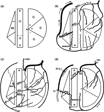
A myocardial score was obtained for the proportion of left ventricular myocardium whose supply was right ventricular dependent.Reference Brandt, Partridge and Wattie4 The scoring system divides the left ventricular myocardium into 15 equal units (Fig. 1).Reference Brandt, Partridge and Wattie4, Reference Kirklin and Barratt-Boyes6 The number of units affected by an upstream coronary arterial lesion is multiplied by a factor, reflecting the severity of the proximal narrowing, ranging between 0 for those with no stenosis to 1 for those with total occlusion. Thus the severity of the stenosis or complete occlusion and the proportion of myocardium affected are combined. A normal coronary arterial circulation would have a score of 0, whereas a total score of 15 would represent proximal occlusion of all major epicardial arteries.Reference Brandt, Partridge and Wattie4
(a) Diagrammatic representation of the segments of the left and right ventricles, including the ventricular septum (modified from Brandt et al.).Reference Brandt, Partridge and Wattie4, Reference Kirklin and Barratt-Boyes6 The heart is viewed from the apex (a). The free wall of the left ventricle is flattened into a semi circle so that the curved perimeter represents the atrioventricular groove. The left ventricular free wall is divided into diagonal area (D), obtuse marginal area (O), and inferior (diaphragmatic) area (I). The septum is drawn as a central oblong with anterior (A) and posterior (p) portions. Each left ventricular myocardial segment is given a numerical value (numbers in circles) for a total of 15. (b–d) Examples of diagrams to illustrate the coronary arterial assessment and derivation of myocardial score for right ventricular dependent circulation in 3 separate patients. Arrows show direction of flow. Dilation of an artery shown by increased thickness of line. X=interruption (100%) or stenosis (%=diameter loss). Wavy line with arrow=fistulas. (b) Several fistulas connect the right ventricle to the right and left anterior descending coronary arteries. The right coronary artery is dominant. Classified as significant fistulas (Grade2). Myocardial score= 0. (c) Multiple fistulas present. Interruptions in left anterior descending and distal right coronary arteries. Although interruptions are present in two coronary arteries, only a small portion of left ventricular circulation is right ventricular dependent, therefore the myocardial score is 2, with the fistulas graded at 4. (d) Absence of connection of the main stem of the left coronary artery to the aorta, plus two interruptions in left anterior descending and mid portion of right coronary artery. The coronary myocardial score is 15, with the fistulas graded as 4. The left coronary artery is dominant.

The coronary artery passing round to the crux of the heart and supplying the inferior interventricular artery was nominated as the dominant artery.
Size of tricuspid valve, mitral valve and right ventricle
The diameters of the tricuspid and mitral valves were measured in preoperative angiograms, using calibration from centimetre grids, or from the diameter of the catheter when no grids were available. Z scores were obtained using the nomograms,Reference Kirklin and Barratt-Boyes7, Reference Rowlatt, Rimoldi and Lev8 and the ratio of the tricuspid to the mitral valve calculated.
The global right ventricular size was subjectively assessed, and assigned a score ranging from −5 for those very severely hypoplastic to +5 for those deemed to be very severely dilated. A normal right ventricle was assigned a score of 0. The size of the tricuspid valve was compared with right ventricular size, presence of coronary fistulas, and the severity of any coronary arterial abnormalities. The presence and amount of any tricuspid valvar regurgitation was assessed subjectively, and categorised as zero when absent, mild in presence of small amounts of contrast medium passing retrogradely but not opacifying the right atrium, moderate when larger amounts of contrast passed back and opacified the whole right atrium, and severe when a large volume of contrast was seen moving retrogradely across the tricuspid valve.
In the post operative patients, survival was assessed in relation to the presence of fistulas, absence of connections between the coronary arteries and the aorta, interruptions or stenosis in coronary arteries with right ventricular-dependent coronary circulation, the myocardial score, the sizes of the tricuspid valve and right ventricle, and age and size in neonates.
Statistical methods
Results are reported as range and median or interquartile range or number and percentage when appropriate. Differences across groups were compared using a Mann-Whitney U test. To assess the association, if any, between the myocardial score and the grade of fistulous communications, a Spearman’s rank correlation coefficient was calculated. Probability of survival in the post operative patients was evaluated with Kaplan-Meier actuarial curves and factors correlating with survival assessed by univariate and multivariate Cox hazard regression.
Results
There were 61 males and 60 females. The median age at presentation was 4 days, with an interquartile range from 2 to 9 days. Major aortopulmonary collateral arteries were found in only two patients. Of these, one died at 10 days of age in 1961; while the other was the sole survivor without surgery, and was alive at 16 years before becoming lost to follow up. All remaining patients were dependent upon patency of the arterial duct, many before the availability of prostaglandin.
Coronary arterial abnormalities
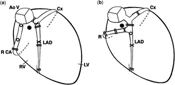
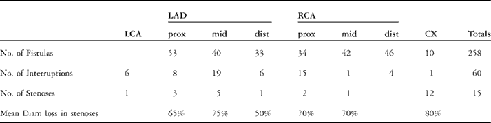
We discovered fistulas in 87 of the 116 patients (75% – Table 1). As expected, they occurred most commonly in the coronary arteries adjacent to the right ventricle (Fig. 2a). Only 4% of the fistulas were found joining the circumflex coronary artery.
In Figure 2a, we show a diagram of the usual origin and pattern of distribution of coronary arteries in pulmonary atresia with intact ventricular septum. The aortic valve (AoV) and atretic pulmonary valve (represented by black circle) are seen in a superior, plan view whereas the ventricles are viewed from a frontal, right anterior projection. Right (RCA) and left coronary arteries arise from the right and left sided aortic sinuses. The left gives origin to the left anterior descending (LAD) and circumflex (Cx) coronary arteries. Note the rightward position of LAD demarcating the hypoplasia of right ventricle (RV). Fistulas found at sites of interruptions (==), or stenoses (X) are not shown in the diagrams. Additional fistulas=O, LV=left ventricle. In Figure 2b, we show single coronary arterial origin, with the right coronary artery (R) arising from the left anterior descending coronary artery (LAD). The RCA passes anteriorly to the pulmonary trunk. If this course were not recognized, the RCA would be at risk from surgical procedures to enlarge the right ventricular outflow. Abbreviations and symbols as in Figure 2a.

A single origin of the coronary arteries was found in six patients (5%), with the right coronary artery arising from the left coronary artery in all cases (Table 1). In the five patients where the right coronary artery arose from the left anterior descending coronary artery, it passed anteriorly to the pulmonary trunk (Fig. 2b). In the other case, in which the right coronary artery arose from the main stem of the left coronary artery, it coursed between the aorta and the pulmonary trunk.Reference Calder, Co and Sage5 In two other patients, the circumflex coronary artery arose from the right coronary artery (Table 1).
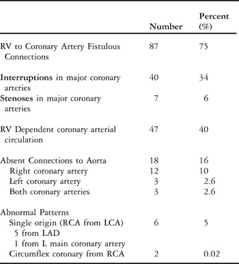
Interruptions in the major coronary arteries occurred in 40 of the 116 patients (34% – Tables 1 and 2). A number of patients also had additional stenoses at other sites in the same artery, or in other arteries, with 7 additional patients having significant stenoses in coronary arteries without any interruptions. The sites are listed in Table 3.
Prevalence of coronary arterial abnormalities in 116 patients.

LAD = left anterior descending coronary artery, LCA = Left coronary artery, RCA = Right coronary artery, RV = Right ventricle.
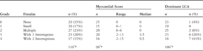
Correlation of severity of abnormalities with dominant left coronary artery.

* = number where data available, LCA = left coronary artery.
Sites of coronary arterial abnormalities in 87 patients with fistulas.

CX = circumflex coronary artery, dist = distal, LAD = left anterior descending coronary artery, LCA = left main coronary artery, No. = number, Prox = proximal, RCA =right coronary artery.
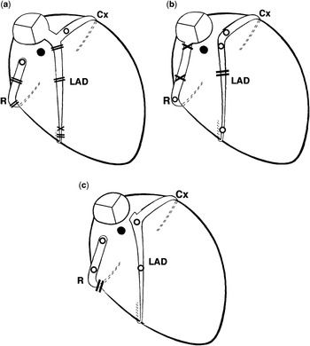
Absence of connection between a proximal coronary artery and the aorta was found in 18 patients (16%), specifically between the right coronary artery and the aorta in 15, including 3 patients with absence of connections of both coronary arteries, and to the left coronary artery in 6, including the 3 with absence of connections of both coronary arteries (Table 1, Fig. 3). The distance between the origin of the coronary arterial branch and the aorta varied from zero in those with an atretic orifice to 10 millimetres.
Diagrams of absent connections between the coronary arteries and the aorta. (a) the right (R) coronary arterial origin is absent. (b) the origin of left main coronary artery is absent. (c) the origins of both right (R) and left main coronary are absent. Symbols and abbreviations as in Figure 2.

The myocardial score, indicating the proportion of left ventricular myocardial flow which is right ventricular-dependent, was normal in 53 of the 96 patients (55%) in whom the whole coronary arterial tree cold be seen on the images available, and abnormal, with a range from 1 to 15, and a mean of 6.4 ± 4.3, in the other 43 patients (45% – Fig. 1). The higher score correlated with the increasing grade of fistulous communications (p<0.001 – Table 2). In 47 patients, the coronary circulation was at least partly right ventricular-dependent, detected in 43 from angiograms and in 4 from post mortem examination.
The left coronary artery was dominant in 16 of 106 patients (15%) in whom the data were available. The prevalence of left coronary arterial dominance increased with the more severe coronary arterial lesions, with 13 of 39 patients with interruptions (33%) having dominant left coronary arteries (p< 0.001 – Table 2).
Size of right ventricle and tricuspid valve
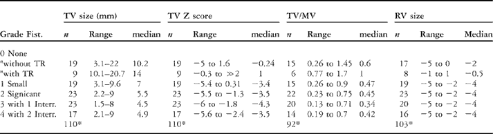
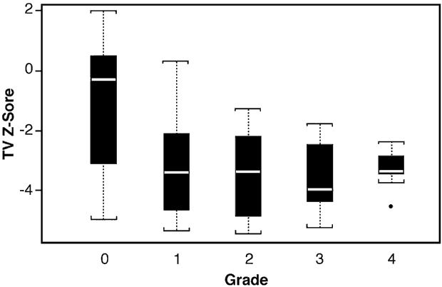
The right ventricular size as estimated from the angiograms correlated with the Z score for the tricuspid valve (p< 0.001, Table 4). The Z scores were in the normal range in only 18% patients, that is 20 of 109, being minus 2 or less in 78%, or 85 patients, and plus 2 or more in 4%, or 4 patients. The ratio of size of the tricuspid and mitral valves was less than normal in 86 of 92 patients (93%). Fistulas were present in 72 of these (84%). The normal ratio is from 1 to 1.4.Reference Calder, Co and Sage5 In patients without significant tricuspid regurgitation, the Z scores for the tricuspid valve ranged from minus 5.6 to plus 1.6, and ratios ranged from 0.13 to 1.4. The smaller tricuspid valve size, Z scores and ratios all correlated with increasing severity of coronary arterial lesions (p< 0.001 – Table 4, Fig. 4).
Correlation of fistulas with hypoplasia of the tricuspid valve and right ventricle.

Fist = fistulas, Interr. = interruptions (s), n = number, MV = mitral valve, TR = significant tricuspid regurgitation, TV = tricuspid valve.
*=number where data available.
Box plot showing the lower Z scores for the tricuspid valve (TV) with the increasing severity of coronary fistulas. p = < 0.001. Grades: 0=none, 1=minor or small, 2=significant, usually multiple, 3=associated with one interruption in a major coronary artery, and 4=interruptions in two coronary arteries.

Significant tricuspid valvar regurgitation, with dilation of the annulus, was found in 11 patients. In these, the regurgitation, as graded from the angiograms was severe in 3. The Z scores ranged from −0.3 to plus 2, with a median of 1, and the ratio relative to the mitral valve ranged from 0.8 to 1.7, again with a median of 1. In 10 patients the tricuspid valve was dysplastic or had Ebstein’s malformation. Although the 11th patient had severe tricuspid regurgitation with dilation of the tricuspid valve and right ventricle at the initial study, she successfully underwent a biventricular repair, and now has mild-to-moderate residual tricuspid regurgitation at 17 years of age.
In one rare patient with severe tricuspid regurgitation and marked dilation of the right atrium and right ventricle on pre-operative echocardiograms, we discovered a fistulous communication from the right ventricle to the right coronary artery on subsequent angiograms.
Effect of coronary arterial abnormalities on surgical results
Surgical procedures
A variety of operations were performed in 88 patients by seven surgeons over more than three decades. The range of ages at the initial procedure was from one day to 9.9 years, with a median of 6 days, and a mean of 87 days plus or minus 469 days. Of the patients, 76 were less than 30 days of age at their first operation. In 47 patients, a systemic-to-pulmonary arterial shunt was performed as the first stage of palliation. In 35 patients, the shunt was complemented with opening of the right ventricular outflow tract, either by a pulmonary valvotomy or a Brock-type operation, this group including 11 patients with tricuspid regurgitation. Of these, 4 had tricuspid valvoplasty, l had closure of the tricuspid valve and 1 had the valve replaced later. In 6, right ventricular decompression was carried out in isolation.
Among the 62 hospital survivors, 19 have had a Fontan procedure, 7 a one and a half ventricular repair, and 17 a biventricular repair. In 17 patients, death occurred before repair could be attempted, and 2 further patients are alive without any subsequent procedures. The median duration of follow-up since the first operation for those surviving was 6.1 years, with a range from 39 days to 36.6 years, and a mean of 7.9 plus or minus 8.5 years. We have lost 3 patients to long term follow up because they returned to their country of origin.
Coronary arterial abnormalities
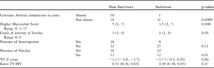
Data about the coronary arteries was available in 83 of the patients who had undergone surgery. Fistulas were present in 58 of these (70%), interruptions in one or two coronary arteries in 26 (31%), and lack of connections of the coronary arteries to the aorta in 11 (13%).
Factors affecting survival
In patients who had undergone surgery as neonates, smaller body weight (p = 0.02), younger age at operation (p = 0.04), and hypoplasia of the right ventricle (p = 0.005) were significant factors relative to survival. Survival was not related to the type of initial surgical procedure.
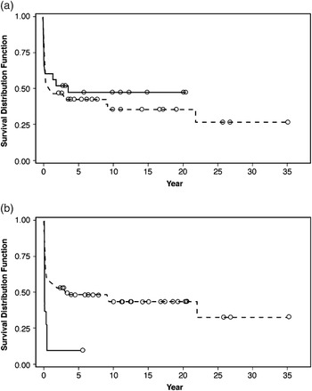
For the total group undergoing surgery, the presence of fistulas did not alter the survival after surgical procedures (Table 5). Although the presence of interruptions was not statistically significant (p = 0.11), the increasing severity of fistulas with associated interruptions was significant (p = 0.05). Survival was reduced by the presence of a right ventricular-dependent circulation with interruptions or stenoses of coronary arteries producing a higher myocardial score (p = 0.008), or absence of connections with the aorta (p = 0.0009 – Fig. 5, Table 5).
Factors affecting survival.

TV = tricuspid valve, MV = mitral valve.
Actuarial survival curves. (a) There is no significant difference in post operative survival for those without fistulous connections from the right ventricle to the coronary arteries (______) compared with those with fistulas (……). (b) The survival in the patients with absence of connections between the coronary arteries and the aorta (____) is significantly worse that those without (….). p = 0.009.

Tricuspid valvar regurgitation
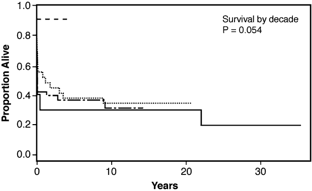
The post-operative survival was similar in patients with or without significant tricuspid regurgitation (p = 0.11), and these patients have been included in the results. For the total group, the survival at 5 years is 42%, with an interquartile range from 33% to 54%. Survival at 10 years is 38%, the interquartile range being from 29% to 51%. If the patients with significant tricuspid regurgitation are excluded, the five year survival is 40%, with interquartile range from 30% to 53%, and 10 years survival is 36%, with interquartile range from 26% to 49%. In the most recent decade, results are much improved (Fig. 6), perhaps related to improved surgical techniques, postoperative care, and better selection of patients.
Actuarial curves comparing survival between the decades in which patients had their first operative procedure. Although the number of patients in the 2000s (- - - - -) is smaller, their survival is significantly improved compared to earlier decades: 1966 to 1979 =_____, 1980 to 1989=……., 1990 to 1999=_ ._. _. .

Discussion
Prevalence of coronary arterial abnormalities
We found fistulas in three-quarters of our patients, this being greater compared to previous reports in series of over 50 patients, with a prevalence of 32 to 56%.Reference Daubeney, Delany and Anderson1, Reference Ashburn, Blackstone and Wells3, Reference Giglia, Mandell, Connor, Mayer and Lock9–Reference Coles, Freedom and Lightfoot14 The prevalence of fistulas in surgical series from multiple institutions of 38 to 45%,Reference Ashburn, Blackstone and Wells3, Reference Hanley, Sade, Blackstone, Kirklin, Freedom and Nanda12 or a single institution of 51 to 56%,Reference Lightfoot, Coles and Dasmahaptra13–Reference Dyamenahalli, McCrindle and McDonald15 was again less than in our patients undergoing surgery, seven-tenths of whom had fistulas.
Similarly, the prevalence of interruptions was greater in our study, being found in one-third, compared to other reports finding them in no more than one-quarter.Reference Daubeney, Delany and Anderson1, Reference Ashburn, Blackstone and Wells3, Reference Coles, Freedom and Lightfoot14, Reference Powell, Mayer, Lang and Lock16, Reference Humpl, Soderberg and McCrindle17 The sites of the abnormalities in the coronary arteries, however, was similar, with only minor differences from the distributions reported by others.Reference Daubeney, Delany and Anderson1, Reference Giglia, Mandell, Connor, Mayer and Lock9 FreedomReference Freedom18 reported absence of proximal connections of the coronary arteries to the aorta in only 4 of 82 patients, whereas we found this anomaly in one-sixth of our cohort. Since the first report in 1972,Reference Lenox and Briner19 we are aware of 8 additional patients reported with lack of connection of both coronary arteries to the aorta.Reference Powell, Mayer, Lang and Lock16, Reference Lenox and Briner19–Reference Selamet, Hsu, Thaker and Gersony25 As far as we are aware, none have survived more than two months of age.
A right ventricular-dependent coronary circulation has been variably defined as an interruption or stenosis in one coronary arteryReference Jahangiri, Zurakowski, Bichell, Mayer and del Nido26 to interruptions in two major coronary arteries.Reference Giglia, Mandell, Connor, Mayer and Lock9, Reference Powell, Mayer, Lang and Lock16, Reference Humpl, Soderberg and McCrindle17 The myocardial score adds a more precise, objective and quantitative assessment of the effect on the left ventricular myocardial arterial supply.
Burrows et alReference Burrows, Freedom and Benson27 reported a single coronary artery in 4 of 29 patients. Only one of these patients, however, had a solitary coronary artery, with two having single origins with the right coronary artery arising from the left coronary artery, and one with lack of connection of both coronary arteries to the aorta. We found such abnormal patterns in 6 of our patients (5% – Table 1). Dominance of the left coronary artery has been found on coronary arterial angiograms in around one-twentieth of otherwise normal adults.Footnote ☆ In selected patients with pulmonary atresia and intact ventricular septum with fistulous communications, Burrows et al.Reference Burrows, Freedom and Benson27 found a dominant left coronary artery in one-quarter, while we discovered such dominance in one-third of our patients with an interrupted coronary arterial tree. We are not aware of any other reports noting the increased prevalence of left coronary arterial dominance with an increasingly right ventricular-dependent coronary arterial circulation.
Correlation with the size of the right heart
We found similar Z scores for the tricuspid valve as those reported previously,Reference Daubeney, Delany and Anderson1, Reference Ashburn, Blackstone and Wells3 and the correlation between fistulous communications and the small size of the tricuspid valve and/or right ventricle has also been reported previously.Reference Calder, Co and Sage5, Reference Satou, Perry, Gauvreau and Geva23, Reference Zuberbuhler and Anderson28, Reference Giglia, Jenkins and Matitiau29
Although FreedomReference Freedom, Moller and Hoffman30 said that tricuspid regurgitation and fistulous connections were mutually exclusive, there are two known cases. The first was reported by Mair et al.Reference Mair, Julsrud, Puga and Danielson31 Our patient with the significant tricuspid regurgitation and a fistula is the second one, as far as we know.
Predictors of survival
In our patients who had surgery as neonates, and as others have reported, there is increased mortality associated with low birth weight,Reference Ashburn, Blackstone and Wells3, Reference Rychik, Levy, Gaynor, DeCampli and Spray11, Reference Hanley, Sade, Blackstone, Kirklin, Freedom and Nanda12, Reference Joshi, Brawn and Mee32 or small right ventricles and small tricuspid valves.Reference Walmsley, Sandor, Potts, Imai and Momma2, Reference Rychik, Levy, Gaynor, DeCampli and Spray11, Reference Joshi, Brawn and Mee32 The correlation between hypoplasia of the right ventricle and tricuspid valve with increasing severity of coronary arterial lesions was associated with increased mortality in our neonatal patients, again similar to other reports.Reference Sauer, Bindl, Pilossoff, Doyle, Engle, Gersony, Rashkind and Talner10, Reference Joshi, Brawn and Mee32
Joshi et alReference Joshi, Brawn and Mee32 stated that risk factors for mortality before definitive repair could be clustered into two groups: those with right ventricular-dependent coronary arterial circulation and those others with tricuspid regurgitation. The severity of interruptions and right ventricular dependent coronary arterial circulation was significant in both our own study and others.Reference Rychik, Levy, Gaynor, DeCampli and Spray11, Reference Hanley, Sade, Blackstone, Kirklin, Freedom and Nanda12
Diagnosis of coronary arterial abnormalities
The most appropriate management for patients depends upon thorough investigation.Reference Dyamenahalli, Hanna and Sharratt33 Formal angiography is still required to image the coronary arteries in neonates at present. Although magnetic resonance imaging has been shown to detect lesions in coronary arteries, this has not been achieved consistently even in adults.Reference Kefer, Coche and Legros34 Computerised tomographic coronary angiography is becoming a reliable clinical technique, but the fast resting heart rate and the small size of a neonate’s arteries makes this technique impractical as yet in this clinical setting. The diameter of the major epicardial vessels is about 1 millimetre in neonates.Reference Oberhoffer, Lang and Fellen35
We recommend that invasive right ventriculography and aortography, with punctilious attention to excellent opacification of the right ventricle and aortic roots respectively, be obtained in all such patients. Imaging should be performed biplane, at high frame rates, and with identical camera angles used for both angiographic runs to allow for comparison between the views. Interpretation of such angiograms is extremely demanding, and should be reported by the most experienced staff available. Some of the apparent differences in incidence of fistulas reported by different authors may well be due to differences in angiographic techniques and interpretation.
Limitations of study
Our retrospective review includes patients seen over a period of 4 decades, and may represent the most severe end of the spectrum of cases. Many of the patients are not from the current era, and thus surgical results cannot be compared directly with recent series.
Conclusion
Abnormalities of the coronary arterial circulation unequivocally affect the outcome of treatment for neonates with pulmonary atresia and intact ventricular septum. The presence of fistulous communications to the coronary arteries is not associated on its own with a higher mortality, but the presence of interrupted coronary arteries, and a higher myocardial score, is related to increased mortality. At present, formal angiography is still required to image the coronary arteries in neonates. Assessment of the coronary circulation requires punctilious attention to detail during the performance and interpretation of invasive angiography, particularly in view of the impracticality of selective coronary angiography at this age. With this in mind, we have used the myocardial score as an objective and quantitative measure of the severity of the effect of the coronary lesions on the arterial supply of the left ventricular myocardium.
Acknowledgements
We thank Barbara J Semb, Research Secretary, Green Lane Research and Education Fund, for secretarial assistance, Diane Stephenson for the illustrations, and Teena West and Irene Zeng for the statistical analysis.