

Introduction
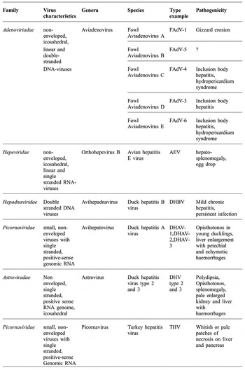
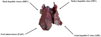
The liver contains great functional reserve capacity, which is important in domestic animals subjected to high production requirements. This organ adapts easily to different conditions by increasing the intensity of its functions. The liver has to cope with many challenges, including high energy level feed, the addition of chemotherapeutics, coccidiostats and other compounds, whereby metabolites must be maintained in equilibrium by hepatic homeostasis. Viral hepatitis in poultry is an important disease caused by a number of viruses including avian hepatitis E virus (avian HEV), duck hepatitis B virus (DHBV), duck hepatitis A virus (DHAV), duck hepatitis virus Types 2 and Type 3, fowl adenoviruses (FAdV), and turkey hepatitis virus (THV) ( Table 1 , Figure 1 ). DHBV or avihepadnaviruses, part of the family Hepadnaviridae, have been isolated from ducks, snow geese, white storks, grey herons, cranes, and parrots. DHBV evolved with the host as a non-cytopathic form without clinical signs and rarely progresses to chronicity. The outcome for DHBV infection varies by the host's ability to elicit an immune response and is dose and age dependent in ducks, thus mimicking the pathogenesis of human hepatitis B virus (HBV) infections and providing an excellent animal model for human HBV. DHAV is a picornavirus that causes a highly contagious virus infection in ducks with up to 100% flock mortality in ducklings less than six weeks of age, while older birds remain unaffected. Resulting high morbidity and mortality has an economic impact on intensive duck production farming. Duck hepatitis virus Types 2 and 3 are astroviruses, belonging to the family of Astroviridae, being similar phylogenetically to turkey astroviruses, and therefore has the potential for cross-species infections between strains. While these avian hepatitis viruses have the same target organ, the liver, they are very different in their biology and pathogenesis, as each belongs to a different family (Yugo et al., Reference YUGO, HAUCK, SHIVAPRASAD and MENG2016). Therefore, understanding the common and unique features of these hepatitis viruses in poultry will aid the diagnosis and differential diagnosis of viral hepatitis in poultry and help develop more effective flock management and preventive and control measures. The focus of this review is on viral hepatitis infections in chicken and turkeys, with emphasis on HEV, FAdV and THV.
Etiology of viral hepatitis in poultry.

Major Liver damaging viruses in poultry.

Fowl adenoviruses (FADV)
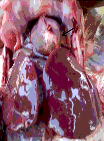
In 1953, Wallace Rowe, while investigating the ‘common cold’ isolated a new virus in human adenoid tissue grown in cell culture (Rowe et al., Reference ROWE, HUEBNER, GILMORE, PARROTT and WARD1953). In 1956, this new virus was formally named Adenovirus (AdV), derived from the Greek word for ‘gland’, the tissue where virus was first isolated (Enders et al., Reference ENDERS, BELL, DINGLE, FRANCIS, HILLEMAN, HUEBNER and PAYNE1956). These non-enveloped, icosahedral, linear and double-stranded DNA-viruses have been detected in every vertebrate class (Harrach et al., Reference HARRACH, BENKÖ, BOTH, BROWN, DAVISON, ECHAVARRIA, HESS, KAJON, LEHMKUHL, MAUTNER, MITTAL and WADELL2011) and cause two important diseases in poultry, namely hydro pericardium syndrome (HPS) and inclusion body hepatitis (IBH). In the late eighties, HPS was reported as a new disease in broilers with some clinical similarities to classical IBH in Pakistan, India, South America and several other countries. The disease was mainly characterised by accumulation of fluid in the pericardial sac and hepatitis ( Figure 2 ). HPS was first reported in broiler birds of three to five weeks of age from Angara Goth, near Karachi, Pakistan, in 1987 and is therefore commonly known as ‘Angara Disease’ (Mahmood et al., Reference MAHMOOD, ALI, HUSSAIN and ASLAM2014). Since then, the disease has spread to Iraq, Mexico, Peru, Chile, South and Central America, Russia, Slovakia and Korea (Asthana et al., Reference ASTHANA, CHANDRA and KUMAR2012).
Inclusion body hepatitis (IBH) was recognised for the first time in the USA by Helmboldt and Frazier (Reference HELMBOLDT and FRAZIER1963). Since then, the disease has been reported in many countries in the world (Rahimi and Minoosh Siavosh Haghighi, Reference RAHIMI and MINOOSH SIAVOSH HAGHIGHI2015). IBH is caused by FAdV of the genus Aviadenovirus (Group I avian adenoviruses) (Harrach et al., Reference HARRACH, BENKÖ, BOTH, BROWN, DAVISON, ECHAVARRIA, HESS, KAJON, LEHMKUHL, MAUTNER, MITTAL and WADELL2011). Group II avian adenoviruses cause haemorrhagic enteritis in turkeys and marble spleen disease in pheasants, and both pathogens belong to the genus Siadenovirus.
Duck adenovirus A causing egg drop syndrome in laying hens is a member of the genus Atadenovirus and designated as Group III avian adenovirus. Within the genus Aviadenovirus, six species (FAdV A-E and goose adenovirus) and three tentative species (duck adenovirus B, pigeon adenovirus, and turkey adenovirus B) are recognised (Harrach et al., Reference HARRACH, BENKÖ, BOTH, BROWN, DAVISON, ECHAVARRIA, HESS, KAJON, LEHMKUHL, MAUTNER, MITTAL and WADELL2011). Twelve serotypes can be distinguished within FAdV A-E (Meulemans et al., Reference MEULEMANS, COUVREUR, DECAESSTECKER, BOSCHMANS and VAN DEN BERG2004; Marek et al., Reference MAREK, GÜNES, SCHULZ and HESS2010). FAdVs causing IBH have predominantly been typed as fowl adenovirus D or E (Ojkic et al., Reference OJKIC, MARTIN, SWINTON, VAILLANCOURT, BOULIANNE and GOMIS2008; Marek et al., Reference MAREK, GÜNES, SCHULZ and HESS2010; Yugo et al., Reference YUGO, HAUCK, SHIVAPRASAD and MENG2016).
Accumulation of fluid in pericardium (arrow) and hepatitis in HPS.

All FAdVs, including those responsible for IBH, are assumed to have similar transmission. Horizontal transmission occurs mainly by the faecal-oral route (Kohn, Reference KOHN1962). The virus is shed in the faeces for up to six weeks (Cook, Reference COOK1974). Adenoviruses are very resistant in the environment, and therefore fomites contaminated with faeces are important in spreading the virus. Airborne transmission is probable, since the virus can be reisolated from the trachea, and experimental intratracheal infection or infection by eye drop have been successful (Cook, Reference COOK1974).
Vertical transmission is an important route of infection. Infected breeder hens shed virus in their eggs, thus infecting progeny, as early as one week after experimental infection, and for at least five weeks afterwards (Toro et al., Reference TORO, GONZÁLEZ, ESCOBAR, CERDA, MORALES and GONZALEZ2001). Several serotypes can be detected sequentially during the production period of a flock, or at the same time, without necessarily being correlated to disease (Cook, Reference COOK1974; Cowen, Reference COWEN1988). In Canada, approximately 77% of 231 randomly selected broiler flocks had been exposed to FAdV, with approximately 39% being positive for FAdV D or E viral DNA (Eregae et al., Reference EREGAE, DEWEY, MCEWEN, OUCKAMA, OJKIĆ and GUERIN2014). Outbreaks of IBH are often epidemic, such as those in the early 1970s in England (Macpherson et al., Reference MACPHERSON, MCDOUGALL and LAURSEN-JONES1974) and Canada (Pettit and Carlson, Reference PETTIT and CARLSON1972), and more recently in Korea and Japan (Choi et al., Reference CHOI, KYE, KIM, JEON, LEE, PARK and SUNG2012).
The clinical course of IBH varies depending on the virulence of the virus, the age of the infected chickens, and the presence of other pathogens, especially immunosuppressive chicken infectious anaemia virus (CIAV) and infectious bursal disease virus (IBDV). IBH has been observed in vertically infected chickens as young as five days, with mortality up to 5%. This has been experimentally reproduced in one day old specific-pathogen-free chickens who subsequently developed signs of the disease three days later (Steer et al., Reference STEER, SANDY, O'ROURKE, SCOTT, BROWNING and NOORMOHAMMADI2015). On the other hand, FAdV has been repeatedly isolated from layers and broiler breeders up to 45 weeks of age and confirmed as having IBH (Lim et al., Reference LIM, LEE, LEE, LEE, PARK, YOUN, KIM, YOUN, LEE, PARK, CHOI and SONG2011).
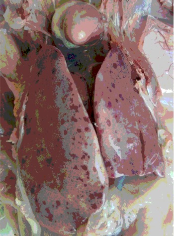
Generally, older chickens seem to be less susceptible than younger birds. Mortalities after experimental infection of two day-old chickens were between 50% and 83%, depending on the infective dose and route of infection, but only between 0% and 43% in two week old chickens (Dar et al., Reference DAR, GOMIS, SHIRLEY, MUTWIRI, BROWNLIE, POTTER, GERDTS and TIKOO2012). Co-infections with IBDV or CIAV aggravate the disease, and it was even doubted that FAdV were primary pathogens for IBH during co-infection studies (Fadly et al., Reference FADLY, WINTERFIELD and OLANDER1976). However, a high number of IBH cases were observed in New Zealand and Asia without the presence of IBDV and CIAV, and there was no correlation between cases of IBH or FAdV antibodies, respectively with antibodies against IBDV and CIAV in Canada and Belgium (Gomis et al., Reference GOMIS, GOODHOPE, OJKIC and WILLSON2006; Eregae et al., Reference EREGAE, DEWEY, MCEWEN, OUCKAMA, OJKIĆ and GUERIN2014). Clinical signs of IBH are nonspecific; chickens are depressed with ruffled feathers and having watery droppings (Alvarado et al., Reference ALVARADO, VILLEGAS, EL-ATTRACHE, JENSEN, ROSALES, PEROZO and PURVIS2007). After experimental oral or ocular infection of chickens, deaths were observed between five and 12 days post-infection, with varying mortality (Steer et al., Reference STEER, SANDY, O'ROURKE, SCOTT, BROWNING and NOORMOHAMMADI2015). Hepatic lesions are characterised by diffusely swollen, friable, and pale liver, sometimes with necrotic or haemorrhagic foci ( Figure 3 ) (Nakamura et al., Reference NAKAMURA, MASE, YAMAMOTO, TAKIZAWA, KABEYA, WAKUDA, MATSUDA, CHIKUBA, YAMAMOTO, OHYAMA, TAKAHASHI, SATO, AKIYAMA, HONMA and IMAI2011; Steer et al., Reference STEER, SANDY, O'ROURKE, SCOTT, BROWNING and NOORMOHAMMADI2015). Jaundice, especially in recovering birds, has been reported.
Enlarged and haemorrhagic liver in adenovirus (inclusion body hepatitis) infection in poultry.

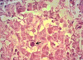
Histologic lesions are characterised by multifocal necrosis of hepatocytes and acute hepatitis. Intranuclear inclusion bodies are located in the nuclei of the hepatocytes (Howell et al., Reference HOWELL, MACDONALD and CHRISTIAN1970; Erny et al., Reference ERNY, BARR and FAHEY1991; Zhao et al., Reference ZHAO, ZHONG, ZHAO, HU and ZHANG2015) ( Figure 4 ). Lesions in other organs have rarely been observed. The mechanism of pathogenesis of IBH is largely unknown. Hepatitis with inclusion bodies caused by adenoviruses has been reported in pigeons, parrots, cockatiels (Nymphicus hollandicus), and falcons, as well as turkeys (Schelling et al., Reference SCHELLING, GARLICK and ALROY1989; Capua et al., Reference CAPUA, LIBERTI, GOUGH, CASACCIA and ASDRUBALI1995; Shivaprasad et al., Reference SHIVAPRASAD, WOOLCOCK and MCFARLAND2001). However, in most cases with a few exceptions, it is unclear if these viruses were FAdV or other adenoviruses. An adenovirus isolated from various psittacine species exhibiting hepatitis with inclusion bodies, was shown to be similar to FAdV D (Capua et al., Reference CAPUA, LIBERTI, GOUGH, CASACCIA and ASDRUBALI1995).
Diagnosis in acute cases is primarily based on pathologic lesions with laboratory confirmation by detection of FAdV, which is required for typing. Since FAdV are almost ubiquitous, mere detection of FAdV does not prove a causal relationship with observed lesions. FAdV can be isolated in embryonated chicken eggs as well as in cell culture (McFerran and Adair, Reference MCFERRAN and ADAIR1977; Cowen, Reference COWEN1988). Serologic methods such as ELISAs, serum neutralisation tests, and agar gel precipitation tests using anti-FAdV antisera can detect either all FAdV or specific serotypes, depending on the test (McFerran and Adair, Reference MCFERRAN and ADAIR1977; Grimes et al., Reference GRIMES, CULVER and KING1977; Junnu et al., Reference JUNNU, LERTWATCHARASARAKUL, JALA, PHATTANAKUNANAN, MOONJIT and SONGSERM2014). Numerous PCR assays have been used to detect FAdV followed by typing (Meulemans et al., Reference MEULEMANS, BOSCHMANS, BERG and DECAESSTECKER2001). Liver samples generally yielded more positive results than faecal samples for the PCR assays (Yugo et al., Reference YUGO, HAUCK, SHIVAPRASAD and MENG2016).
Biosecurity practices are the primary and essential step to prevent infection. Proper management, cleaning and disinfection of premises and equipment, restricted entry of visitors and vaccination crews in poultry houses play a significant role in prevention of the disease (Iqbal et al., Reference IQBAL, JAVED, HASSAN, KHAN and MUNIR2015; Ali et al., Reference ALI, ALI, AZEEM, UMAR, KHAN, ALI, SHAH and UMAR2014; Azeem et al., Reference AZEEM, REHMAN, UMAR, ASIF, ARIF and RAHMAN2014; Ahmad, Reference AHMAD2014; Shah, Reference SHAH2013). In countries with high infection exposure, the disease has been brought under control by formalin-inactivated vaccines prepared from liver homogenates from infected birds or by inactivated cell culture (Mahmood et al., Reference MAHMOOD, ALI, HUSSAIN and ASLAM2014). Several studies have reported the control of FAdv infection and revealed successful protection by immunisation with either attenuated or inactivated vaccines (Alvarado et al., Reference ALVARADO, VILLEGAS, EL-ATTRACHE, JENSEN, ROSALES, PEROZO and PURVIS2007; Kim et al., Reference KIM, LIM, LEE, YOUN, YUK, KIM, CHOI, JUNG and HAN2014; Junnu et al., Reference JUNNU, LERTWATCHARASARAKUL, JALA, PHATTANAKULANAN, ANGKASIYA MONKONG, KULPRASERTSRI and THIVALAI2015). An inactivated vaccine against FAdV is commercially available in China, and a live vaccine is available in Australia. Since IBH is of greatest concern in younger broilers, and maternal antibodies are capable of protecting progeny against IBH, broiler breeders are vaccinated with inactivated FAdV to induce maternal antibodies, which are then passed on to their progeny (Alvarado et al., Reference ALVARADO, VILLEGAS, EL-ATTRACHE, JENSEN, ROSALES, PEROZO and PURVIS2007). In Australia, a live FAdV E vaccine has been used to control IBH in parent flocks since 1989 (Steer et al., Reference STEER, O'ROURKE, GHORASHI and NOORMOHAMMADI2011). However, in spite of the vaccination, there are still sporadic outbreaks of IBH in Australia.
Liver, large eosinophilic intra-nuclear inclusion body (Arrow), (Rahimi and Minoosh Siavosh Haghighi, Reference RAHIMI and MINOOSH SIAVOSH HAGHIGHI2015).

Avian hepatitis E virus (Avian HEV)
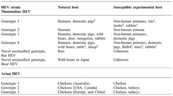
Avian HEV is a small, non-enveloped, positive-sense single stranded RNA virus of approximately 7.2 kb in genomic size that belongs to the Hepeviridae family. The HEV nomenclature system has been in constant change due the frequent identification of novel strains of the virus in various animal species (Pavio et al., Reference PAVIO, MENG and DOCEUL2015). Recently, a novel classification has been proposed that divides this family into two genera Orthohepevirus and Piscihepevirus. Orthohepevirus A includes all genotype 1-4 strains that infect humans, camels, wild boars, deer, rabbits, rats, and mongooses. Orthohepevirus B includes all avian HEV strains, whereas Orthohepevirus C consists of all ferret and most rat HEV strains and Orthohepevirus D includes variants of bat HEV strains; and Piscihepevirus includes all strains of cutthroat trout hepeviruses (Yugo et al., Reference YUGO, HAUCK, SHIVAPRASAD and MENG2016) ( Table 2 ).
Host range and cross-species infection of the hepatitis E virus (Meng, Reference MENG2011).

a Reported but not independently confirmed.
Payne et al. (Reference PAYNE, ELLIS, PLANT, GREGORY and WILCOX1999) reported a HEV-related virus associated with ‘big liver and spleen disease’ (BLS) in chickens from Australia. BLS affects commercial broiler breeder flocks and causes decreased egg production and a slight increase in mortality. Based upon the sequence of a 523 bp fragment of the BLS virus (BLSV), it was found that BLSV shared approximately 62% nucleotide sequence identity with human HEV (Payne et al., Reference PAYNE, ELLIS, PLANT, GREGORY and WILCOX1999).
In the United States and Canada, a disease known as Hepatitis-Splenomegaly (HS) syndrome with no known cause was also reported. Bacteria could not be routinely isolated from affected livers, and toxins or bacterins were ruled out as the cause. Haqshenas et al. (Reference HAQSHENAS, SHIVAPRASAD, WOOLCOCK, READ and MENG2001) first isolated and characterised a virus genetically and antigenically related to human HEV from bile of chickens with HS syndrome in the United States. Based upon the similar genomic organisation and significant sequence identities with human and swine HEVs, the novel virus in chickens was designated as avian HEV to distinguish it from the mammalian HEV. Avian HEV shared approximately 80% nucleotide sequences with the Australian BLSV (Haqshenas et al., Reference HAQSHENAS, SHIVAPRASAD, WOOLCOCK, READ and MENG2001), suggesting that BLS in Australia and HS syndrome in North America are likely caused by variant strains of the same virus. The estimated impact of BLS on Australia chicken producers, with an average loss of eight eggs per hen in affected broiler-breeder flocks, was AU$2.8 million loss to the industry with nearly 50% of all flocks affected (Yugo et al., Reference YUGO, HAUCK, SHIVAPRASAD and MENG2016). HS syndrome presented as a decrease in overall mortality and egg production, while field veterinarians reported serosanguinous abdominal fluid or clotted blood in the abdomen and enlarged livers and spleens at necropsy (Meng et al., Reference MENG, SHIVAPRASAD and PAYNE2008). Initially, attempts to link the syndrome to any specific agent were unsuccessful (Ritchie and Riddell, Reference RITCHIE and RIDDELL1991; Meng et al., Reference MENG, SHIVAPRASAD and PAYNE2008; Sun et al., Reference SUN, CHEN, CAO, LIU, LEI, LI, SONG, LU, LIU, QIN and LI2014), and only one outbreak was able to be linked to a strain of Campylobacter spp. (Meng et al., Reference MENG, SHIVAPRASAD and PAYNE2008). Avian HEV was first identified from chickens with HS syndrome in the United States in 2001 (Haqshenas et al., Reference HAQSHENAS, SHIVAPRASAD, WOOLCOCK, READ and MENG2001). HS syndrome and BLS are caused by variants of the same avian HEV (Payne et al., Reference PAYNE, ELLIS, PLANT, GREGORY and WILCOX1999; Haqshenas et al., Reference HAQSHENAS, SHIVAPRASAD, WOOLCOCK, READ and MENG2001; Huang et al., Reference HUANG, HAQSHENAS, SHIVAPRASAD, GUENETTE, WOOLCOCK, LARSEN, PIERSON, ELVINGER, TOTH and MENG2002; Marek et al., Reference MAREK, GÜNES, SCHULZ and HESS2010). Chickens are the only known reservoir for avian HEV infection under natural conditions. Approximately 71% of chicken flocks within the United States are seropositive for avian HEV based on a survey of 1276 chickens from 76 flocks within five states (Huang et al., Reference HUANG, HAQSHENAS, SHIVAPRASAD, GUENETTE, WOOLCOCK, LARSEN, PIERSON, ELVINGER, TOTH and MENG2002). The virus appears to spread easily within and between flocks via the faecal-oral route of transmission (Meng et al., Reference MENG, SHIVAPRASAD and PAYNE2008). In an age-dependent manner, 17% of young chickens (under 18 weeks of age) are positive for anti-HEV antibodies, while 36% of adult chickens are seropositive (Huang et al., Reference HUANG, HAQSHENAS, SHIVAPRASAD, GUENETTE, WOOLCOCK, LARSEN, PIERSON, ELVINGER, TOTH and MENG2002; Sun et al., Reference SUN, CHEN, CAO, LIU, LEI, LI, SONG, LU, LIU, QIN and LI2014).
Avian HEV infections in chickens have been described throughout the United States (Haqshenas et al., Reference HAQSHENAS, SHIVAPRASAD, WOOLCOCK, READ and MENG2001), Canada (Huang et al., Reference HUANG, HAQSHENAS, SHIVAPRASAD, GUENETTE, WOOLCOCK, LARSEN, PIERSON, ELVINGER, TOTH and MENG2002), the United Kingdom, Australia, Taiwan, China, and Russia (Yugo et al., Reference YUGO, HAUCK, SHIVAPRASAD and MENG2016). Caged leghorn chickens are frequently affected with reoccurrence on many farms due to circulation of the virus within the flock (Ritchie and Riddell, Reference RITCHIE and RIDDELL1991). Broiler-breeder hens, dual-purpose hens, and smaller flocks may be infected by avian HEV with sporadic mortality especially when housed on litter, which allows for the accumulation and ease of transmission between birds (Huang et al., Reference HUANG, HAQSHENAS, SHIVAPRASAD, GUENETTE, WOOLCOCK, LARSEN, PIERSON, ELVINGER, TOTH and MENG2002). In a prospective transmission study monitoring 14 seronegative chickens beginning at 12 weeks of age, all birds seroconverted to anti-HEV antibodies by 21 weeks old. Large amounts of virus are shed in faeces from chickens infected experimentally by avian HEV; therefore, faeces likely serve as the main source for virus spread within the flock (Meng et al., Reference MENG, SHIVAPRASAD and PAYNE2008; Meng, Reference MENG2011). Other routes of transmission, including aerosol, vertical, vector-borne, or mechanical carriers, have not been demonstrated in natural or experimental avian models (Meng, Reference MENG2011).
The mortality rate for chickens infected with avian HEV genotypes 1-3 ranges from 0.3% to 1.0% of the overall flock (Meng et al., Reference MENG, SHIVAPRASAD and PAYNE2008); therefore, many birds carry subclinical avian HEV infections, thus expediting and masking transmission between birds. Few birds show clinical signs prior to acute death (Ritchie and Riddell, Reference RITCHIE and RIDDELL1991; Huang et al., Reference HUANG, HAQSHENAS, SHIVAPRASAD, GUENETTE, WOOLCOCK, LARSEN, PIERSON, ELVINGER, TOTH and MENG2002; Meng et al., Reference MENG, SHIVAPRASAD and PAYNE2008); however, egg drop, where up to 20% of hens show with reduced production and hepato-splenomegaly, are described in some outbreaks (Ritchie and Riddell, Reference RITCHIE and RIDDELL1991), while others do not appear to be affected. The incubation period ranges between one and three weeks in birds infected via the oral-nasal route in an experimental setting (Billam et al., Reference BILLAM, HUANG, SUN, PIERSON, DUNCAN, ELVINGER, GUENETTE, TOTH and MENG2005). In the field, broiler-breeder and laying hens experience the highest incidence of mortality at 30-72 weeks of age, with mortality typically lasting several weeks during the mid-production period for these flocks (Huang et al., Reference HUANG, HAQSHENAS, SHIVAPRASAD, GUENETTE, WOOLCOCK, LARSEN, PIERSON, ELVINGER, TOTH and MENG2002; Meng, Reference MENG2011).
Clinical signs in BLS virus-infected flocks in Australia are more severe, with birds having pale wattles and combs, pasty droppings, soiled vents, anorexia, depression, and elevations in egg drop and mortality for three to four weeks. While the eggs produced in infected chickens retain their commercial value based on comparable quality, fertility, and hatchability, the shells themselves lack pigment and are thin (Meng et al., Reference MENG, SHIVAPRASAD and PAYNE2008). The appearance of IgG antibodies following avian HEV infection characterises the humoral immune response in chickens (Billam et al., Reference BILLAM, HUANG, SUN, PIERSON, DUNCAN, ELVINGER, GUENETTE, TOTH and MENG2005), although knowledge is lacking regarding the cell mediated immune response. Following inoculation via intravenous or oral-nasal route, chickens seroconvert between one and four weeks post-infection (wpi) (Sun et al., Reference SUN, CHEN, CAO, LIU, LEI, LI, SONG, LU, LIU, QIN and LI2014). The capsid protein (open reading frame 2) of avian HEV shares conserved antigenic epitopes with swine and human HEV capsids and elicits a protective immune response against avian HEV infection (Yugo et al., Reference YUGO, HAUCK, SHIVAPRASAD and MENG2016). In vitro analysis of recombinant HEV capsid proteins demonstrates cross-reactivity between avian HEV capsid and antisera raised against human HEV (Sar-55 genotype 3 strain), swine HEV (Meng strain), and human HEV (US-2 genotype 2 strain) (Guo et al., Reference GUO, ZHOU, SUN and MENG2008). Clinico-pathological parameters of liver disease including elevations in aspartate aminotransferase, albumin/globulin ratios or bile acids, are not present in chickens experimentally infected with avian HEV. However, lactate dehydrogenase (LDH) levels varied with progression of virus infection with intravenously infected chickens exhibiting peak LDH levels one wpi, and oral-nasally infected chickens exhibiting peak LDH elevations from one to four and six wpi prior to returning to baseline level by seven wpi (Billam et al., Reference BILLAM, HUANG, SUN, PIERSON, DUNCAN, ELVINGER, GUENETTE, TOTH and MENG2005).
At necropsy, birds infected with avian HEV display regressed ovaries, enlarged, haemorrhagic, friable, and necrotic livers with subcapsular or capsular haematomas, enlarged spleens, and serosanguinous fluid within the abdominal cavity (Haqshenas et al., Reference HAQSHENAS, SHIVAPRASAD, WOOLCOCK, READ and MENG2001; Huang et al., Reference HUANG, HAQSHENAS, SHIVAPRASAD, GUENETTE, WOOLCOCK, LARSEN, PIERSON, ELVINGER, TOTH and MENG2002; Meng et al., Reference MENG, SHIVAPRASAD and PAYNE2008). Experimental avian HEV infections in chickens show similar gross lesions at necropsy, with nearly 25% presenting with enlarged haemorrhagic livers (Billam et al., Reference BILLAM, HUANG, SUN, PIERSON, DUNCAN, ELVINGER, GUENETTE, TOTH and MENG2005). Microscopic evaluations of natural and experimental infections in chickens consistently show inflammatory cellular infiltrations within the parenchyma of the liver and a lymphocytic periphlebitis and phlebitis in the liver (Billam et al., Reference BILLAM, HUANG, SUN, PIERSON, DUNCAN, ELVINGER, GUENETTE, TOTH and MENG2005). Lesions vary from multifocal to extensive areas of necrosis, haemorrhages, heterophillic and mononuclear inflammatory infiltrates, as well as segmental lymphocytic and plasma cell infiltrates around the portal veins. Commonly, hepatocytes are separated by interstitial accumulation of eosinophilic amyloid deposits (Billam et al., Reference BILLAM, HUANG, SUN, PIERSON, DUNCAN, ELVINGER, GUENETTE, TOTH and MENG2005). The pathogenesis of avian HEV in chickens remains mostly unknown. Presumably, the virus enters the host by direct contact with contaminated faeces via oral-nasal inoculation and travels to the gastrointestinal tissues where virus replication takes place at the primary site (Billam et al., Reference BILLAM, HUANG, SUN, PIERSON, DUNCAN, ELVINGER, GUENETTE, TOTH and MENG2005).
The detection of avian HEV in experimentally infected chickens in the colon and caecum as early as five days post-infection (dpi), jejunum by 20 dpi, ileum by seven dpi, duodenum by 20 dpi, and caecal tonsils by 35 dpi further confirms a gastrointestinal site of primary virus replication. Following entry and replication in the gastrointestinal tissues, the virus travels to the liver as a secondary site of virus replication, based on experimental infections of mammalian HEV in primates and swine, and is released from hepatocytes into the gallbladder (Yugo et al., Reference YUGO, HAUCK, SHIVAPRASAD and MENG2016). The gallbladder expels contents containing infectious avian HEV particles during normal digestion into the small intestines, where the virus is capable of travelling through the gut to be expelled in excrement. The exact mechanisms of avian HEV replication and release remain largely unknown, however, and studies so far rely solely upon the ability to detect infectious virus as confirmation of replication (Meng et al., Reference MENG, SHIVAPRASAD and PAYNE2008).
Understanding the importance of these extra-hepatic sites of virus replication and their roles in transmission is crucial in understanding and diminishing the spread of virus. Avian HEV is limited to chickens under field conditions; however, turkeys (Meleagris gallopavo) have been successfully infected with avian HEV under experimental conditions as evidenced by seroconversion to anti-HEV antibodies, faecal virus shedding, and viremia (Sun et al., Reference SUN, CHEN, CAO, LIU, LEI, LI, SONG, LU, LIU, QIN and LI2014). The inoculated turkeys seroconverted at four to six wpi with viremia detectable by two to six weeks. In addition, contact between control and infected turkeys allowed for transmission of the virus, demonstrating the ease of transmission in birds (Sun et al., Reference SUN, LARSEN, DUNLOP, HUANG, PIERSON, TOTH and MENG2004). Subsequent attempts to infect mice (Meng et al., Reference MENG, SHIVAPRASAD and PAYNE2008) and rhesus macaques with avian HEV were unsuccessful. Experimentally, all ages of chickens are susceptible to infection with avian HEV by intravenous and oral-nasal routes of inoculation (Billam et al., Reference BILLAM, HUANG, SUN, PIERSON, DUNCAN, ELVINGER, GUENETTE, TOTH and MENG2005; Sun et al., Reference SUN, CHEN, CAO, LIU, LEI, LI, SONG, LU, LIU, QIN and LI2014). Embryonic chicken eggs; however, were only susceptible to avian HEV infection when inoculated intravenously (Yugo et al., Reference YUGO, HAUCK, SHIVAPRASAD and MENG2016).
Humoral immune responses play an important role in anti-HEV immunity and are mostly induced by the ORF2 capsid protein. Several studies have demonstrated that the ORF2 capsid protein can induce efficient neutralising antibodies to prevent HEV infection (Guo et al., Reference GUO, ZHOU, SUN and MENG2007). A vaccine against avian HEV is not yet available. Avian HEV capsid protein has been used to induce protective immunity when challenged with avian HEV in chickens. Chickens immunised with keyhole limpet haemocyanin conjugated avian HEV capsid peptides did not have increased protection when challenged with avian HEV (Guo et al., Reference GUO, ZHOU, SUN and MENG2008). However, chickens immunised with recombinant avian HEV capsid antigens had complete protection against avian HEV challenge. Identifying specific B-cell epitopes within avian HEV capsid proteins is useful in future vaccine design and development of diagnostic immunoassays (Guo et al., Reference GUO, ZHOU, SUN and MENG2008). As currently there is no viable vaccine control, strict biosecurity and hygiene practices on the farm help prevent the spread of avian HEV infection in flocks. A better understanding of the different routes of transmission and pathogenesis of HEV will contribute to the implementation of efficient prevention and control measures against HEV infection.
Turkey hepatitis virus (THV)
The recent discovery of two distinct picornavirus species in turkeys (megrivirus from livers of turkeys with viral hepatitis in the United States and gallivirus from both healthy and sick turkeys from the United States and Hungary) has renewed the interest in studying such diseases (Ng et al., Reference NG, CHEUNG, WONG, LAGER, KONDOV, CHA, MURPHY, POGRANICHNIY and DELWART2013). Picornaviruses are non-enveloped vertebrate-infecting viruses with a single linear positive-sense RNA genome. The family Picornaviridae consists of 12 formal genera, but potential new genera infecting humans and domestic and wild animals have been recently described. For a long time, a picornavirus had been suspected as possible causative agent for turkey viral hepatitis (TVH) (Snoeyenbos et al., Reference SNOEYENBOS, BASCH and SEVOIAN1959; MacDonald et al., Reference MACDONALD, RANDALL, DAGLESS and GRAY1982; Klein et al., Reference KLEIN, CASTRO, METEYER, REYNOLDS, SWARTZMAN-ANDERT, COOPER, CHIN and SHIVAPRASAD1991). In 2011, a novel picornavirus was identified by pyrosequencing as the likely causative agent of TVH and tentatively named ‘turkey hepatitis virus’ (THV) (Honkavuori et al., Reference HONKAVUORI, SHIVAPRASAD, BRIESE, STREET, HIRSCHBERG, HUTCHISON and LIPKIN2011). Two strains of the novel picornavirus were identified with 96.8% amino acid and 89.9% nucleotide sequence being identical. Phylogenetic analyses revealed that the novel picornavirus is distinct from other known picornaviruses, including avian encephalomyelitis virus (AEV) (Honkavuori et al., Reference HONKAVUORI, SHIVAPRASAD, BRIESE, STREET, HIRSCHBERG, HUTCHISON and LIPKIN2011; Yugo et al., Reference YUGO, HAUCK, SHIVAPRASAD and MENG2016).
Experimental infections using the oral, intravenous, intraperitoneal, intraocular, and intratracheal routes were successful (Snoeyenbos et al., Reference SNOEYENBOS, BASCH and SEVOIAN1959; Klein et al., Reference KLEIN, CASTRO, METEYER, REYNOLDS, SWARTZMAN-ANDERT, COOPER, CHIN and SHIVAPRASAD1991; Honkavuori et al., Reference HONKAVUORI, SHIVAPRASAD, BRIESE, STREET, HIRSCHBERG, HUTCHISON and LIPKIN2011). Since the virus is shed in the faeces and resistant to inactivation, it can be assumed that the faecal-oral route is the main route of transmission. Since AEV is transmitted vertically, it is assumed that THV may transmit similarly, although experimental evidence is still lacking so far (Hauck et al., Reference HAUCK, CHIN, SENTÍES-CUÉ, CHARLTON and SHIVAPRASAD2014). The disease seems to be most prevalent in winter, presumably due to the fact that cold weather favours the survival of the virus in the environment (Hauck et al., Reference HAUCK, CHIN, SENTÍES-CUÉ, CHARLTON and SHIVAPRASAD2014).
Eleven of seventeen THV cases in turkey flocks had been diagnosed in flocks younger than five weeks, and only two in flocks of six to eight weeks of age (Snoeyenbos et al., Reference SNOEYENBOS, BASCH and SEVOIAN1959). In a more recent study, TVH was observed in turkey flocks between seven and 61 days old, with a median age of 28 days (Hauck et al., Reference HAUCK, CHIN, SENTÍES-CUÉ, CHARLTON and SHIVAPRASAD2014). Experimentally, the same virus inoculum caused the disease in two day old turkeys but not in four week old turkeys, suggesting potential age resistance against THV (MacDonald et al., Reference MACDONALD, RANDALL, DAGLESS and GRAY1982). In field cases, there was no correlation between the age of diseased flocks and the presence of gross lesions in the liver, and the disease has been experimentally reproduced in turkeys as old as 11 weeks in one study (Snoeyenbos et al., Reference SNOEYENBOS, BASCH and SEVOIAN1959).
Under experimental conditions, clinical signs or mortality are lacking in infected birds, even though necropsies demonstrated pathologic lesions (Snoeyenbos et al., Reference SNOEYENBOS, BASCH and SEVOIAN1959; MacDonald et al., Reference MACDONALD, RANDALL, DAGLESS and GRAY1982; Klein et al., Reference KLEIN, CASTRO, METEYER, REYNOLDS, SWARTZMAN-ANDERT, COOPER, CHIN and SHIVAPRASAD1991). Under field conditions, mortality was reported to be 1-25% in the late 1950s and early 1960s (Snoeyenbos and Basch, Reference SNOEYENBOS and BASCH1960), and 0.03-5% daily mortality in the 2000s (Hauck et al., Reference HAUCK, CHIN, SENTÍES-CUÉ, CHARLTON and SHIVAPRASAD2014). Other diseases were frequently observed in addition to TVH. Whitish or tan foci in livers of affected birds are typical post-mortem (Snoeyenbos et al., Reference SNOEYENBOS, BASCH and SEVOIAN1959; Snoeyenbos and Basch, Reference SNOEYENBOS and BASCH1960; Mongeau, Reference MONGEAU1961; Klein et al., Reference KLEIN, CASTRO, METEYER, REYNOLDS, SWARTZMAN-ANDERT, COOPER, CHIN and SHIVAPRASAD1991). However, analysis of 76 cases, in which TVH was diagnosed mainly based on histopathological lesions, showed that whitish or tan foci were only present in 46% of cases. In 28% of the cases, gross lesions were not present in the liver, while in the rest, dark, congested, enlarged, or greenish livers were observed (Hauck et al., Reference HAUCK, CHIN, SENTÍES-CUÉ, CHARLTON and SHIVAPRASAD2014). Histopathological liver lesions include mostly coagulative necrosis of hepatocytes and different stages of inflammation characterised by an influx of mononuclear inflammatory cells primarily composed of lymphocytes, macrophages, and a few plasma cells and heterophils. Sometimes, in subacute or chronic cases, multifocal hepatitis without necrosis is present. Other hepatic lesions that have been described include biliary hyperplasia and the presence of giant cells or syncytia as well as vacuolation of hepatocytes (Snoeyenbos et al., Reference SNOEYENBOS, BASCH and SEVOIAN1959; Snoeyenbos and Basch, Reference SNOEYENBOS and BASCH1960; Mongeau, Reference MONGEAU1961; Klein et al., Reference KLEIN, CASTRO, METEYER, REYNOLDS, SWARTZMAN-ANDERT, COOPER, CHIN and SHIVAPRASAD1991; Hauck et al., Reference HAUCK, CHIN, SENTÍES-CUÉ, CHARLTON and SHIVAPRASAD2014).
In addition to liver lesions, pale patchy areas in the pancreas with focal necrosis of acinar cells with different stages of inflammation were observed. Gross lesions in the pancreas were observed in 28% of cases, and histopathological lesions in 46% of cases (Hauck et al., Reference HAUCK, CHIN, SENTÍES-CUÉ, CHARLTON and SHIVAPRASAD2014). Virus was reisolated from liver, bile, faeces, kidney, spleen and blood of experimentally infected birds. THV genome was detected by PCR in liver, pancreas, intestines, faeces, and serum of naturally infected birds (Snoeyenbos and Basch, Reference SNOEYENBOS and BASCH1960; Honkavuori et al., Reference HONKAVUORI, SHIVAPRASAD, BRIESE, STREET, HIRSCHBERG, HUTCHISON and LIPKIN2011). It has been speculated that, after faecal-oral transmission, THV initially replicates in the intestine, and subsequently the birds become viremic, and the virus eventually localises in the liver and pancreas causing lesions in these organs (Hauck et al., Reference HAUCK, CHIN, SENTÍES-CUÉ, CHARLTON and SHIVAPRASAD2014).
Until recently, diagnosis was based mainly on the identification of gross and characteristic histopathological lesions. The causative agent, assumed to be THV, can be isolated in the yolk sac of embryonated chicken eggs causing high mortality of the embryos. The virus does not multiply after inoculation into the allantoic sac (Mongeau, Reference MONGEAU1961). More recently, a PCR assay and in situ hybridisation have been reported for the detection of THV (Honkavuori et al., Reference HONKAVUORI, SHIVAPRASAD, BRIESE, STREET, HIRSCHBERG, HUTCHISON and LIPKIN2011). Transmission electron microscopy can also be used to detect the virus with a low sensitivity. Immunohistochemistry using convalescent sera from naturally infected turkeys can detect virus antigen in livers of diseased poults (Honkavuori et al., Reference HONKAVUORI, SHIVAPRASAD, BRIESE, STREET, HIRSCHBERG, HUTCHISON and LIPKIN2011).
Conclusions
Biosecurity practices are the primary and essential step to prevent viral hepatitis infection in birds (Elahi et al., Reference ELAHI, JALEEL, USMAN, SARWAR, HASSAN, ALI, ALI, MUSHTAQ, AQIL and QAYYUM2016; Yadav et al., 2016; Rampersad et al., Reference RAMPERSAD, ZHANG, LI and QU2016; Munir et al., Reference MUNIR, CHOWDHURY and AHMED2016,). The proper management, cleaning and disinfection of premises and equipment, restricted entry of visitors and vaccination crews in the poultry houses play a significant role in prevention of the disease, but the increasing number of reported cases of IBH and HPS during the last five years indicates a need for better control of the disease. Currently there are very good tools in place to diagnose the disease and type the causative viruses, although there is a lack of understanding of the pathogenesis and of virulence factors. Therefore, future research is necessary to delineate the basis of viral pathogenesis and understand the structural and functional relationship of viral genes in order to develop better vaccines. Even though avian HEV was discovered more than a decade ago, there remain many unanswered questions. Owing to the lack of a cell culture system to propagate avian HEV, the biology and pathogenesis of the virus are still poorly understood. The lack of a cell culture system significantly hinders the development of an effective and practical vaccine against avian HEV. The extent of infection and genetic variations of the disease from chickens worldwide is also unknown, as only three genotypes have been identified from chickens in just a few countries. Although most infections are subclinical, the effect of co-infection between avian HEV and other poultry pathogens on disease manifestation is unknown. A commercial diagnostic assay for avian HEV is still lacking, even though reverse transcriptase (RT)-PCR and ELISA assays have been used for research purposes. Future research needs to identify suitable cell culture systems that can efficiently propagate avian HEV, develop specific diagnostic assays, understand the effect of co-infection with other agents and to develop an effective vaccine. Currently there are more questions than answers about TVH, despite the fact that the disease was first described more than five decades ago. While there is strong evidence that THV is the causative agent, the Koch postulates have not yet been fulfilled. Pathogenesis is unknown; and syncytia in the liver require further study, as there does not seem to be a relationship between a picornavirus and syncytia. It remains to be explored if TVH is in any way related to enteritis. Since the signs of TVH are subtle and can be easily missed, the prevalence of the diseases is unknown. Lesions in the liver are nonspecific and can be easily confused with bacterial infections, ascarid larval migration, and occasionally with histomoniosis, and specific PCR and serology would aid in diagnosis. Variant strains or genotypes may exist. Such viruses may become a serious threat to poultry and humans in the near future, and efficient virus propagation system, modified diagnostic tools and development of animal models are needed to enhance our understanding of the virus biology and pathogenesis and develop an affordable vaccine against these hepatitis causing viruses in poultry.