INTRODUCTION
The family Eudendriidae L. Agassiz, Reference Agassiz1862 is a monophyletic taxon of filiferan hydroids characterized by pedunculate hypostome (Bouillon et al., Reference Bouillon, Gravili, Pagès, Gili and Boero2006). The life cycle of Eudendriidae, as in many other progenetic hydroid taxa, is characterized by the absence of a medusa stage and the structural simplification of the gonophores. The fertilized eggs develop in groups, fixed on the female blastostyle, generally a rudimentary polyp (Mergner, Reference Mergner1957). The embryonic development occurs on the mother colony and at the end the larva planula is released. The larva lives only a few hours, it is freely crawling on the bottom or close to it, and then settles and metamorphoses into a primary polyp that will give rise to a new colony (Sommer, Reference Sommer1988, Reference Sommer1990). Besides the genus Myrionema with its best known species Myrionema amboinense, Pictet, Reference Pictet1893, this family is represented by the large genus Eudendrium Ehrenberg, Reference Ehrenberg1834 with a worldwide distribution, comprising more than 70 nominal species (Bouillon et al., Reference Bouillon, Gravili, Pagès, Gili and Boero2006). Species identification in Eudendrium is often a troublesome task, because of the gross morphological uniformity and the large phenotypical variability. Several authors pointed out that a considerable number of identifications based on taxonomically unreliable characters, such as colony size, should be considered as doubtful (Fraser, Reference Fraser1912, Reference Fraser1944; Millard Reference Millard1975; Calder Reference Calder1988). The first review of Mediterranean eudendriid species dates back to the early 20th Century, when Motz-Kossowska (Reference Motz-Kossowska1905) first recognized the need for combining different morphological features including cnidocyst size and distribution to achieve specific determination of Eudendriidae. It is now generally accepted that description of the cnidome (ideally based on discharged nematocysts) is essential for an accurate Eudendrium species identification, in addition to observations on colony, polyp and gonophore morphologies (Bavestrello & Piraino, Reference Bavestrello and Piraino1991). Following Weill's (Reference Weill1934) morphological analysis of Eudendrium racemosum cnidocysts, Picard (Reference Picard1951, Reference Picard1955, Reference Picard1958) adopted the cnidome as the most important character to distinguish eleven eudendriid Mediterranean species. Marinopoulos (Reference Marinopoulos1990) first published a taxonomic key to the species largely based on Picard's observations of cnidocyst size and structure, enlarging the number of Mediterranean Eudendriidae to twelve. Soon after, the findings of an Indo-Pacific Eudendrium species (Eudendrium merulum) in the Tyrrhenian Sea increased the number of Eudendriidae to thirteen (Bavestrello & Piraino, Reference Bavestrello and Piraino1991). More recently, a revision of the Mediterranean species of Eudendrium (Marques et al., Reference Marques, Peña Cantero and Vervoort2000) based on integrated cnidome and morphological data added a small species from the Gulf of Naples, Eudendrium elsaeoswaldae Stechow Reference Stechow1921, and a new species, the zooxanthellate Eudendrium moulouyensis Marques, Peña Cantero and Vervoort Reference Marques, Peña Cantero and Vervoort2000 from the Chafarinas Island (south-western Mediterranean, Alboran Sea). The finding of a Eudendrium species hosting zooxanthellae in the gastrodermis and tentacle endodermis demonstrated that the presence of symbiotic algae cannot be considered—within the Eudendriidae lineage—as an autoapomorphy of Myrionema (Marques, Reference Marques1996; Marques et al., Reference Marques, Peña Cantero and Vervoort2000). The distinction between the two genera is usually based on the occurrence of two or more whorls of tentacles in Myrionema, whereas a single whorl of tentacles is found in Eudendrium (Millard & Bouillon, Reference Millard and Bouillon1973; Millard, Reference Millard1975; Calder, Reference Calder1988). Nevertheless, the generic limits may not always be clear as sometimes the number of tentacle whorls seems to depend on the total tentacle number, with higher numbers gradually being distributed in separate whorls. Tentacle numbers are somewhat variable, even within a species: they are fewer in Eudendrium, but diagnoses of Myrionema or Eudendrium in different monographs are not consistent to define a genus-specific tentacle number (Myrionema: ⩾60 Millard, Reference Millard1975; ⩾40 Calder, Reference Calder1988 and Bouillon et al., Reference Bouillon, Medel, Pagès, Gili, Boero and Gravili2004. Eudendrium: ⩽40 Millard, Reference Millard1975; usually ⩽ 35 Calder, Reference Calder1988 and Bouillon et al., Reference Bouillon, Medel, Pagès, Gili, Boero and Gravili2004). In general, eudendriid polyps with more than 40 tentacles should be ascribed to Myrionema.
Here we report the finding of several zooxanthellate Eudendrium moulouyensis colonies from two new sites in the Mediterranean Sea, collected on the shallow rocky coasts of the Otranto Channel (southern Adriatic Sea) and of the south-eastern Spanish coast (Andalucia). We provide here basic information on its life cycle and several biological features missing from the original description of the species, namely the morphology and number of male gonophores per reproductive polyp, the development of the female gonophores until oocyte maturity and embryonic development, the formation of planula larvae and primary polyps, as well as the observation of discharged cnidocysts and their distribution in the polyp colony and hydranth. The finding of larger, multi-branched colonies allowed to refine the diagnosis of this species (Marques et al., Reference Marques, Peña Cantero and Vervoort2000), formerly based on smaller specimens, and to provide a detailed iconography of all stages of its life cycle.
MATERIALS AND METHODS
Hydroid colonies were collected in the framework of an 18-month survey carried out from February 2004 to August 2005 near Otranto, Italy (Otranto Channel, south-eastern Adriatic Sea) to investigate the structure and dynamics of the hydrozoan community occurring on the rocky shallow coasts at two different sites (Punta Facì, N 40° 08′ E 18° 30′, and Punta Palascìa N 40° 06′ E 18° 30′) with the similar exposure to the main north-eastern hydrodynamic regimes, but different vertical profiles (semi-horizontal versus nearly vertical). Collections were carried out by SCUBA diving along the two transects at six bathymetric levels from 0 to –30 m depth. Recently, new specimens of the same species were found during an inventory of the shallow water hydrozoans of the European and African coasts of the Strait of Gibraltar. Observations of living colonies of Eudendrium moulouyensis were also made in Croatia (Vis, July 2006), Sicily Channel (Pantelleria and Lampedusa Islands, May 2006) and western Sardinia (Costa Paradiso, October 2006).
After collection, specimens of E. moulouyensis were quickly transferred in the laboratory where the rearing of sexual colonies was carried out in glass dishes (200 ml) with filtered seawater to follow the development of immature gonophores, fertilization, embryo development, larval settlement and metamorphosis. Some specimens were preserved either in ethanol 90% or formalin 4% for further analyses. Measurements of polyp and cnidocysts sizes were carried out on 20 polyps from each colony or on sets of 20 capsules from five different polyps.
RESULTS
DESCRIPTION
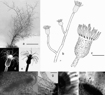
Polyp colonies 20–90 mm high, arborescent, monosiphonic, with erect and irregularly branched hydrocladia in several planes (Figure 1a). Perisarc delicate and thin, brownish to yellowish, often with 3–4 annulations at the origins of hydrocladia, sometimes 1–2 perisarc rings beneath the hydranth (Figure 1b). Annulations can be found also in large colonies along the main stem (Figure 1b) and throughout the whole length of hydrocladia. Colonies collected from the deepest levels of the species distribution (25–30 m depth) have few or no perisarc annulations along the main stem. Hydranths whitish to brown, 0.60–0.95 mm height, 0.30–0.55 mm width (at hydranth base), with 22–28 filiform tentacles arranged in one whorl at the base of a large, trumpet-shaped hypostome (Figure 1b–e). A ring of glandular cells is detectable at the base of large hydranths (Figure 1f), this region is without nematocysts.
Eudendrium moulouyensis: (a) colony; (b) drawing of terminal hydrocladia with three hydranths. Annulations occur at the base of each branching hydrocladium, sometimes also before the branching points; (c) drawing of gastrozooid polyp, showing the homogeneous dense distribution of large microbasic euryteles (capsule size not in scale) on the hydranth body, and on the hypostome, and the presence of concretions between tentacle bases, hydranth body, and perisarc; (d–e) hydranth in dark and white field, showing the amphicoronate pattern of tentacles, the flask-shaped hydranth body and the trumpet hypostome; (f) the basal glandular ring, deprived of large cnidocysts; (g–h) variable density of zooxanthellae in tentacles of winter (g) and summer (h) specimens; (i) unidentified intertentacular bodies, presumably protistan cases. Scale bars: (a) 2 cm; (c) 300 µm; (f, g) 50 µm; (h) 150 µm; (i) 100 µm.

Symbiotic intracellular zooxanthellae are present in the gastrodermis throughout the whole coenosarc, from the hydrorhiza and colony stem to hydranth body and tentacles, hypostome, and gonophores (Figure 1d–h). The density of zooxanthellae within tentacles sharply increases from winter to summer (Figure 1g–h). Most polyps from the Otranto Channel, as well as from Croatia, Sicily, Sardinia and the Gibraltar Strait possessed concretions of unknown nature, packed in the space between bases of each tentacle pair, or scattered along the hydranth body (Figure 1c, i) or on the perisarc of the stem. These bodies resemble large, spumous cells.
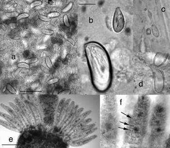
The cnidome was studied in living specimens in order to obtain discharged capsules and guarantee a secure identification of the cnidocyst types. One category of nematocysts—heterotrichous microbasic euryteles (Mariscal, Reference Mariscal, Muscatine and Lenhoff1974)—can be found in two sizes: numerous large microbasic euryteles (15–16 × 8–10 µm), bean-shaped, densely distributed on the whole hydranth body, loosely occurring also on hypostome (Figure 2a–c), and small microbasic euryteles (6–8 × 3–4 µm), oval, on tentacles and hydranth (Figure 2b, d). Both types can be also found scattered throughout the coenosarc in the stem and the hydrorhiza, sites of cnidoblast differentiation. As noted by Puce et al. (Reference Puce, Tazioli and Bavestrello2005), the appearance of the tentacles surface is spiny (Figure 2e). However, the arrangement of cnidocytes in the tentacles seems to be nearly regular, at least in the distal half of the tentacles, where the capsules are arranged in rings. This is especially visible in winter specimens, when algal density is low (Figure 2f).
Eudendrium moulouyensis cnidome: (a) enlargement of central part of hydranth body, filled by dense concentrations of cnidocysts (in the ectoderm) and zooxanthellae (in the endoderm); (b) two sizes of heterotrichous microbasic euryteles (undischarged capsules); (c) small heterotrichous microbasic euryteles, discharged; (d) large heterotrichous microbasic euryteles, discharged; (e) spiny aspect of tentacle surfaces, due to the angled disposition of cnidocysts on the tentacle ectoderm; (f) enlargement of distal tip of tentacles showing a nearly regular, ring-like arrangement (arrows) of the small microbasic euryteles. Scale bars: (a) 25 µm; (b) 5 µm; (e) 200 µm.

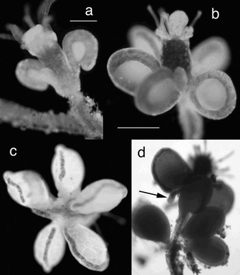
The species is gonochoristic. Female gonophores are oval, up to 5–6, born originally in a single whorl in the middle of a hydranth body (gonozooids) with a distinct hypostome but fewer and shorter tentacles than the gastrozooids. The gonophoral spadix is simple, thick and unbranched, entirely clasped around the egg until a late stage of differentiation (Figure 3a, b). Fertilization occurs in situ. During gonophore and embryonic development, the spadix gradually atrophies, but it does not disappear completely. The same progressive reduction can be observed for the tentacles. As observed in several other eudendriid species (including Myrionema amboinense), perhaps being a common feature of the whole family, embryos develop in capsules covered by thin perisarc, which remain attached to the blastostyle (Figure 3c) or, due to the concomitant growth and elongation of the blastostyle, it can get fused to the perisarc of the hydrocladium (pedicel), at a certain distance below the blastostyle (Figure 3d). Hydrocladia with developing embryos and no recognizable blastostyle remnant (as drawn in Marques et al., Reference Marques, Peña Cantero and Vervoort2000) represent the final stage of development of female gonozooids.
Eudendrium moulouyensis female gonophores and embryos: (a) early stage of development of female gonophores developing on the reproductive zooid with partly reduced tentacles. At this stage, the differentiating oocytes are not infected yet by zooxanthellae; (b) mature female gonophores with thick spadices on a reproductive zooid with reduced tentacles. Migration of zooxanthellae from the spadix endoderm into the egg cytoplasm is already visible; (c) proximal (basal) view of female gonophores bearing developing embryos. The spadices reduce progressively with embryo development; (d) blastostyle with remnants of tentacles, bearing six embryos. Three embryos detached from their spadix (arrow) and remained attached by a thin chitinous envelope to the hydrocladial perisarc at the base of the blastostyle. Scale bars: (a) 200 µm; (b) 300 µm.

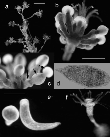
Male gonophores develop in high number (8–10 per gonozooid), usually with two spermatic chambers, sometimes three, on partially reduced zooids (Figure 4a–c). The larva directly inherits zooxanthellae from the maternal tissues. The symbionts are incorporated inside the egg cytoplasm after early differentiation of the female gonophore (Figure 3a, b), and they will be later incorporated in the tissues of the developing embryo (Figure 3c–d), the larva (Figure 4d, e) and the primary polyp (Figure 4f).
Eudendrium moulouyensis. (a) Male colony with reproductive polyps; (b) polyp with 9 male bi-chambered gonophores; (c) magnification of a three-chambered male gonophore; (d) planula larva with zooxanthellae mostly concentrated in the endoderm; (e) two settling planula larvae; (f) primary polyp. Scale bars: (a) 2 mm; (b) 400 µm; (e) 250 µm.

ECOLOGY
Seasonality, zonation and reproduction: Eudendrium mouloyensis was recorded throughout the whole year, with a marked preference for shallow sites characterized by sub-horizontal substrates, inhabited by the infralittoral biocenosis of photophilic algae or by a pre-coralligenous community. All colonies were recorded between 0.5 m to 30 m depth. The largest number and highest frequency of colonies of E. mouloyensis were found between −5 to −20 m depth. All colonies of E. moulouyensis were found epilithic or epibiontic of different organisms with variable calcium carbonate content, such as red coralline algae (Peyssonnelia spp.), green algae (Udothea sp. and Halimeda sp.) and calcified bryozoans, but also on artificial substrates such as fish lines, plastic pieces and iron.
Only unfertile specimens were observed from summer samples from the Alboran Sea (July 2007) whereas sexual reproduction in the Adriatic populations was recorded in winter–spring months, from December to May.
When released from the mother colony, planulae are usually covered by a thin mucous layer. The larvae crawl on the substrate, showing a positive phototropic behaviour, or swim close to the bottom in the search for stimuli for settlement and metamorphosis. In our laboratory rearing, larvae were able to settle and metamorphose in primary polyps only in presence of perisarc fragments from the parent or conspecific colonies.
During the winter months, the number of symbiotic algae, especially in the tentacles, was markedly reduced (Figure 1g, h). A low concentration of zooxanthellae can be detected also in specimens below 20 m depth.
Distribution: Eudendrium moulouyensis seems to be endemic to the Mediterranean, the geographical distribution being limited, according to present knowledge, to the western and central basin of the Mediterranean Sea, from the Alboran Sea (Chafarinas Islands and Andalucia coasts) (Marques et al., Reference Marques, Peña Cantero and Vervoort2000; present study), Western Sardinia (present study), Ligurian Sea (Portofino) (Puce, Reference Puce2003; Puce et al., Reference Puce, Tazioli and Bavestrello2005), Sicily Channel (present study) and Adriatic Sea (Otranto, Italy and Vis, Croatia; present study). It is worth noting, however, that Castric et al. (Reference Castric, Girard and Michel1991) included in their list of hydroids from Brittany a zooxanthellate Eudendrium hargitti colony, which could also belong to this species, based on identifications made by Picard.
DISCUSSION
The first record of Eudendrium moulouyensis dates back to 1991, when Alvaro Peña Cantero surveyed the summer hydrozoan fauna of the Chafarinas Islands (Alboran Sea, south-western Mediterranean Sea), collecting the type material for the description of this species (Marques et al., Reference Marques, Peña Cantero and Vervoort2000). The species diagnosis indicates the occurrence of symbiotic zooxanthellae, a character that was previously considered as an autapomorphy for the genus Myrionema (Marques, Reference Marques1996), but later recognized as inappropriate for generic distinction after the discovery of E. moulouyensis (Marques et al., Reference Marques, Peña Cantero and Vervoort2000). In their review of Mediterranean Eudendrium species, Marques et al. (Reference Marques, Peña Cantero and Vervoort2000) also considered Picard's (1950) identification of Myrionema specimens as doubtful (as Eudendrium hargitti = M. hargitti, considered a junior synonym of M. amboinense by Calder, Reference Calder1988). Indeed, Picard's identification (1950) of E. hargitti was based on unsound characters: ‘...caractérisée par le grand nombre (une trentaine) des tentacules ...et par l'hypostome très protactile et moins volumineux que chez les autres Eudendrium connus des cotes d'Europe’ (Picard, Reference Picard1950). The given tentacle number overlaps with that of E. mouloyensis, whereas size and extensibility of the hypostome have never been placed under a rigourous quantitative scrutiny to be used as valid diagnostic characters. Considering Picard's identification of E. hargitti (=Myrionema amboinense) not reliable, all records of E. hargitti/M. amboinense in the Mediterranean Sea should be regarded as doubtful and a re-examination of available material would be required. Unfortunately, specimens from Picard's collection are no more available (P. Schuchert, personal communication). The main distinction of the two genera of Eudendriidae, Eudendrium and Myrionema is currently based on the presence of a single whorl of tentacles in Eudendrium, and of two or more whorls in Myrionema. However, the high number of tentacles in E. moulouyensis, arranged in an amphicoronate fashion, may render the clear distinction of one or two whorls of tentacles difficult or impossible. The distinction between E. mouloyensis and M. amboinense is easy by the observation of the cnidome. Millard & Bouillon (Reference Millard and Bouillon1973) and Calder (Reference Calder1988) described two types of cnidocysts from M. amboinense: small microbasic euryteles in tentacles, hydranth, and elsewhere; and large macrobasic euryteles (length: 22–23 µm) on hydranth base and hypostome, whereas E. moulouyensis has only microbasic euryteles (Figure 2a–d). Moreover, a different number of male gonophores per gonozooids can be recorded in the two species, as follows:
— reproductive polyps with (partly reduced) tentacles and hypostome, bearing up to 9–10 gonophores (with 2–3 sperm chambers)…………… E. moulouyensis;
— reproductive polyps with (partly reduced) tentacles and hypostome, bearing up to 2–3 (rarely 4) gonophores (with 3–4 sperm chambers)………… M. amboinense.
Some authors consider the number of gonophores in Eudendriidae as a variable character and therefore not useful for genus or species identification. The gonophore number, in fact, may increase in time during gonozooid maturation. However, descriptions of M. amboinense never exceed 4 male gonophores per gonozooid, whereas the finding of 10 male gonophores per gonozooid in E. moulouyensis is a common feature in every fertile colony.
The search for diagnostic characters within the genus Eudendrium led Puce et al. (Reference Puce, Tazioli and Bavestrello2005) to suggest that the arrangement of the small microbasic euryteles on tentacles may have species-specific diagnostic value among seven Eudendrium species (Puce et al., Reference Puce, Tazioli and Bavestrello2005). However, the cnidocyst arrangements on the tentacles of specimens of E. moulouyensis from the south Adriatic and southern Spain was more regular than in specimens collected in the Tyrrhenian Sea (Puce et al., Reference Puce, Tazioli and Bavestrello2005), even if the same ‘spiny’ aspect of tentacles was recorded. The available data, however, seem too scant to establish tentacle cnidocyst arrangement as a reliable identification tool for the whole genus. Further investigations on the cnidocyst arrangements in a larger set of samples are needed. The new analysis must include representatives of different populations and a larger number of species to make clear: (a) whether observed arrangements retain a species-specific discriminatory value; and (b) whether phenotypical variations do not compromise the usefulness of this character. A disadvantage of this character is also that it can only be reliably observed in living material, and not for preserved samples.
The nature of the concretions observed on specimens collected from different sites in the Mediterranean Sea (Otranto and Croatia, Adriatic Sea; Sicily Channel; Western Sardinia; Gibraltar Strait) remains to be elucidated. Specimens from all these different sites showed the same type of concretions (Figure 1c, i). They could be outer cases of protists colonizing the colonies at different positions. Their finding in colonies sampled from different locations of the Mediterranean Sea indicates a possible species-specific association with wide distribution. Their preferential location (at the base of tentacles) might be related to microscale hydrodynamic forces, conveying feeding currents in between the hydroid tentacles. The finding of a scattered distribution of such concretions on the colony perisarc demonstrates that they are not dependent on direct contact with hydroid cells. The morphology and origin of these epibiotic structures are currently under investigation by means of scanning electron microscopy.
Year-round observations demonstrated that E. moulouyensis represents a constant component of the hard-bottom sessile zoobenthos of the south Adriatic Sea. Eudendrium moulouyensis is not characterized by a marked seasonality, as colonies are present either in the South Adriatic or in the Alboran Sea throughout the year, ranging from 0.5 to 30 m depth. It will be worth investigating if the increase of records of this zooxanthellate species might be a biological marker of the climatic changes occurring in the Mediterranean Sea. In fact, associations between marine invertebrates and endosymbiotic dinoflagellates (zooxanthellae) are abundant in nutrient-poor tropical seas, where zooxanthellae supply photosynthetically fixed carbon to their hosts and facilitate the conservation and recycling of essential nutrients (Muscatine, Reference Muscatine and Dubinsky1990; Davies, Reference Davies, John, Hawkins and Price1992). Associations between invertebrates and zooxanthellae are less common at temperate latitudes (Turner, Reference Turner1988; Davy et al., Reference Davy, Lucas and Turner1996; Müller-Parker & Davy, Reference Muller-Parker and Davy2001). Nevertheless, also in temperate waters abundances of symbiotic algae within animal tissues are controlled by temperature and irradiance (Saunders & Müller-Parker, Reference Saunders and Müller-Parker1997), as reflected also by the different algal densities found in winter and summer specimens of E. moulouyensis. Eudendrium moulouyensis shows a wide distribution in the Mediterranean Sea, from the Alboran and Ligurian Seas to the Adriatic Sea. The presence of zooxanthellae may represent an advantage for the hydroid providing a higher competitive ability and favouring its wide diffusion.
Acquisition of zooxanthellae in tropical coral symbioses is mainly from the surrounding waters rather than from maternal inheritance, whereas transmission patterns in temperate symbioses have been rarely identified (Davy & Turner, Reference Davy and Turner2003). In hydrozoans, both zooxanthellate eudendriids (E. moulouyensis and M. amboinense) acquire their symbionts by vertical transmission from the mother colony. However, in Myrionema, the algae were shown to enter the oocytes at a very early stage, during their migration from the stalk ectoderm to the endoderm, and from the polyp endoderm to the ectoderm of the gonophore bud (Fraser Reference Fraser1931). From our observations (Figure 3a, b), eggs in early developing gonophores of E. moulouyensis are still deprived of algae, entering the egg cytoplasm only later by migration from the spadix gastrodermis. Investigations on symbioses between zooxanthellae and hydroids are still scant, and many open questions remain unsolved, especially related to the diversity and life cycle of zooxanthellae.
The repeated findings of large and fertile colonies of E. moulouyensis allowed a refinement of the description of this species, which is now becoming one of the most common components of the shallow-water hydroid assemblages in the Mediterranean Sea. With this work, the full life cycle of E. moulouyensis has been described for the first time and the composition of cnidome has been properly elucidated by observation of discharged cnidocysts. The Mediterranean Eudendriidae most likely do not comprise the genus Myrionema. It seems likely that Picard's records of Myrionema (in Marinopoulous, Reference Marinopoulos1992) were in fact based on E. moulouyensis. However, we urgently need a new revision of the genus Eudendrium that also uses DNA sequence information in order to assess the validity of our currently used nominal species. With their limited dispersal abilities, it seems likely that the speciation rate in this family is high, much higher than can be detected by the limited available character set.
ACKNOWLEDGEMENTS
The authors are grateful to Dr P. Schuchert (Geneva) and Dr S. Puce (Ancona) for their critical reading and valuable comments on the original manuscript. Financial supports by MURST (60%, COFIN and FIRB Projects), the Administration of the Province of Lecce, ICRAM (Project ‘Cnidarian exotic species in the Mediterranean Sea’), the European Commission (Marie Curie contract No. HPMD-CT-2001-00099, IASON, SESAME and the MARBEF network, the SESAME project–EC Contract No. GOCE-036949, funded by the Sixth Framework Programme under the priority ‘Sustainable Development, Global Change and Ecosystems’), the NSF of the USA (PEET project on the Hydrozoa), the Centro EuroMediterraneo per i cambiamenti climatici.